
from Guyton and Hall textbook of medical physiology, 4th South Asia edition, write a 5-mark short note on Neuron-diagram, Nerve Impulse-define.
neuron diagram dendrites axon cell body soma synapse Guyton physiology
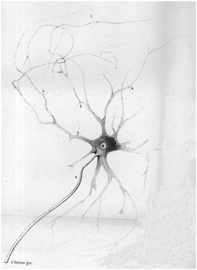
This historical anatomical diagram, originally published by Otto Deiters in 1865, provides a foundational illustration of a multipolar neuron. The illustration depicts the fundamental components of a nerve cell with significant morphological detail. Centrally located is the soma (cell body), which contains a distinct, circular nucleus. Two primary types of cytoplasmic processes extend from the soma: (a) a single, long, relatively smooth and thicker projection representing the axon, and (b) multiple, highly branched, and tapered projections representing dendrites. The dendrites exhibit characteristic arborization, labeled with '1' and 'b' to highlight their complex, radiating structure. This diagram is a seminal piece of neuroanatomical history, illustrating the early conceptualization of the neuron as the functional unit of the nervous system. It serves as an educational resource for understanding basic cellular neurobiology, demonstrating the structural polarity of neurons and the distinction between information-receiving dendrites and information-sending axons.
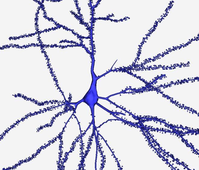
This anatomical diagram presents a high-resolution 3D digital reconstruction of a human cortical pyramidal neuron, likely based on data from the cingulate cortex. The visualization highlights the primary structural components of a neuron: the central soma (cell body) and the extensive dendritic arbor. The soma is depicted as a smooth, pear-shaped central volume. Radiating from the soma are several primary dendrites that taper as they extend and undergo multiple bifurcations to form a complex, tree-like network. A key educational feature is the detailed rendering of dendritic spines—the small, mushroom-like or stalk-like protrusions densely distributed along the length of the dendritic shafts. These spines represent the post-synaptic sites of excitatory synapses. The model demonstrates high morphological fidelity, showcasing the smooth integration between the soma and the first-order dendrites. This type of 3D modeling is essential in neurobiology for studying neuronal connectivity, surface area calculations, and the structural plasticity of synaptic components in the human brain.
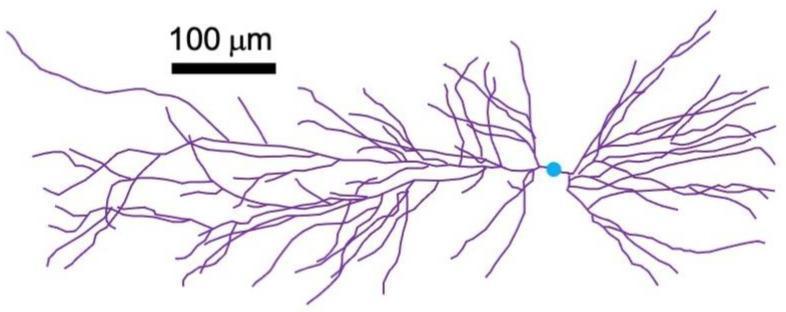
This anatomical diagram illustrates a morphological reconstruction of a hippocampal CA1 pyramidal neuron. The visualization distinguishes key cellular components using color-coding: the central soma (cell body) is represented as a small blue circle, while the extensive dendritic arborization is depicted in purple. The dendrites exhibit a complex, asymmetrical branching pattern, radiating horizontally and vertically from the cell body with a higher density of branching on one side, characteristic of pyramidal cell architecture. A scale bar indicating 100 micrometers (µm) is provided, demonstrating that the total span of the dendritic tree extends several hundred micrometers. This image serves as a structural model for neurobiological studies, specifically regarding neuronal geometry, synaptic integration, and the biophysical properties of hippocampal circuits in the brain. The precise mapping of these structures is essential for understanding the spatial distribution of postsynaptic potentials and their contribution to neural magnetic fields.
"Figure 46.1" in /textbooks/9780443111013
- The neuron diagram image URL:
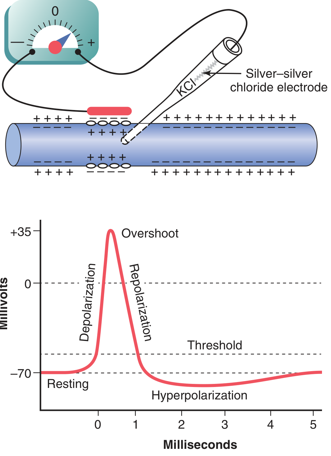
https://cdn.orris.care/cdss_images/7cfc8b27f693760bcfb269232679dcc3d78869e98afdc00e763ce9d6e6e50644.png - The action potential image URL:
https://cdn.orris.care/cdss_images/d94d1eff4e1e7deebae6a16401025c4414dfcf32575f90b2e8587903be7295e4.png - All content on neuron structure and nerve impulse (action potential) from Guyton and Hall.
Short Note: Neuron (Diagram) and Nerve Impulse (Definition)
THE NEURON
Structure of a Typical Neuron
| Part | Description |
|---|---|
| Cell Body (Soma) | Contains the nucleus and organelles; the metabolic center of the neuron. Resting membrane potential is approximately -65 mV. |
| Dendrites | Multiple, branching processes that receive incoming signals. Most synaptic inputs arrive here (up to 200,000 synaptic connections in some neurons). |
| Axon | A single output process that conducts the signal away from the cell body. It may branch to reach multiple targets in the nervous system or peripheral body. |
| Axon Hillock | The initial segment of the axon; the lowest threshold point where action potentials are generated. |
| Synapses | Functional junctions on the dendrites and cell body, through which signals pass only in the forward direction (from axon of one neuron to dendrites/cell body of the next). |
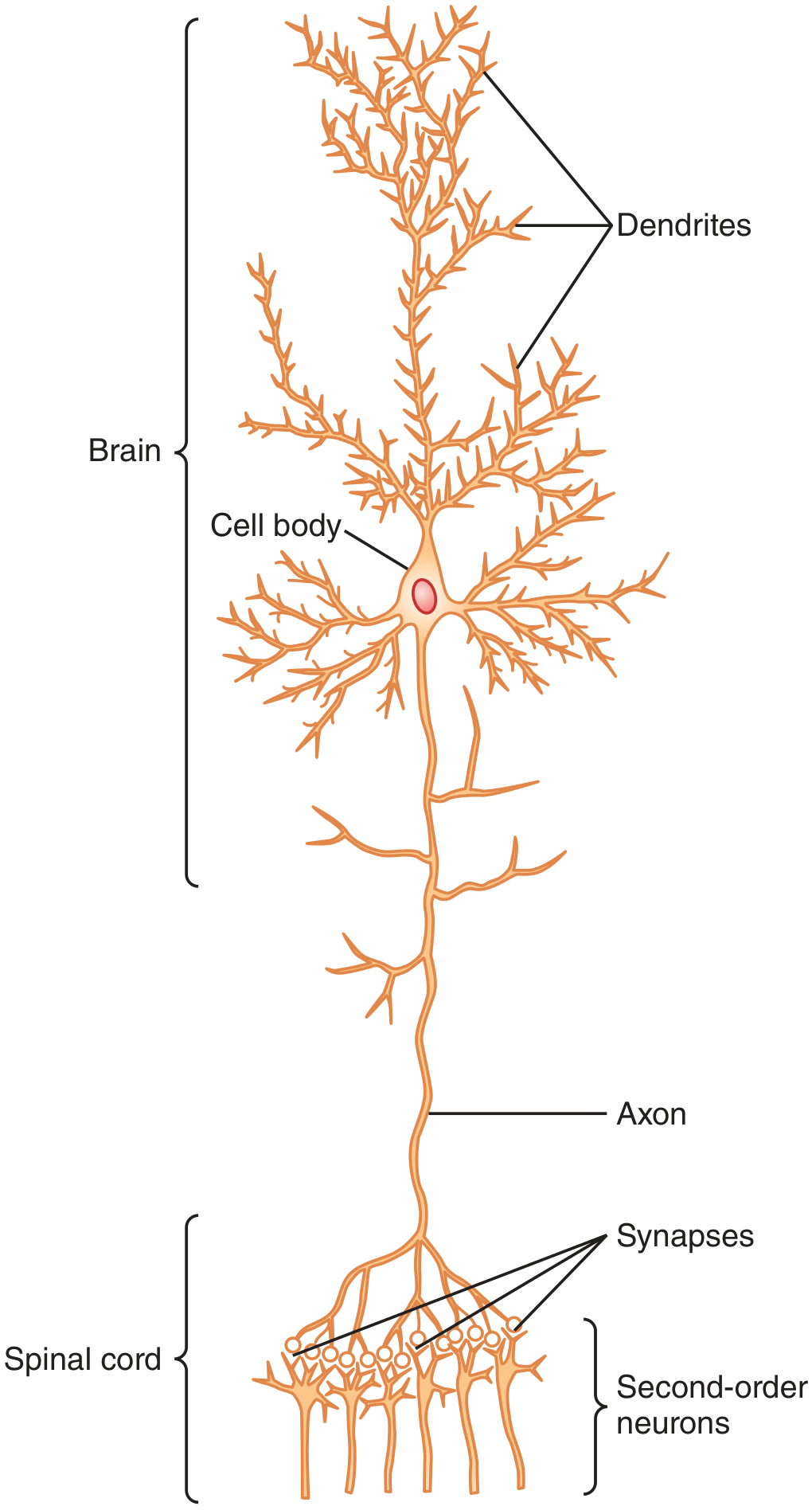
- Guyton and Hall Textbook of Medical Physiology, Ch. 46
NERVE IMPULSE (ACTION POTENTIAL) - DEFINITION
"Nerve signals are transmitted by action potentials, which are rapid changes in the membrane potential that spread rapidly along the nerve fiber membrane."
- Guyton and Hall, Ch. 5
Stages of the Nerve Impulse (Action Potential)
-
Resting Stage
- Membrane potential: -70 mV (polarized)
- Sodium channels closed; membrane at rest.
-
Depolarization Stage
- A stimulus depolarizes the membrane to the threshold level (~-55 mV).
- Voltage-gated Na⁺ channels open suddenly → massive Na⁺ influx → membrane potential rises rapidly toward +35 mV (overshoot).
- This process is called depolarization.
-
Repolarization Stage
- Na⁺ channels begin to close (inactivation gate closes).
- Voltage-gated K⁺ channels open → rapid K⁺ efflux restores the negative resting potential.
- Membrane potential returns to -70 mV (repolarization).
- Transient hyperpolarization (undershoot) may occur if K⁺ channels remain open briefly longer than needed.
Key Ion Channels Involved
- Voltage-gated Na⁺ channel: Has two gates - an activation gate (opens rapidly on depolarization) and an inactivation gate (closes a few ten-thousandths of a second later, terminating Na⁺ influx).
- Voltage-gated K⁺ channel: Opens during repolarization (delayed compared to Na⁺ activation), rapidly restoring the negative membrane potential.
Propagation
Summary Table
| Feature | Detail |
|---|---|
| Resting membrane potential | -70 mV (nerve fiber) / -65 mV (neuronal soma) |
| Threshold potential | ~-55 mV |
| Peak of action potential | ~+35 mV |
| Duration | ~1 millisecond |
| Ion for depolarization | Na⁺ (influx) |
| Ion for repolarization | K⁺ (efflux) |
| Nature of impulse | All-or-none, self-propagating |
- Guyton and Hall Textbook of Medical Physiology, Ch. 5 (Membrane Potentials and Action Potentials) and Ch. 46 (Organization of the Nervous System, Basic Functions of Synapses, and Neurotransmitters)