
Medicine postings- abdomen case explanation 1. Proforma with significance and definition Also add images
Reading File
Now let me search the medical library for abdominal examination proforma content in parallel:
Finding Sources
Finding Sources
Searching the Web
medicine posting abdomen case proforma clinical examination significance MBBS
Now let me read the most relevant sources - S Das Clinical Surgery (best for surgical/clinical proforma) and Goldman-Cecil Medicine for internal medicine perspective, in parallel:
Reading File
Reading File
Finding Sources
Excellent content retrieved. Let me now get more detail from S Das on palpation/percussion/auscultation plus look for relevant images:
Reading File
Finding Sources
Finding Sources
Reading File
Finding Sources
Finding Sources
Now let me fetch the clinical images found in the textbooks:
Loading Image
Finding Sources
Reading File
I now have comprehensive content to build a detailed medicine posting abdomen case proforma. Let me compile the full response:
Medicine Posting - Abdomen Case Proforma
Complete Clinical Proforma with Significance, Definitions & Images
PART I: HISTORY TAKING
A. Biodata
| Item | Details |
|---|---|
| Name | |
| Age / Sex | |
| Occupation | |
| Address | |
| Date of Admission | |
| Informant |
Significance: Occupation can point to hepatic (alcohol-related), infectious, or malignant causes. Age and sex influence disease prevalence (e.g., peptic ulcer commoner in males; cholelithiasis in females).
B. Chief Complaints
List in chronological order with duration.
- e.g., Pain abdomen - 2 weeks
- Abdominal distension - 1 week
- Jaundice - 3 days
C. History of Presenting Illness (HOPI)
For each complaint, ask: SOCRATES - Site, Onset, Character, Radiation, Associations, Timing, Exacerbating/Relieving factors, Severity.
Key GI symptoms to elaborate:
| Symptom | Key Enquiries | Significance |
|---|---|---|
| Pain abdomen | Site, type (colicky/constant), radiation, relation to food, relieving factors | Colicky = hollow viscus; constant = solid organ or peritoneal irritation |
| Vomiting | Timing, contents (blood, bile, food), forceful or effortless | Haematemesis = upper GI bleed; bilious = intestinal obstruction |
| Jaundice | Colour of urine/stool, pruritus, fever | Pre-hepatic/hepatic/post-hepatic |
| Distension | Onset, gradual or sudden, associated passage of flatus/stool | 5 Fs: Fat, Fluid (ascites), Flatus, Faeces, Foetus |
| Dysphagia | Progressive, solid vs liquid | Carcinoma oesophagus; achalasia |
| Altered bowel habits | Constipation, diarrhoea, alternating | Colorectal carcinoma, IBS, IBD |
| Haematochezia / Melena | Colour, quantity | Upper vs lower GI bleed |
| Appetite/Weight loss | Amount, duration | Malignancy, malabsorption, chronic liver disease |
D. Past History
- Similar complaints
- Previous surgeries (note scar type)
- Peptic ulcer disease, tuberculosis, hepatitis, diabetes, hypertension
- Blood transfusions (hepatitis B/C risk)
Significance: Prior abdominal surgeries cause adhesions - a leading cause of intestinal obstruction.
E. Personal History
- Diet (vegetarian/non-vegetarian)
- Alcohol intake - quantity, duration (cirrhosis, pancreatitis)
- Smoking (peptic ulcer, malignancy)
- Drug history - NSAIDs (GI bleed), steroids
- Travel history (amoebiasis, hydatid disease)
F. Family History
- Malignancy, inflammatory bowel disease, polyposis syndromes
G. Menstrual / Obstetric History (females)
- LMP, parity - to rule out ectopic pregnancy, ovarian pathology
PART II: GENERAL PHYSICAL EXAMINATION
Patient Position: Supine, arms at sides, head on one pillow.
"The nonabdominal examination should assess nutritional status and any signs of systemic conditions that may cause GI symptoms." - Goldman-Cecil Medicine
| Sign | Method | Significance |
|---|---|---|
| Build & Nutrition | Visual survey | Cachexia suggests malignancy or chronic disease |
| Pallor | Conjunctiva, palms | Anaemia - GI bleed, malabsorption, malignancy |
| Jaundice | Sclera (best site), skin | Pre/hepatic/post-hepatic; visible when bilirubin >2 mg/dL |
| Clubbing | Distal finger phalanges | IBD, cirrhosis, malabsorption |
| Cyanosis | Lips, tongue (central), fingertips | Hepatopulmonary syndrome |
| Oedema | Ankle, sacral | Hypoalbuminaemia (cirrhosis, nephrotic syndrome), CCF |
| Lymphadenopathy | All nodal groups especially Virchow's node (left supraclavicular) | Virchow's node = gastric carcinoma (Troisier's sign) |
| Spider naevi | Upper body, face, arms | Chronic liver disease (>5 pathological) |
| Palmar erythema | Hypothenar eminence | Chronic liver disease |
| Leuconychia | White nails | Hypoalbuminaemia (cirrhosis) |
| Dupuytren's contracture | Palmar fascia | Alcoholic liver disease |
| Gynaecomastia | Breast tissue in males | Cirrhosis (excess oestrogen) |
| Caput medusae | Dilated veins around umbilicus | Portal hypertension |
| Flapping tremor (asterixis) | Wrist extension held 15 sec | Hepatic encephalopathy |
| Pulse, BP, Temperature | Routine vitals | Fever >38.5°C = infection/peritonitis; tachycardia = sepsis/haemorrhage |
PART III: SYSTEMIC / LOCAL EXAMINATION OF THE ABDOMEN
Definition of Abdomen: The cavity bounded above by the diaphragm, below by the pelvic inlet (and pelvic floor functionally), anterolaterally by the abdominal wall muscles, and posteriorly by the vertebral column and posterior abdominal wall muscles. For clinical purposes, it extends from the level of the nipples above to the saphenous openings below.
Surface Divisions
9-Region Division (preferred for localizing pathology):
| Right Hypochondrium | Epigastrium    | Left Hypochondrium |
|---------------------|----------------|--------------------|
| Right Lumbar        | Umbilical      | Left Lumbar        |
| Right Iliac Fossa   | Hypogastrium   | Left Iliac Fossa   |
4-Quadrant Division: Right Upper, Left Upper, Right Lower, Left Lower - divided by lines through umbilicus.
STEP 1: INSPECTION
"It cannot be overemphasised how rewarding it is to spend time on inspection. Examination should be carried out in good light (preferably daylight) looking first from the side, then tangentially and finally from either end of the bed." - S. Das, Manual on Clinical Surgery
1. Skin and Subcutaneous Tissue
| Finding | Significance |
|---|---|
| Erythema over abdomen | Hot-water bottle application - indicates site and chronicity of pain |
| Surgical scars - linear | Healed by first intention (no wound infection) |
| Surgical scars - broad, irregular | Wound infection in previous surgery |
| Hard subcutaneous nodules at umbilicus | Sister Mary Joseph's nodule - intra-abdominal malignancy (especially gastric carcinoma) |
| Striae (silvery-white) | Previous stretching - pregnancy, obesity, ascites |
| Striae (purple) | Cushing's syndrome |
| Hernia bulge | Umbilical, inguinal, incisional hernias |
2. Superficial Veins (Caput Medusae)
Method to determine direction of flow: Place two index fingers on the vein, milk a segment empty, release one finger - note direction of refilling.
| Pattern | Direction of Flow | Significance |
|---|---|---|
| Periumbilical (caput medusae) | Away from umbilicus (radially) | Portal hypertension |
| Lateral abdominal wall veins | From below upwards | Inferior vena cava obstruction |
3. Umbilicus
| Finding | Significance |
|---|---|
| Displaced upwards | Pelvic mass (ovarian cyst, fibroid) |
| Displaced downwards | Tanyol's sign - ascites |
| Everted | Ascites, large umbilical hernia |
| Inverted/deeply tucked | Obesity |
| Pushed to opposite side | Unilateral abdominal swelling |
4. Contour of Abdomen
| Finding | Significance |
|---|---|
| Scaphoid/retracted | Starvation, dehydration, peritonitis |
| Symmetrical distension | 5 Fs - Fat, Fluid, Flatus, Faeces, Foetus |
| Fullness of right iliac fossa | Chronic intestinal obstruction (distended caecum - "better seen than felt") |
| Upper abdominal distension | Gastric, pancreatic, hepatic enlargement |
| Lower abdominal distension | Bladder, uterine, ovarian masses |
| Visible peristalsis | Ladder pattern in intestinal obstruction; seen in thin patients |
Obesity vs Ascites on inspection: In obesity, umbilicus is deeply inverted; in ascites, umbilicus shows varying degrees of eversion. - S. Das
5. Respiratory Movement
- Normally abdominal movement is seen with respiration
- Absent respiratory movement = peritonitis (board-like rigidity, patient "splints" the abdomen)
STEP 2: AUSCULTATION
Perform BEFORE palpation to avoid altering bowel sounds artificially.
| Finding | Characteristics | Significance |
|---|---|---|
| Normal bowel sounds | Intermittent, soft gurgling every 5-10 sec | Normal gut motility |
| Absent/silent bowel sounds | No sound after listening 2 minutes | Paralytic ileus, peritonitis |
| High-pitched tinkling sounds | Tinkling or metallic rushes | Early intestinal obstruction |
| Hyperactive sounds (borborygmi) | Loud, prolonged rumbles | Gastroenteritis, early obstruction |
| Succussion splash | Splashing sound on shaking abdomen | Gastric outlet obstruction (>4 hrs after meal) |
| Hepatic bruit | Systolic bruit over liver | Hepatocellular carcinoma, alcoholic hepatitis |
| Renal bruit | Over renal artery | Renal artery stenosis (hypertension) |
STEP 3: PERCUSSION
General Percussion
| Finding | Significance |
|---|---|
| Tympanic (resonant) | Gas-filled bowel - normal; excessive = obstruction/ileus |
| Dull | Solid organ, fluid, mass |
| Obliteration of liver dullness | Perforation of hollow viscus (free gas under diaphragm) |
Liver Percussion
- Upper border: percuss down from 2nd intercostal space in right midclavicular line - normal at 5th intercostal space
- Lower border: percuss upward from right iliac fossa - normally at right costal margin
- Normal liver span: 6-12 cm in midclavicular line
Splenic Percussion
- Traube's space (left 6th-10th ribs, midaxillary line): normally tympanic (due to stomach)
- Dullness in Traube's space = splenomegaly
- Nixon's method: percuss from left midaxillary line downward
- Dullness >8 cm = splenomegaly
Ascites Detection
Shifting Dullness:
- Definition: The demonstration of peritoneal fluid by percussing the flanks with the patient supine (dull), then rolling the patient to one side - the dullness shifts to the dependent flank.
- Method: Percuss from umbilicus toward flank until dullness is detected. Keep finger at that point. Ask patient to roll toward you - reassess in 30 seconds. If note changes from dull to resonant = shifting dullness positive.
- Significance: Positive when >500 mL of free fluid in peritoneal cavity. Distinguishes ascites (shifts) from ovarian cyst (central dullness, does not shift).
Fluid Thrill (Fluid Wave):
- Method: Ask assistant to place edge of hand firmly at midline. Examiner places one hand on one flank and flicks/taps the other flank - an impulse conducted across fluid is felt.
- Significance: Positive only with large quantities of ascites (>1.5 L). Less reliable than shifting dullness; false positive in obese patients (the assistant's hand prevents fat transmission).
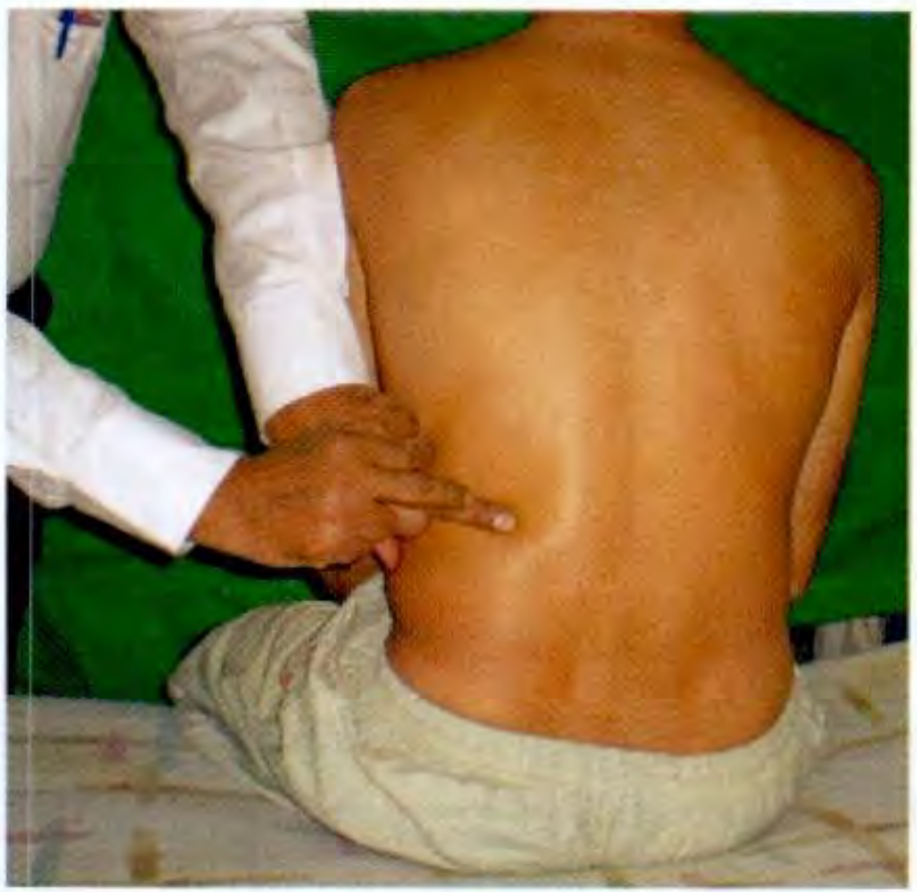
Renal vs Splenic Swelling - Percussion Differentiation
| Feature | Renal Swelling | Splenic Swelling |
|---|---|---|
| Loin percussion (outside erector spinae) | Dull (colon displaced) | Resonant (colon in normal position) |
| Overlying bowel | Resonant unless very large | Dull (no bowel overlying spleen) |
STEP 4: PALPATION
Preliminary
- Warm hands, short nails, patient relaxed with knees slightly flexed
- Begin away from the site of pain
- Watch patient's face throughout for signs of pain
Light Palpation (Superficial)
- Purpose: detect tenderness, guarding, superficial masses
| Finding | Definition | Significance |
|---|---|---|
| Tenderness | Pain on palpation | Localised = specific organ pathology |
| Guarding (voluntary) | Patient tenses muscles when examiner approaches | Anticipatory, conscious |
| Rigidity (involuntary guarding) | Persistent board-like hardness regardless of distraction | Peritoneal irritation/peritonitis |
| Rebound tenderness (Blumberg's sign) | Pain worse on sudden release of pressure than on pressing | Peritoneal inflammation (appendicitis, peritonitis) |
| Rovsing's sign | Palpation of left iliac fossa causes pain in right iliac fossa | Appendicitis |
| Murphy's sign | Inspiratory arrest on deep palpation at right costal margin | Acute cholecystitis |
Deep Palpation - Organ Assessment
Liver Palpation:
- Start from right iliac fossa, use radial border of index finger or flat of hand, press during expiration, feel during inspiration
- A palpable liver edge is present in: hepatomegaly, Riedel's lobe (normal variant), pushed down by emphysema
| Liver Characteristic | Significance |
|---|---|
| Smooth, tender enlargement | Hepatitis, congestive cardiac failure, early cirrhosis |
| Smooth, non-tender enlargement | Fatty liver, infiltrative disease |
| Nodular, hard | Cirrhosis (irregular), secondary metastases (hard nodules) |
| Pulsatile liver | Tricuspid regurgitation |
| Hepatic bruit on auscultation | Hepatocellular carcinoma |
Spleen Palpation:
- Start from right iliac fossa, move toward left costal margin
- Cannot palpate between spleen and costal margin (no notch palpable in early enlargement)
- Ask patient to roll to right lateral position if not felt in supine
Causes of splenomegaly - graded:
- Mild (<3 cm below costal margin): Typhoid, SBE, hepatitis, early portal hypertension
- Moderate (3-8 cm): Malaria, haemolytic anaemia, lymphoma
- Massive (>8 cm / to umbilicus or beyond): Kala-azar, CML, myelofibrosis, Gaucher's disease
Kidney Palpation (Bimanual Ballottement):
- One hand behind flank, other on anterior abdominal wall
- Ballot (flick) from behind - kidney is felt anteriorly
- Ballottement = kidney is felt returning to posterior hand after being displaced
- Normal kidneys usually not palpable (except in thin adults, right kidney slightly lower)
| Feature | Renal Swelling | Splenic Swelling |
|---|---|---|
| Bimanual ballottement | Positive | Negative |
| Can get above swelling | Cannot (retroperitoneal) | Can get above spleen |
| Notch | Absent | Present on medial border (if enlarged) |
| Movement with respiration | Downward | Downward + medially |
| Percussion | Resonant (bowel overlies) | Dull |
Gallbladder Palpation:
- Felt below tip of 9th costal cartilage in right midclavicular line (Courvoisier's sign area)
- Courvoisier's Law: A palpable, non-tender gallbladder in the presence of jaundice is unlikely to be due to gallstones (stones cause fibrosis and non-distensible gallbladder); suggests malignant obstruction of the common bile duct (pancreatic head carcinoma)
Aortic Pulsation:
- Normally palpable in thin patients midline above umbilicus
- Expansile pulsation = Aortic aneurysm (pulsates outward in all directions)
- Transmitted pulsation = Overlying mass transmits pulsation from aorta
PART IV: SPECIAL / FOCUSED EXAMINATIONS
| Examination | When to Do | What You Find |
|---|---|---|
| Hernial orifices | All abdominal cases | Irreducible hernia, strangulation |
| External genitalia | Male patients with groin/scrotal swelling | Hydrocele, varicocele, testicular mass |
| Per Rectal (PR) examination | Anorectal complaints, pelvic mass, rectal bleeding | Rectal carcinoma, faecal impaction, pelvic abscess |
| Per Vaginal (PV) examination | Pelvic pain, mass in females | Ovarian pathology, PID |
| Flank / Loin | Renal cases | Renal angle tenderness = pyelonephritis |
PART V: PROVISIONAL DIAGNOSIS AND DIFFERENTIAL DIAGNOSIS
Based on:
- History + HOPI
- General examination findings
- Systematic abdominal examination
Write: "This is a case of _________ most likely due to _________ based on ..."
PART VI: INVESTIGATIONS REQUIRED
| Level | Tests |
|---|---|
| Routine | CBC, ESR, Blood group, Urine R/E, RFT |
| Liver | LFT (bilirubin, AST, ALT, ALP, GGT, proteins, PT/INR) |
| Infection | Blood culture, Widal, malaria smear, HBsAg, anti-HCV |
| Imaging | USG abdomen (first-line), X-ray abdomen (erect + supine for obstruction/perforation) |
| Advanced | CT abdomen (CECT), MRI, MRCP for biliary disease |
| Ascitic tap | Cell count, protein (transudate vs exudate), SAAG, AFB, cytology |
| Endoscopy | OGD, colonoscopy for mucosal lesions |
SAAG (Serum-Ascites Albumin Gradient):
- SAAG ≥1.1 g/dL = Portal hypertension (cirrhosis, CCF, Budd-Chiari)
- SAAG <1.1 g/dL = Non-portal (TB peritonitis, malignancy, nephrotic syndrome)
QUICK REFERENCE: KEY CLINICAL SIGNS SUMMARY
| Sign | Definition | Disease |
|---|---|---|
| Murphy's sign | Inspiratory arrest on palpation of right hypochondrium | Acute cholecystitis |
| Courvoisier's sign | Palpable non-tender gallbladder + jaundice | Pancreatic/CBD carcinoma |
| Sister Mary Joseph's nodule | Hard subcutaneous umbilical nodule | Intra-abdominal malignancy |
| Virchow's (Troisier's) node | Left supraclavicular lymphadenopathy | Gastric carcinoma |
| Cullen's sign | Periumbilical bruising | Acute haemorrhagic pancreatitis |
| Grey Turner's sign | Flank bruising | Acute haemorrhagic pancreatitis |
| Rovsing's sign | LIF palpation causes RIF pain | Appendicitis |
| Blumberg's sign | Rebound tenderness | Peritonitis |
| Tanyol's sign | Umbilical displacement downward | Ascites |
| Shifting dullness | Dependent dullness shifting on rolling | Ascites (>500 mL) |
| Fluid thrill | Wave transmitted across peritoneal fluid | Massive ascites |
| Succussion splash | Splashing on shaking | Gastric outlet obstruction |
| Caput medusae | Periumbilical dilated veins, flow away from umbilicus | Portal hypertension |
| Hydatid thrill | Three-finger percussion thrill | Hydatid cyst of liver |
Sources: S. Das - A Manual on Clinical Surgery, 13th Ed. | Goldman-Cecil Medicine | Bailey & Love's Short Practice of Surgery, 28th Ed. | Yamada's Textbook of Gastroenterology, 7th Ed.
This is a shared conversation. Sign in to Orris to start your own chat.