
Functional types of blood vessels for mbbs physiology To get full marks in physiology practical exam.
Reading File
Now let me search the medical library for functional types of blood vessels.
Finding Sources
Finding Sources
Finding Sources
Finding Sources
Found it in Costanzo Physiology. Let me read those sections now.
Reading File
Reading File
Reading File
Reading File
Reading File
Loading Image
Now let me also get the cross-sectional area/blood volume figure and fetch one more supporting image:
Loading Image
Loading Image
Excellent! I now have everything needed for a thorough, exam-ready answer. Here it is:
Functional Types of Blood Vessels - MBBS Physiology
Source: Costanzo Physiology 7th Ed. | THIEME Atlas of General Anatomy
Overview
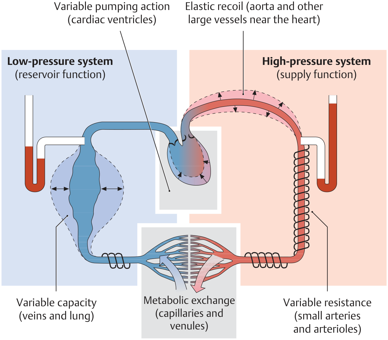
Blood is transported through a pressure gradient: ~100 mmHg in the arterial (high-pressure) system down to <20 mmHg in the venous (low-pressure) system. Based on their structure and function, blood vessels are classified into 5 functional types.
The 5 Functional Types
1. Elastic Arteries (Conducting / Windkessel Vessels)
- Examples: Aorta and large arteries near the heart
- Wall: Rich in elastic tissue, thick-walled
- Function: Receive blood ejected during systole and expand to store it. During diastole, they recoil elastically to maintain continuous blood flow (Windkessel effect - converting pulsatile cardiac output into steady flow)
- Pressure: Highest in the vasculature (systolic ~120 mmHg)
- Blood volume contained: ~13% of total
- Key exam point: Called "pressure reservoirs" or "conducting vessels"
2. Muscular Arteries (Distributing Vessels)
- Examples: Medium and small arteries (femoral, radial, etc.)
- Wall: Predominantly smooth muscle in the tunica media; thicker walls relative to lumen
- Function: Distribute blood to specific organs. Can constrict/dilate to redirect flow
- Key exam point: Called "distributing vessels" - they determine which organ gets how much blood
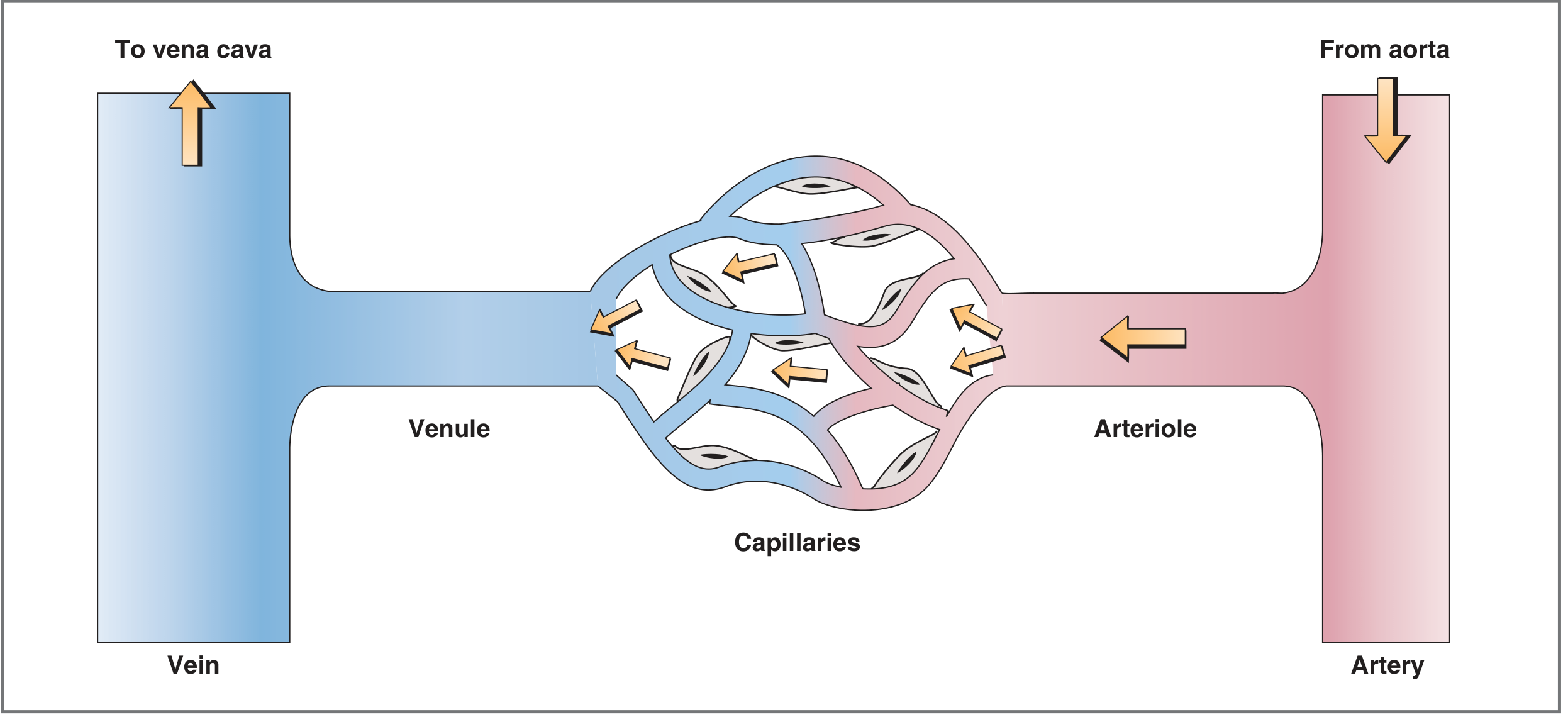
3. Arterioles (Resistance Vessels) - MOST IMPORTANT
- Examples: Terminal arterioles, precapillary arterioles
- Wall: Extensive smooth muscle; heavily innervated by sympathetic adrenergic fibers
- Function: Site of highest resistance in the entire vasculature - they are the main controllers of:
- Blood pressure (systemic vascular resistance)
- Blood flow distribution to individual organs
- Capillary hydrostatic pressure
- Innervation:
- alpha-1 adrenergic receptors (skin, splanchnic) → vasoconstriction → ↑ resistance
- beta-2 adrenergic receptors (skeletal muscle) → vasodilation → ↓ resistance
- Key exam point: Called "resistance vessels" or "tap of the circulation" - diameter changes here have the greatest effect on blood flow (Poiseuille's law: R ∝ 1/r⁴)
4. Capillaries (Exchange Vessels)
- Examples: Systemic capillaries, fenestrated capillaries (kidney, GI), sinusoidal capillaries (liver, spleen)
- Wall: Single layer of endothelial cells + basal lamina only - no smooth muscle, no elastic tissue
- Function: Exchange of O2, CO2, nutrients, water, and waste between blood and tissues
- Mechanisms of exchange:
- Lipid-soluble substances (O2, CO2): diffuse directly through endothelial cell membrane
- Water-soluble substances (ions, glucose): pass through intercellular clefts or fenestrations
- Cross-sectional area: Largest of all vessel types (~2500 cm²) due to enormous number (~10¹⁰) → slowest velocity of flow (favorable for exchange)
- Not all capillaries perfused at once: Controlled by arterioles and precapillary sphincters
- Blood volume: ~5% (smallest of all segments)
- Key exam point: Called "exchange vessels" - their thin wall is the structural basis of function
5. Veins and Venules (Capacitance / Reservoir Vessels)
- Wall: Thin - endothelium + modest elastic tissue + smooth muscle. Contain venous valves (prevent backflow)
- Function: Reservoir/capacitance function - hold the largest volume of blood
- Veins contain ~64% of total blood volume (called the "unstressed volume" - blood under low pressure)
- Capacitance: High - veins can hold large volumes with minimal pressure change
- Innervation: Sympathetic alpha-1 receptors → venoconstriction → reduces capacitance → shifts blood volume to arterial side (important in hemorrhage response and exercise)
- Venous return to right atrium = cardiac output from left ventricle (in steady state)
- Key exam point: Called "capacitance vessels" or "reservoir of circulation"
Summary Table (Exam-Ready)
| Vessel | Functional Name | Key Feature | Main Function |
|---|---|---|---|
| Elastic arteries (aorta) | Conducting / Windkessel | High elastic tissue | Smooth pulsatile flow, pressure reservoir |
| Muscular arteries | Distributing vessels | Thick smooth muscle wall | Distribute blood to organs |
| Arterioles | Resistance vessels | Highest resistance, sympathetic control | Control BP & local flow |
| Capillaries | Exchange vessels | Single endothelial layer, huge cross-section | O2/CO2/nutrient exchange |
| Veins/venules | Capacitance vessels | Thin wall, venous valves | Blood reservoir (~64% volume) |
Area, Volume & Number Graph
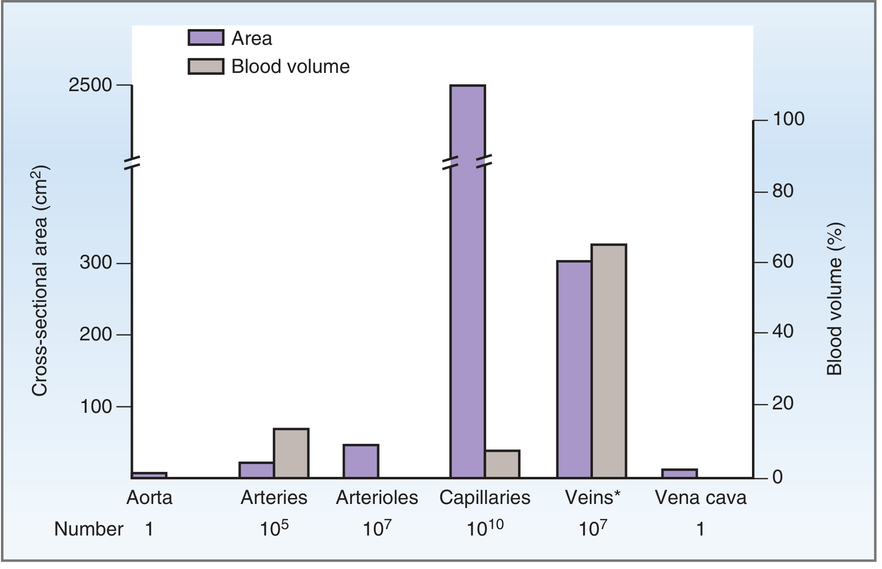
Key observations from this graph (frequently asked in viva):
- Largest cross-sectional area → Capillaries (2500 cm²) → slowest velocity
- Largest blood volume → Veins/venules (~64%)
- Number of vessels → Capillaries (10¹⁰) > Arterioles (10⁷) = Veins (10⁷) > Arteries (10⁵) > Aorta (1)
Velocity of Blood Flow (Linked Concept)
v = Q/A (velocity = flow / cross-sectional area)
- Aorta: fastest velocity (~40 cm/s) - smallest total cross-section
- Capillaries: slowest velocity (~0.07 cm/s) - largest total cross-section
- This slowest capillary velocity is physiologically ideal for exchange
High-Yield Exam Points (Viva/Practical)
- Arterioles are the most important site for regulation of blood pressure and local blood flow - "taps of the circulation"
- Precapillary sphincters (not arterioles) control perfusion of individual capillaries
- Windkessel effect: elastic arteries convert pulsatile cardiac output into steady capillary flow
- Venoconstriction during haemorrhage: sympathetic activation constricts veins → mobilizes unstressed volume → raises venous return → compensates for blood loss
- Total blood volume distribution: Veins 64% > Arteries 13% > Capillaries 5% > Heart + pulmonary ~18%
- Capillary wall has no smooth muscle - it cannot constrict; flow is regulated upstream by arterioles and precapillary sphincters
Costanzo Physiology 7th Ed., pp. 125-127 | THIEME Atlas of General Anatomy, pp. 83-84
This is a shared conversation. Sign in to Orris to start your own chat.