
Give a note on syphilis
syphilis
syphilis chancre skin lesion rash
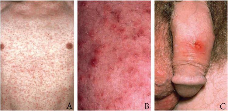
This composite of three clinical photographs illustrates the dermatological and urogenital manifestations of early syphilis (Treponema pallidum infection). Panel A shows a wide-field view of the anterior torso featuring a dense, generalized maculopapular rash, typical of secondary syphilis. The lesions are symmetric, non-confluent, and reddish-pink. Panel B provides a high-magnification view of the same rash, highlighting individual erythematous papules with distinct borders and underlying skin inflammation. Panel C depicts a primary syphilis lesion: a solitary, well-demarcated chancre on the dorsal penile shaft. The chancre presents as a round, reddish-brown ulcer with indurated edges, a clean base, and associated localized erythema. This educational visual is designed to assist in the clinical identification of primary versus secondary stages of syphilis, focusing on the transition from a localized inoculation site (chancre) to systemic cutaneous dissemination (maculopapular rash). It is intended for medical education in infectious diseases, dermatology, and sexual health.
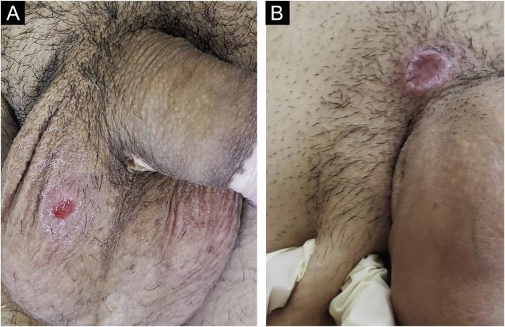
Clinical photograph consisting of two panels (A and B) showing primary syphilis manifestations (hard chancres) in the male genital region. Panel A displays a single, well-defined, round ulcer on the scrotal skin. The lesion features a characteristic 'clear' reddish base and smooth sloping edges. The surrounding scrotal and penile skin also exhibits depigmented patches consistent with comorbid vitiligo. Panel B shows a larger, cupuliform lesion located on the pubis near the base of the penis. This lesion demonstrates significant induration (infiltration) at the base, resulting in a raised, crater-like morphology. These visuals serve as educational examples of Treponema pallidum infection sites, highlighting the classic morphology of the hard chancre: a generally painless, indurated ulcer with a clean base. The clinical context notes that such primary syphilitic lesions are often accompanied by regional lymphadenopathy, representing the initial stage of systemic infection.
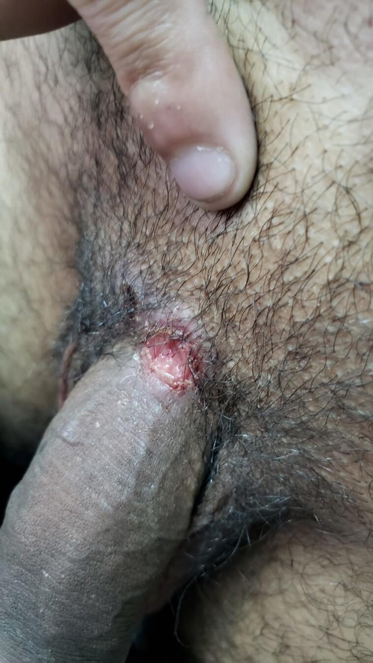
This clinical photograph shows a single, well-defined ulcerated lesion located at the base of the penile shaft, near the junction with the pubic region. The lesion is roughly circular with irregular borders and features a raw, erythematous base with a moist appearance, suggesting potential serous exudate. The surrounding tissue demonstrates mild perilesional inflammation and erythema. Anatomically, the site is characterized by overlying pubic hair and standard genital skin architecture. The visual presentation is highly characteristic of a primary syphilis chancre (Treponema pallidum infection), though the context indicates a coinfection with herpes simplex virus type 1 (HSV-1). This image serves as an educational example of genital ulcer disease (GUD) and primary syphilis manifestations in a clinical setting, highlighting the morphology of a syphilitic chancre which is typically firm, indurated, and painless.
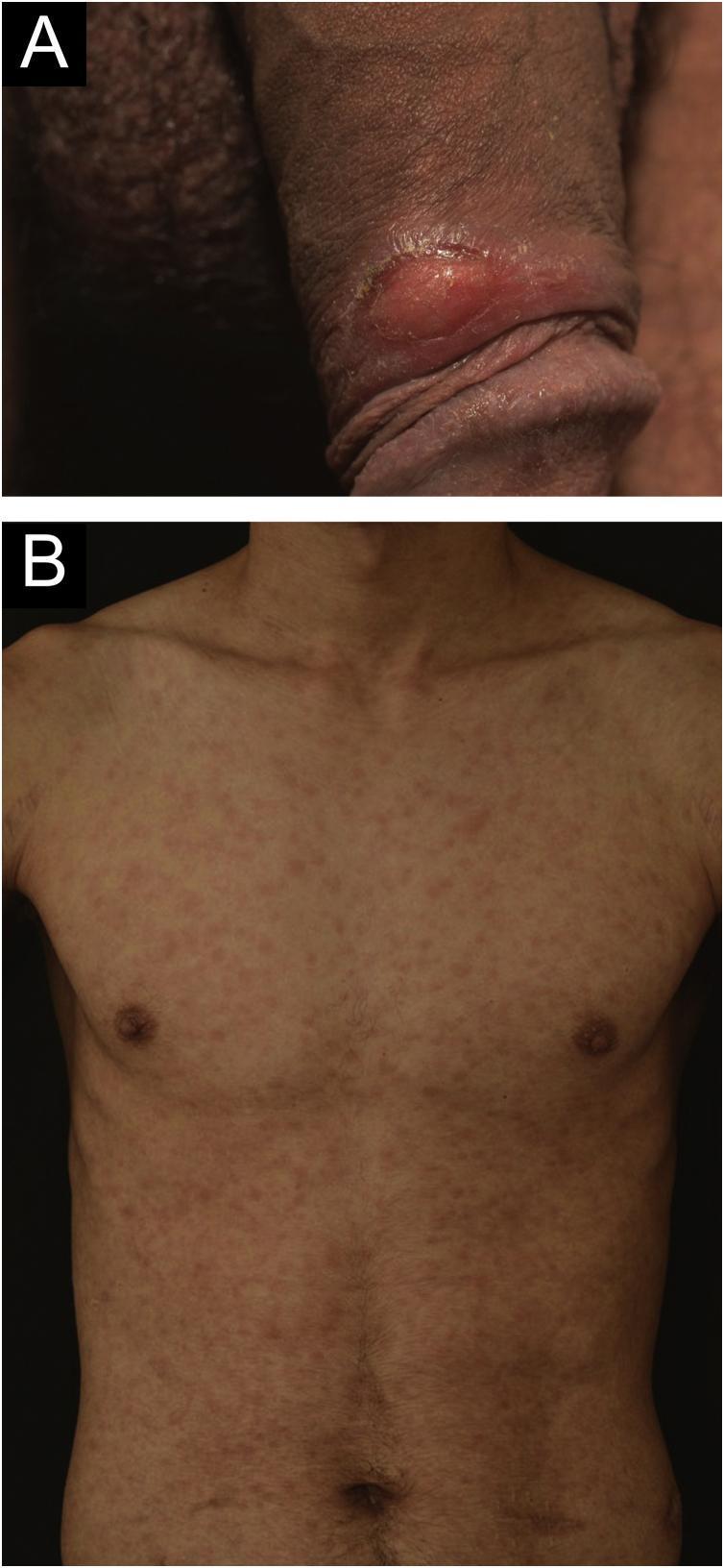
Two-panel clinical photograph illustrating manifestations of syphilis in a 24-year-old male. Panel A (top) is a close-up clinical photograph of the male genitalia showing a solitary, well-demarcated genital ulcer (chancre) located on the foreskin. The lesion exhibits clean, well-defined edges and an erythematous, indurated base, characteristic of primary syphilis. Panel B (bottom) is an anterior clinical photograph of the patient's trunk demonstrating a diffuse, symmetrical cutaneous eruption. The rash consists of multiple, discrete, erythematous maculopapular lesions distributed across the chest and abdomen. These findings represent secondary syphilis occurring concurrently with the resolving primary lesion. The images serve as an educational resource for identifying the clinical progression of Treponema pallidum infection, particularly in immunocompromised patients such as kidney transplant recipients.
Syphilis
Definition and Causative Organism
- Andrews' Diseases of the Skin, p. 4720
Etiology and Microbiology
-
Length: 5-20 µm, with 4-14 spiral coils
-
Demonstrated in lesion exudate by darkfield microscopy or fluorescent antibody techniques
-
Characteristic motility: projection along its long axis, rotation on its long axis, and side-to-side bending
-
Darkfield examination of oral lesions is unreliable (cannot be distinguished from commensal oral treponemes)
-
Cannot be cultured in vitro - its genome is small (~25% of most bacteria) with minimal metabolic capacity
-
Disseminates widely and rapidly: reaches the bloodstream within hours of infection, and the brain within 18 hours of inoculation
-
Evades immune clearance by expressing very few surface antigens and rapidly mutating outer membrane proteins
-
Andrews' Diseases of the Skin, p. 4724-4728
Modes of Transmission
- Sexual contact - direct contact with an active lesion (primary route)
- Transplacental - congenital syphilis; the treponeme crosses the placenta to infect the fetus
- Blood-borne - rarely via transfusion or shared needles
Stages of Syphilis
1. Primary Syphilis
- Incubation period: 2-6 weeks after exposure
- Classic lesion: chancre - a painless, indurated ulcer with well-defined borders and a clean base
- Usually on the genital mucosa, but may appear on oral or anorectal mucosa
- Accompanied by painless regional lymphadenopathy
- Heals spontaneously in 3-6 weeks even without treatment
2. Secondary Syphilis
- Develops in 60%-90% of persons with untreated primary syphilis
- Results from hematogenous dissemination of treponemes
- Systemic features:
- Rash - the hallmark; classically a symmetric, non-itchy maculopapular rash involving the palms and soles (characteristic)
- Fever, malaise, generalized lymphadenopathy
- Headache, sore throat, arthralgias
- Condylomata lata - broad, flat, warty lesions in moist areas
- Mucous patches - grayish erosions on mucous membranes
- Less commonly: CNS involvement, uveitis, hepatitis, immune-complex glomerulonephritis
3. Latent Syphilis
- Defined as reactive serologic tests without clinical signs or symptoms
- Early latent: acquired within the past year
- Late latent: all other cases of latent syphilis
- Patients are not infectious (except pregnant women who can transmit transplacentally)
4. Tertiary (Late) Syphilis
| Form | Features |
|---|---|
| Cardiovascular syphilis | Aortic aneurysm (classically ascending aorta), aortic regurgitation, coronary ostial stenosis |
| Neurosyphilis | General paresis, tabes dorsalis, Argyll Robertson pupil, CNS gumma |
| Gummatous syphilis | Granulomatous inflammatory masses with necrotic center; can affect skin, bone, viscera |
- Textbook of Family Medicine 9e, p. 2592-2594
Congenital Syphilis
- Early congenital (within 2 years): rhinitis ("snuffles"), maculopapular rash, hepatosplenomegaly, osteochondritis
- Late congenital (after 2 years): Hutchinson's teeth, interstitial keratitis, 8th nerve deafness ("Hutchinson's triad"), saddle nose, saber shins
Clinical Images
Diagnosis
Direct Methods (Primary Syphilis)
- Darkfield microscopy - demonstrates motile spirochetes from chancre exudate; gold standard for primary syphilis
- Direct immunofluorescence (DFA-TP) - can confirm oral lesions where darkfield is unreliable
Serologic Tests
| Test Type | Examples | Notes |
|---|---|---|
| Nontreponemal (screening) | RPR, VDRL | Quantitative; used to monitor treatment response; can give false positives |
| Treponemal (confirmatory) | MHA-TP, FTA-ABS, TPPA | Remain positive for life; not useful for monitoring treatment |
-
RPR: 99% sensitive in secondary syphilis
-
MHA-TP: 100% sensitive in secondary syphilis
-
For neurosyphilis: CSF-VDRL is used
-
Indications for CSF evaluation: neurologic/auditory/ophthalmic/cardiovascular signs, treatment failure (nontreponemal titers fail to decrease 4-fold within 6 months), or latent syphilis with HIV
-
Textbook of Family Medicine 9e, p. 2596
Treatment
| Stage | Regimen |
|---|---|
| Primary, secondary, early latent | Benzathine penicillin G 2.4 million units IM, single dose |
| Late latent / latent of unknown duration | Benzathine penicillin G 2.4 million units IM weekly x 3 doses |
| Neurosyphilis / cardiovascular syphilis | Aqueous crystalline penicillin G IV for 10-14 days |
| Penicillin-allergic (non-pregnant) | Doxycycline 100 mg PO twice daily for 14 days (early) or 28 days (late) |
| Penicillin-allergic, pregnant | Penicillin desensitization then penicillin (no alternatives safe in pregnancy) |
- Textbook of Family Medicine 9e, p. 2597-2598
Follow-Up and Treatment Response
- Monitor nontreponemal titers (RPR) at 6 and 12 months
- Treatment success = 4-fold (2-dilution) drop in titer (e.g., 1:32 to 1:8)
- Treponemal antibodies (MHA-TP, FTA-ABS) remain reactive for life and cannot be used to assess cure
Syphilis and HIV Co-infection
- HIV-infected persons may have atypical clinical presentations
- Serologic tests may be unreliable (both false negatives and unusually high titers possible)
- Neurosyphilis is more common and more aggressive
- Disproportionately high rates of syphilis occur in HIV-infected persons
- Some guidelines recommend CSF examination in all HIV-positive patients with syphilis