
Lung recruitment manuvers 5 mark answer
lung recruitment maneuver PEEP atelectasis mechanical ventilation
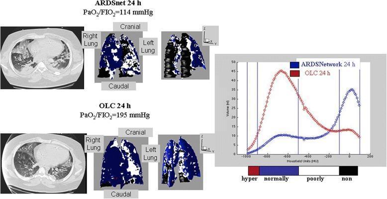
This medical comparison chart displays axial CT scans, 3D lung reconstructions, and voxel distribution histograms comparing two mechanical ventilation strategies in Acute Respiratory Distress Syndrome (ARDS): ARDSnet-24h and Open Lung Concept (OLC)-24h. The top panel (ARDSnet) shows significant bilateral dorsal atelectasis, represented in the 3D model by large black regions (non-aerated tissue) and poorly aerated white areas. Its corresponding histogram shows a bimodal distribution with a prominent peak in the non-aerated range (> -100 Hounsfield Units). The bottom panel (OLC) demonstrates improved alveolar recruitment, evidenced by a shift in the 3D model to predominantly blue (normally aerated) tissue and a reduction in black dorsal collapse. The OLC histogram shows a unimodal distribution with a primary peak in the normally-aerated range (-900 to -500 HU) and a higher PaO2/FiO2 ratio (195 vs 114 mmHg). This clinical imaging study illustrates the efficacy of recruitment maneuvers and PEEP titration in achieving more homogeneous lung aeration and reducing intrapulmonary shunt in critically ill patients.
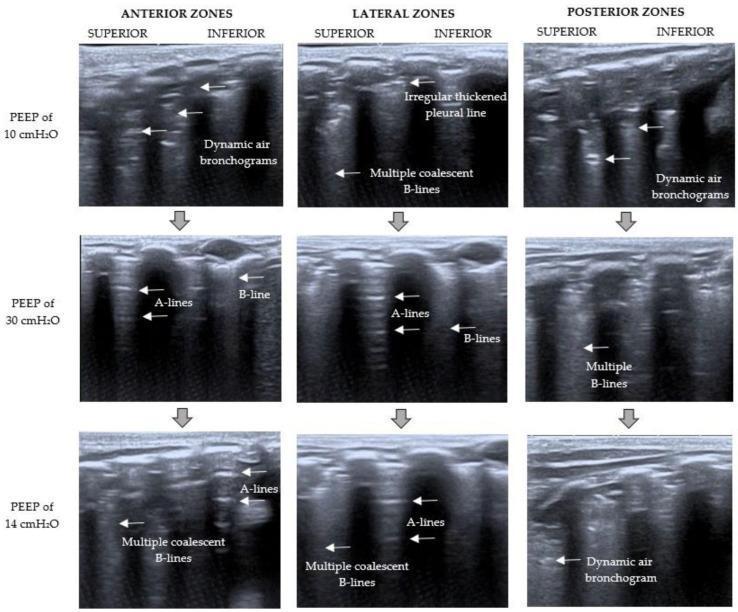
This diagnostic image matrix displays Lung Ultrasound (LUS) frames arranged by anatomical region (Anterior, Lateral, and Posterior zones) and positive end-expiratory pressure (PEEP) levels (10, 30, and 14 cmH2O) to demonstrate recruitment maneuver outcomes. At baseline (PEEP 10 cmH2O), the images show a 'C-pattern' characterized by severe loss of aeration with dynamic air bronchograms in anterior/posterior zones and an irregular thickened pleural line with multiple coalescent B-lines laterally. During recruitment (PEEP 30 cmH2O), there is visible reaeration characterized by a transition to a 'B1-pattern' with horizontal A-lines and well-defined, discrete B-lines. At the final titrated PEEP (14 cmH2O), the 'B2-pattern' is evident, showing some recruitment maintenance via A-lines but recurring multiple coalescent B-lines and persistent dynamic air bronchograms posteriorly. This comparison illustrates the dynamic nature of lung aeration, pleural line morphology, and ultrasound artifacts (A-lines vs. B-lines) in response to mechanical ventilation adjustments in patients with respiratory distress.
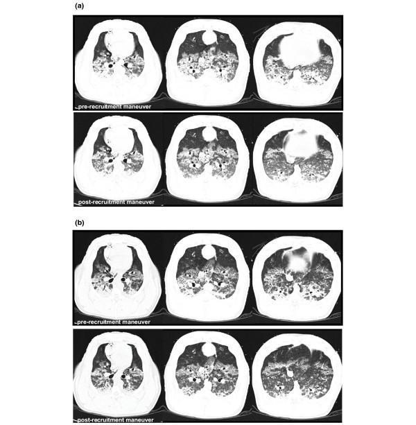
A series of axial thoracic CT scans illustrating lung recruitment in an animal model, likely used for studying Acute Respiratory Distress Syndrome (ARDS) or mechanical ventilation. The image is divided into two main panels: (a) Expiratory occlusion and (b) Inspiratory occlusion. Each panel contains a 2x3 grid comparing 'pre-recruitment maneuver' (top row) and 'post-recruitment maneuver' (bottom row) states at three anatomical levels: apical, middle, and basal. In the 'pre-recruitment' images, there are significant bilateral areas of high density (ground-glass opacities and consolidation) predominately in the dependent (posterior) lung regions, indicating atelectasis and non-aerated lung tissue. Post-recruitment, there is a visible reduction in these dense areas and a concomitant increase in radiolucency (darker regions), signifying improved alveolar aeration and lung volume (Vgas). The changes are most pronounced in the basal levels during both expiratory and inspiratory occlusions, demonstrating the effectiveness of the maneuver in opening collapsed alveoli and homogenizing lung parenchyma density.
Lung Recruitment Maneuvers
Definition
Rationale / Physiology
- Loss of respiratory muscle tone
- Absorption atelectasis (high FiO2)
- Compression from the diaphragm, abdominal contents, or fluid
- Increased intrapulmonary shunt → hypoxemia
- Cyclic opening and closing (atelectrauma) → VILI
- Inhomogeneous distribution of stress → barotrauma in open alveoli
Types of Recruitment Maneuvers
1. Sustained Inflation (SI) - Most Common
- Airway pressure held at 30-40 cmH2O for 30-40 seconds
- In obese patients or those with low chest wall compliance: 40-55 cmH2O for 10 seconds
- Simple to perform; requires only pressure control or CPAP mode
- Most studied method
2. Incremental / Stepwise PEEP (Staircase RM)
- PEEP is increased in steps of 2-5 cmH2O every 3-5 minutes
- Continued until respiratory system compliance worsens or hemodynamic compromise occurs
- Considered gentler than sustained inflation
- Used in conjunction with decremental PEEP trials
3. Pressure-Controlled RM
- Multiple breaths delivered at elevated peak inspiratory pressure (e.g., 40 cmH2O) with increased PEEP
- Allows intermittent sigh breaths
4. Sigh Breaths
- Periodic delivery of breaths 1.5-2x the normal tidal volume
- Older technique; less commonly used alone
Open Lung Strategy
- After a recruitment maneuver, PEEP is titrated to prevent end-expiratory alveolar collapse
- Decremental PEEP trial: PEEP raised to 20-25 cmH2O after RM, then decreased stepwise; final PEEP is set at the level that maximizes lung compliance
- Esophageal manometry: PEEP titrated to maintain positive transpulmonary pressure (prevents collapse at end-expiration); especially useful in obesity and ARDS
Indications
- ARDS (moderate to severe)
- Perioperative atelectasis (especially in obese, post-intubation, laparoscopic surgery)
- One-lung ventilation
- Post-extubation atelectasis
- VILI prevention in lung-protective ventilation
Complications / Adverse Effects
| Complication | Mechanism |
|---|---|
| Transient hypotension | Increased intrathoracic pressure → reduced venous return |
| Oxygen desaturation (transient) | Redistribution of blood flow during maneuver |
| Ventilator dyssynchrony | Patient-ventilator asynchrony |
| Barotrauma / Tension pneumothorax | High airway pressures (rare but serious) |
| Hemodynamic instability | Elevated mean airway pressure |
- Hemodynamic instability
- Unilateral lung disease (risk of overdistension of healthy lung)
- Bullous emphysema
- Elevated ICP
Evidence and Clinical Outcomes
- Despite physiologic benefit (improved oxygenation, compliance), large RCTs have not consistently shown improved mortality in ARDS
- The ART trial (largest RCT of open lung protocol) found increased 6-month mortality, ventilator days, and pneumothorax risk with high-pressure recruitment, likely due to barotrauma and hemodynamic instability
- Current consensus: RMs may be used selectively in ARDS and peri-operative settings, but should be applied cautiously with individualized PEEP titration
- Prone positioning is an adjunct strategy with proven mortality benefit in severe ARDS (PaO2:FiO2 < 150 mmHg)
Summary Table
| Feature | Detail |
|---|---|
| Primary goal | Re-aerate collapsed alveoli; reduce shunt |
| Most common method | Sustained inflation: 30-40 cmH2O × 30-40 s |
| Combined with | PEEP titration (decremental PEEP trial) |
| Key complication | Hemodynamic instability, pneumothorax |
| Evidence in ARDS | Physiologic benefit; no consistent mortality benefit |