
Anatomy & Course of main portal vein Bro I want points for this based on year 1 MBBS student on GIT system . Then I want summarise points of u given for me to add in my eposter in an area of it. And if can pls use the points based on what I asked from this slide
cd /tmp/workspace && mkdir -p portal_vein && python3 -c " import pdfplumber with pdfplumber.open('/tmp/workspace/attachments/ada65072-711a-422d-974e-e244c96535ca/L9.pdf') as pdf: print('pages', len(pdf.pages)) for i, page in enumerate(pdf.pages): text = page.extract_text() print(f'--- PAGE {i+1} ---') print(text) "
hepatic portal vein formation tributaries splenic superior mesenteric vein diagram
Portal Vein — Anatomy & Course (Year 1 MBBS, GIT System)
1. Detailed Study Points
- The hepatic portal vein is the main vessel draining venous blood from the abdominal GIT, spleen, pancreas, and gallbladder into the liver.
- Carries nutrient-rich (but deoxygenated) blood — supplies about 70% of liver blood flow (the remaining 30% is oxygenated blood from the hepatic artery).
- Functional significance: because gut venous blood drains directly into the liver via this vein, the liver is a common site for secondary spread of gut infections and cancers (portal metastasis) - L9.pdf, p. 44.
- Formed by the union of the splenic vein and the superior mesenteric vein (SMV), behind the neck of the pancreas.
- The inferior mesenteric vein usually joins the splenic vein (at a right angle) before this union, rather than joining the portal vein directly - L9.pdf, p. 34.
- Textbook detail: formation occurs at the level of vertebra L2 - Gray's Anatomy for Students, p. 416.
- From its origin behind the pancreatic neck, it ascends posterior to the superior (first) part of the duodenum.
- Enters the right free margin of the lesser omentum, running within the portal triad.
- Within the lesser omentum it lies posterior to (deep to) both the bile duct and the hepatic artery proper - L9.pdf, p. 33:
- Bile duct - lies to its right (anterior)
- Hepatic artery proper - lies to its left (anterior)
- Portal vein - lies posterior/deep to both
- It is also anterior to the epiploic (omental) foramen of Winslow.
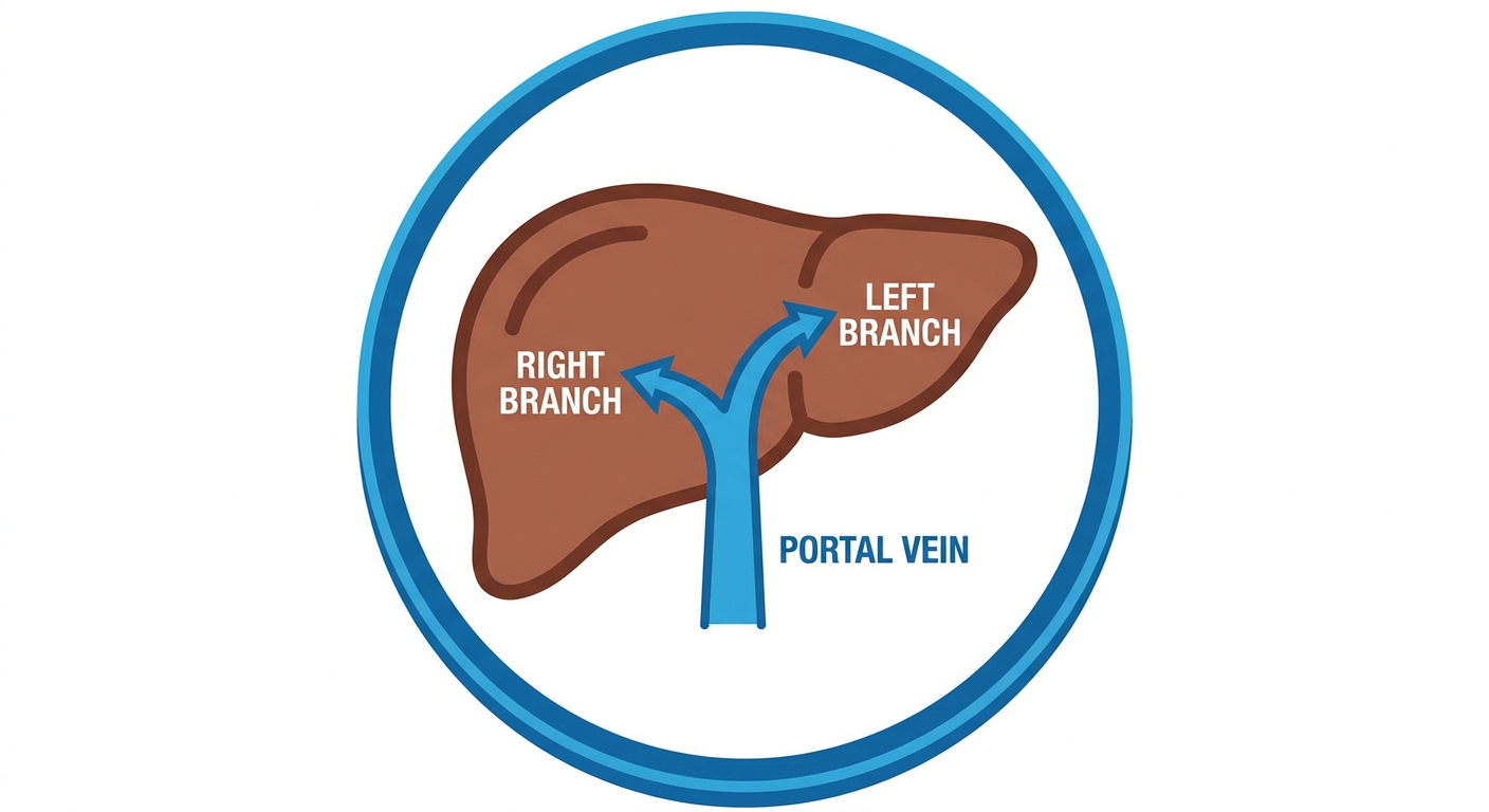
- On reaching the porta hepatis (hilum of the liver), it divides into right and left branches that enter the liver substance along with branches of the hepatic artery and bile duct, supplying the functional right and left lobes respectively.
- Splenic vein
- Superior mesenteric vein
- Left and right gastric veins (draining the lesser curvature of stomach/oesophagus)
- Cystic vein (from gallbladder)
- Para-umbilical veins (linked to the obliterated umbilical vein/ligamentum teres — clinically important in portal hypertension, e.g. caput medusae)
- Portal vein forms part of the portal triad (portal vein + hepatic artery proper + bile duct) within the porta hepatis.
- Site of portosystemic anastomoses important in portal hypertension (oesophageal varices, caput medusae, rectal varices).
- Relevant to portal vein thrombosis and to surgical procedures like the Pringle manoeuvre (temporary occlusion of the portal triad to control bleeding).
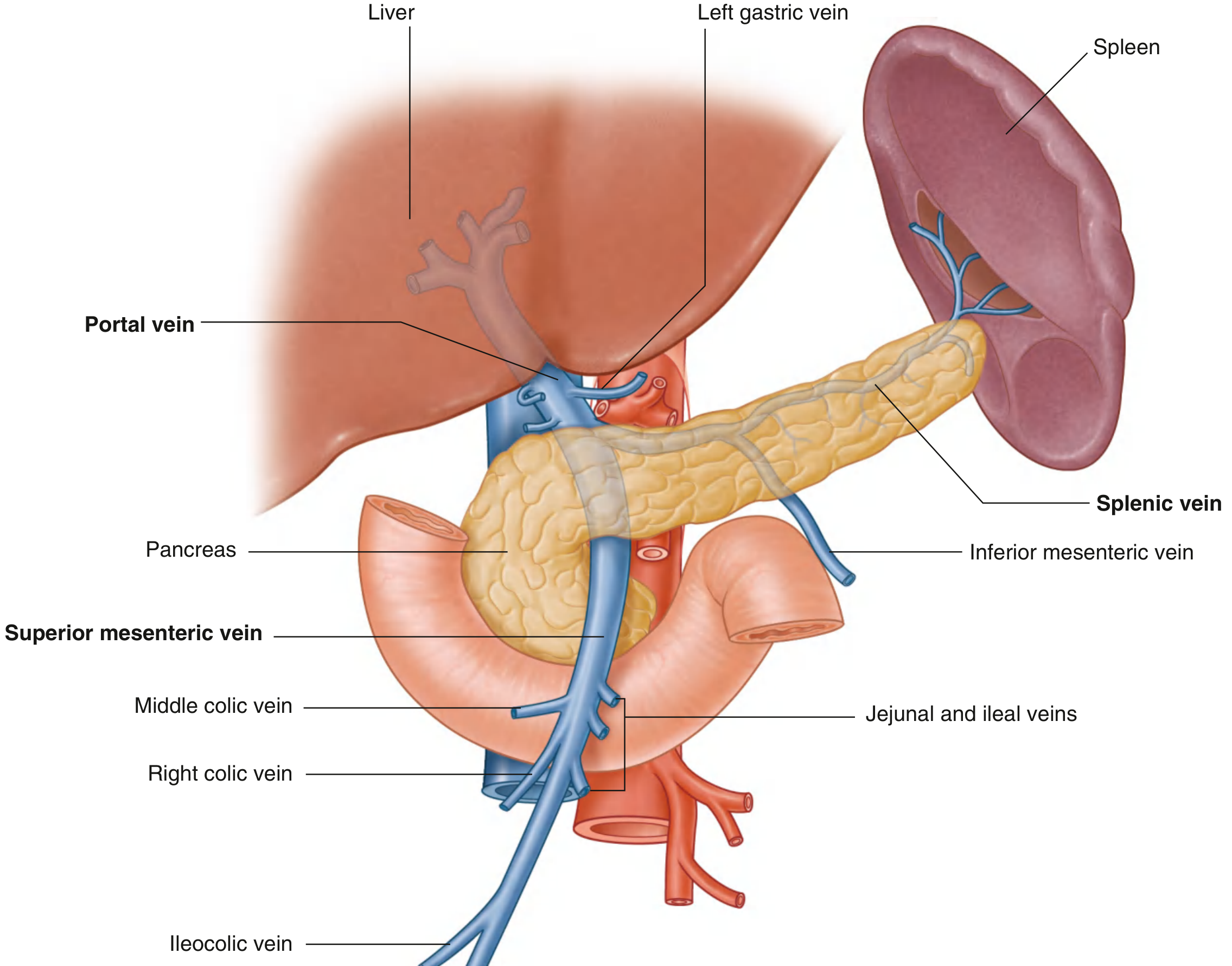
2. Condensed Summary for Your E-Poster
- Drains blood from GIT, spleen, pancreas, gallbladder → liver (~70% of liver's blood supply)
- Formed by union of splenic vein + superior mesenteric vein, behind the neck of pancreas (level of L2)
- Course: ascends behind 1st part of duodenum → enters right free margin of lesser omentum → lies posterior to bile duct (right) and hepatic artery (left) in the portal triad
- At porta hepatis, divides into right & left branches entering the liver
- Tributaries: splenic vein, SMV, left/right gastric veins, cystic vein, para-umbilical veins
- Clinical relevance: portal hypertension → varices/caput medusae; route for gut infection/cancer spread to liver; portal vein thrombosis
Bro can u give me more diagrams based on this
portal vein anatomy diagram liver
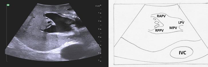
This diagnostic visual includes a B-mode grayscale ultrasound image and a corresponding labeled line diagram illustrating a portal vein trifurcation, an anatomical variant of the liver. The image demonstrates the main portal vein (MPV) dividing simultaneously at a single point into three branches: the left portal vein (LPV), the right anterior portal vein (RAPV), and the right posterior portal vein (RPPV), indicating the absence of a common right portal vein trunk. The vascular structures exhibit characteristic hyperechoic walls due to Glisson's capsule and are surrounded by homogeneous, medium-echogenicity liver parenchyma. Posterior and inferior to the portal confluence, the inferior vena cava (IVC) is visible as an anechoic oval structure. This content is intended for surgical planning and radiological education, highlighting variations in hilar anatomy crucial for performing segmentectomies or hepatic resections. The labels facilitate identification of segmental portal supply in the context of intraoperative or diagnostic ultrasound.
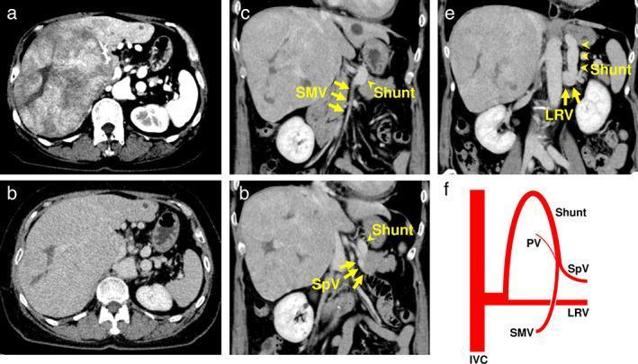
This composite figure includes contrast-enhanced computed tomography (CECT) scans and a corresponding anatomical diagram illustrating Congenital Absence of the Portal Vein (CAPV) Type II associated with hepatocellular carcinoma (HCC). (a) Axial arterial phase image shows a large, inhomogeneous hyperenhancing mass occupying the right lobe of the liver with a central hypoattenuating area suggestive of necrosis. (b) Axial portal venous phase image demonstrates the mass becoming isoattenuating relative to the liver parenchyma. (c-e) Coronal reconstruction images highlight a portosystemic shunt. Yellow arrows indicate the superior mesenteric vein (SMV) and splenic vein (SpV) joining to form a large shunt that drains directly into the left renal vein (LRV). (f) A schematic diagram summarizes the venous anatomy, showing the SMV and SpV bypassing the liver via a shunt to the LRV/IVC, with a hypoplastic portal vein (PV) branching toward the liver, characteristic of CAPV Type II. This case highlights the radiological features of hepatoma in the context of rare congenital vascular anomalies.
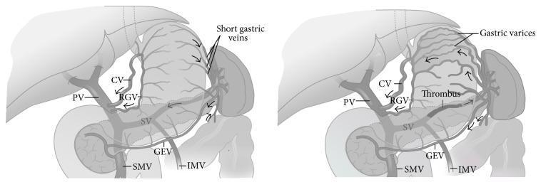
This medical illustration consists of two side-by-side comparison diagrams depicting the pathophysiology of sinistral (left-sided) portal hypertension due to splenic vein thrombosis. The diagrams show the hepatobiliary and gastrointestinal venous anatomy, including the liver, stomach, spleen, and pancreas. The left diagram represents normal venous anatomy where the splenic vein (SV), portal vein (PV), superior mesenteric vein (SMV), and inferior mesenteric vein (IMV) are patent with physiological blood flow. The right diagram illustrates the effects of a thrombus within the splenic vein. This obstruction leads to retrograde flow and increased pressure in the collateral venous pathways, specifically causing the development of prominent gastric varices, dilatation of the short gastric veins, coronary vein (CV), and gastroepiploic vein (GEV). Notably, in both the normal and pathological states, the portal vein, SMV, and IMV remain patent, highlighting that the hypertension is localized to the splenic drainage system. Arrows indicate the direction of venous blood flow, shifting toward the gastric wall in the presence of the thrombus.
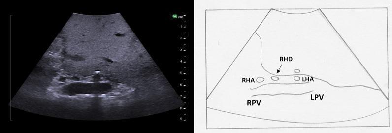
This diagnostic image consists of an intraoperative ultrasound of the liver (left) paired with an anatomical line diagram (right) illustrating the hepatic hilum in a transverse plane. The ultrasound displays normal liver parenchyma with medium echogenicity and a homogeneous texture. The central focus is the bifurcation of the portal vein and the overlying hepatic artery. Key anatomical structures labeled in the diagram and visible on ultrasound include the right portal vein (RPV) and left portal vein (LPV), which appear as larger, hypoechoic vascular structures. Positioned superficial to the portal bifurcation are the smaller, circular cross-sections of the right hepatic artery (RHA) and left hepatic artery (LHA). Additionally, the right hepatic duct (RHD) is identified as a small anechoic structure adjacent to the RHA. This visual material is used in surgical education to demonstrate the 'Mickey Mouse' sign and the spatial relationships within the hepatoduodenal ligament, essential for identifying segmental anatomy during hepatobiliary procedures.
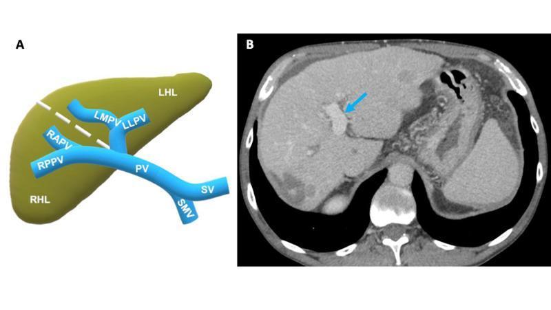
This medical figure consists of two panels focusing on hepatic portal anatomy and pathology. Panel A is an anatomical diagram illustrating the portal venous system in relation to the liver lobes. It depicts the convergence of the splenic vein (SV) and superior mesenteric vein (SMV) into the main portal vein (PV), which bifurcates into the right hepatic lobe (RHL) branches—specifically the right anterior (RAPV) and right posterior (RPPV) portal veins—and the left hepatic lobe (LHL) branches, including the left medial (LMPV) and left lateral (LLPV) portal veins. Panel B is a contrast-enhanced axial abdominal CT scan of a patient with a history of hepatocellular carcinoma (HCC). A blue arrow indicates a widely patent portal vein with uniform contrast opacification, showing no evidence of tumor thrombus at this baseline. The liver parenchyma exhibits a heterogeneous, coarse texture consistent with cirrhotic changes. This comparison serves as an educational reference for normal portal venous flow prior to the development of macrovascular invasion or tumor thrombosis.
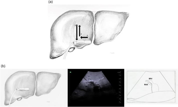
This composite educational graphic illustrates the technique for intraoperative ultrasound (IOUS) of the right portal vein. Panel (a) contains an anatomical diagram showing a T-style ultrasound probe placed on the surface of the liver. Numbered arrows (1-3) demonstrate the systematic scanning maneuvers (sliding and rolling) used to visualize the segmental branches of the right portal vein. Panel (b) correlates a specific probe placement over the right liver with a corresponding transverse ultrasound image and its labeled schematic. The ultrasound clearly identifies the right portal vein (RPV) and the right hepatic artery (RHA). The RHA is seen as a smaller tubular structure positioned inferior and slightly anterior to the RPV. The image highlights the use of hyperechoic portal vein walls and proximity to the hepatic hilum to define segmental anatomy. This visual serves as a procedural guide for surgeons to perform complete parenchymal and vascular exploration during hepatobiliary surgery.
portal triad bile duct hepatic artery portal vein lesser omentum
splenic vein superior mesenteric vein union pancreas portal vein formation
portosystemic anastomosis caput medusae esophageal varices diagram
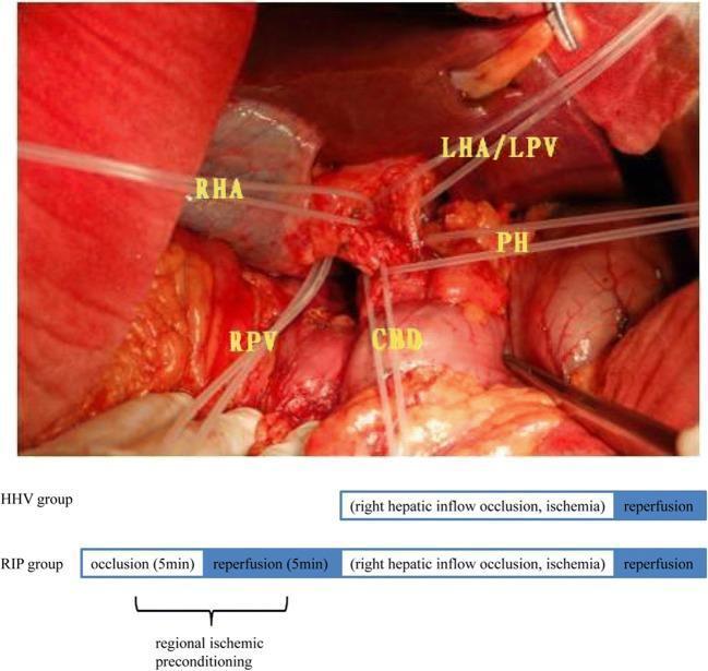
hepatic portal system veins draining stomach spleen pancreas intestine diagram
lesser omentum contents hepatoduodenal ligament cross section
caput medusae portosystemic collateral circulation varices diagram anatomy
liver hilum porta hepatis anatomy labeled diagram textbook
portal vein bifurcation right left branch liver segments simple line diagram
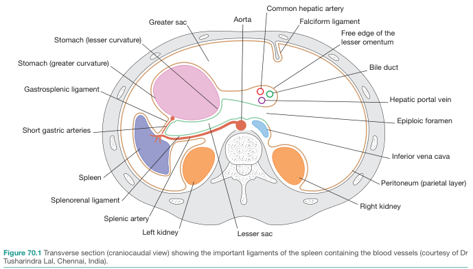
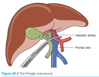
- Diagram 1 or 4 → main "Formation & Tributaries" box
- Diagram 2 → "Course & Relations" box (shows the lesser omentum/portal triad relationship clearly)
- Diagram 3 → "Applied Anatomy / Clinical Correlation" box (Pringle manoeuvre ties directly to your point about the portal triad being occluded during liver surgery)
Bro i need like small pictures based on my title for me to add in eposter
Simple labeled diagram showing formation of the hepatic portal vein by union of splenic vein and superior mesenteric vein behind the neck of pancreas
Simple labeled diagram of the course of the portal vein ascending behind the duodenum into the lesser omentum, showing its relation to the bile duct and hepatic artery in the portal triad
Small simple icon-style diagram of the liver at the porta hepatis showing the portal vein dividing into right and left branches entering the liver
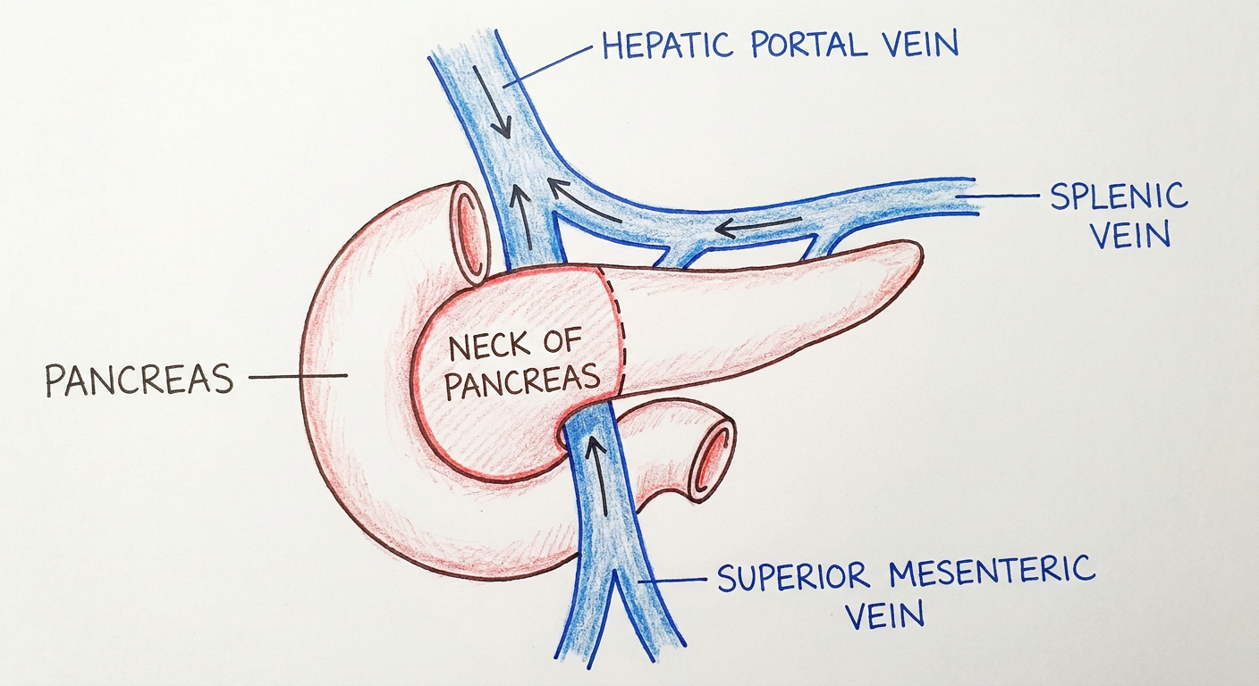
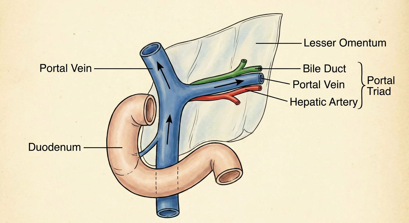
PORTAL VEIN — Anatomy & Course Drains blood from GIT, spleen, pancreas, gallbladder → liver (~70% of liver's blood supply) Formed by union of splenic vein + superior mesenteric vein, behind the neck of pancreas (level of L2) Course: ascends behind 1st part of duodenum → enters right free margin of lesser omentum → lies posterior to bile duct (right) and hepatic artery (left) in the portal triad At porta hepatis, divides into right & left branches entering the liver Tributaries: splenic vein, SMV, left/right gastric veins, cystic vein, para-umbilical veins Clinical relevance: portal hypertension → varices/caput medusae; route for gut infection/cancer spread to liver; portal vein thrombosis With this info can u like generate me a generated slide for me to add in my eposter but in a small thing and add the pictures u sent me in it
mkdir -p /tmp/workspace/portal_vein_poster/assets && ls -la /tmp/workspace/
cd /tmp/workspace/portal_vein_poster/assets && curl -s -o img1.png "https://cdn.orris.care/image-gen/915133b927f04f1e9e64c580361f26a7.png" && curl -s -o img2.png "https://cdn.orris.care/image-gen/47cc68110b364794a1107dec5168943f.png" && curl -s -o img3.png "https://cdn.orris.care/image-gen/75f0931db56447e8a57f3b12c419e6a4.png" && ls -la
fc-list | grep -i dejavu | head -20; python3 -c "import PIL; print(PIL.__version__)"
python3 -c " import matplotlib.font_manager as fm for f in fm.fontManager.ttflist[:20]: print(f.name, f.fname) " 2>/dev/null | sort -u | head -30 find / -iname "*.ttf" 2>/dev/null | grep -v conda | head -20
~/portal_vein_poster/build_panel.py
from PIL import Image, ImageDraw, ImageFont

FONT_DIR = "/opt/amazon/genesis1p-tools/venv/lib64/python3.12/site-packages/matplotlib/mpl-data/fonts/ttf"
bold = lambda sz: ImageFont.truetype(f"{FONT_DIR}/DejaVuSans-Bold.ttf", sz)
reg = lambda sz: ImageFont.truetype(f"{FONT_DIR}/DejaVuSans.ttf", sz)

W, H = 1200, 1500
BG = (255, 255, 255)
NAVY = (13, 59, 102)
DARK = (25, 25, 25)
ACCENT = (176, 30, 30)
LIGHT_BG = (235, 244, 250)

img = Image.new("RGB", (W, H), BG)
d = ImageDraw.Draw(img)

# Header bar
d.rectangle([0, 0, W, 130], fill=NAVY)
d.text((40, 25), "PORTAL VEIN", font=bold(46), fill=(255, 255, 255))
d.text((40, 82), "Anatomy & Course", font=reg(26), fill=(210, 225, 240))

def wrap_text(text, font, max_width, draw):
    words = text.split(" ")
    lines, cur = [], ""
    for w in words:
        test = (cur + " " + w).strip()
        if draw.textlength(test, font=font) <= max_width:
            cur = test
        else:
            lines.append(cur)
            cur = w
    if cur:
        lines.append(cur)
    return lines

bullets = [
    ("Drainage", "Drains blood from GIT, spleen, pancreas, gallbladder \u2192 liver (~70% of liver's blood supply)"),
    ("Formation", "Formed by union of splenic vein + superior mesenteric vein, behind the neck of pancreas (level of L2)"),
    ("Course", "Ascends behind 1st part of duodenum \u2192 enters right free margin of lesser omentum \u2192 lies posterior to bile duct (right) and hepatic artery (left) in the portal triad"),
    ("Termination", "At porta hepatis, divides into right & left branches entering the liver"),
    ("Tributaries", "Splenic vein, SMV, left/right gastric veins, cystic vein, para-umbilical veins"),
    ("Clinical relevance", "Portal hypertension \u2192 varices/caput medusae; route for gut infection/cancer spread to liver; portal vein thrombosis"),
]

y = 150
left_margin = 40
text_width = W - 80
label_font = bold(22)
body_font = reg(20)
line_gap = 6
block_gap = 14

for label, body in bullets:
    # bullet marker + label
    d.ellipse([left_margin, y+6, left_margin+10, y+16], fill=ACCENT)
    d.text((left_margin+22, y), f"{label}:", font=label_font, fill=NAVY)
    label_w = d.textlength(f"{label}: ", font=label_font)
    y += 30
    lines = wrap_text(body, body_font, text_width - 22, d)
    for ln in lines:
        d.text((left_margin+22, y), ln, font=body_font, fill=DARK)
        y += body_font.size + line_gap
    y += block_gap

# separator
y += 6
d.line([left_margin, y, W-left_margin, y], fill=(200, 200, 200), width=2)
y += 20

d.text((left_margin, y), "Key Diagrams", font=bold(28), fill=NAVY)
y += 44

# Load and paste the 3 generated images side by side (small thumbnails)
paths = ["assets/img1.png", "assets/img2.png", "assets/img3.png"]
captions = ["Formation", "Course in Portal Triad", "Branching at Porta Hepatis"]

thumb_w = (W - 80 - 2*20) // 3
thumb_h = thumb_w  # square-ish

x = left_margin
for p, cap in zip(paths, captions):
    im = Image.open(p).convert("RGB")
    im.thumbnail((thumb_w, thumb_h), Image.LANCZOS)
    # center within thumb box
    box = Image.new("RGB", (thumb_w, thumb_h), LIGHT_BG)
    ox = (thumb_w - im.width) // 2
    oy = (thumb_h - im.height) // 2
    box.paste(im, (ox, oy))
    img.paste(box, (x, y))
    d.rectangle([x, y, x+thumb_w, y+thumb_h], outline=NAVY, width=2)
    x += thumb_w + 20

y += thumb_h + 8
x = left_margin
cap_font = reg(16)
for cap in captions:
    tw = d.textlength(cap, font=cap_font)
    d.text((x + (thumb_w-tw)/2, y), cap, font=cap_font, fill=DARK)
    x += thumb_w + 20

y += 30
img_final = img.crop((0, 0, W, y+20))
img_final.save("/tmp/workspace/portal_vein_poster/portal_vein_panel.png")
print("saved", img_final.size)
cd /tmp/workspace/portal_vein_poster && python3 build_panel.py
portal vein panel
Image · PNG
- Header: "PORTAL VEIN - Anatomy & Course"
- All 6 bullet points from your summary (Drainage, Formation, Course, Termination, Tributaries, Clinical relevance)
- The 3 diagrams in a row underneath, each labeled (Formation / Course in Portal Triad / Branching at Porta Hepatis)