Explain the carcinoid tumor and carcinoid sequence from 2nd year mbbs pathology Use table flowchart images as much as possible

Reading File
Finding Sources
Finding Sources
Searching PubMed

carcinoid tumor AND sequence

Searching the Web

carcinoid tumor pathology enterochromaffin-like cells carcinoid sequence

Searching Images

carcinoid tumor pathology histology organoid nesting salt and pepper chromatin

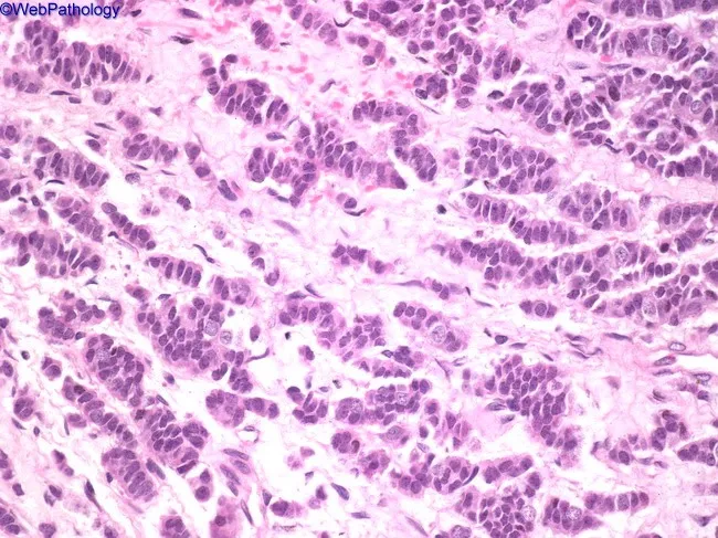
This is a thymic neuroendocrine tumor (thymic carcinoid) represented on a hematoxylin and eosin stained histology slide prepared from thymic tissue. The specimen is a thymic lesion in the anterior mediastinum. The tumor displays organoid nesting and ribbon-like arrangements (zellballen/perivascular rosettes) of uniform small cells with round to oval nuclei, finely dispersed chromatin (salt-and-pepper appearance), and scant cytoplasm. Cells are densely packed in interlacing cords and fascicles with delicate fibrovascular stroma; mitotic figures are infrequent in this field; necrosis is not conspicuous. The image shows a cohesive neuroendocrine morphology with minimal pleomorphism and absence of overt squamous differentiation. Immunophenotype in practice would be positive for neuroendocrine markers (chromogranin A, synaptophysin) and cytokeratins, supporting thymic carcinoid. The differential diagnosis includes spindle cell thymoma (type A) when spindle cells predominate, paraganglioma, medullary thyroid carcinoma with amyloid stroma, metastatic mucinous adenocarcinoma, and angiomatoid variants mimicking hemangioma. Clinically, thymic carcinoids are aggressive, may recur or metastasize, and require surgical excision with possible adjuvant therapy. This image is relevant for educational purposes in surgical pathology, mediastinal tumors, and neuroendocrine tumor biology, enabling recognition of organoid nesting, ribbon/festoons growth, salt-and-pepper chromatin, and differential with thymic spindle tumors and paragangliomas. High clinical relevance for diagnosis and management.

This is a thymic neuroendocrine tumor (thymic carcinoid) represented on a hematoxylin and eosin stained histology slide prepared from thymic tissue. The specimen is a thymic lesion in the anterior mediastinum. The tumor displays organoid nesting and ribbon-like arrangements (zellballen/perivascular rosettes) of uniform small cells with round to oval nuclei, finely dispersed chromatin (salt-and-pepper appearance), and scant cytoplasm. Cells are densely packed in interlacing cords and fascicles with delicate fibrovascular stroma; mitotic figures are infrequent in this field; necrosis is not conspicuous. The image shows a cohesive neuroendocrine morphology with minimal pleomorphism and absence of overt squamous differentiation. Immunophenotype in practice would be positive for neuroendocrine markers (chromogranin A, synaptophysin) and cytokeratins, supporting thymic carcinoid. The differential diagnosis includes spindle cell thymoma (type A) when spindle cells predominate, paraganglioma, medullary thyroid carcinoma with amyloid stroma, metastatic mucinous adenocarcinoma, and angiomatoid variants mimicking hemangioma. Clinically, thymic carcinoids are aggressive, may recur or metastasize, and require surgical excision with possible adjuvant therapy. This image is relevant for educational purposes in surgical pathology, mediastinal tumors, and neuroendocrine tumor biology, enabling recognition of organoid nesting, ribbon/festoons growth, salt-and-pepper chromatin, and differential with thymic spindle tumors and paragangliomas. High clinical relevance for diagnosis and management.

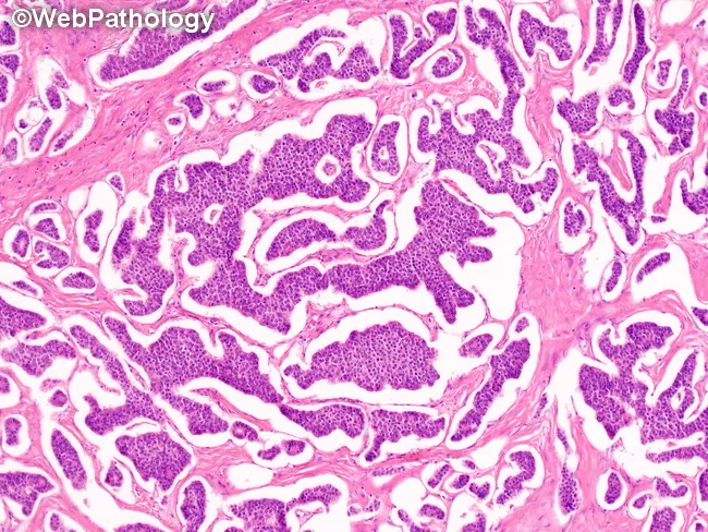
Imaging modality: Bright-field light microscopy of hematoxylin and eosin–stained ovarian tissue sections. Anatomical location: ovary (pelvic region), primary tumor in ovarian parenchyma. Visual features: orderly nests and cords of uniform neuroendocrine cells with round to oval nuclei, scant to moderate cytoplasm, and characteristic finely stippled (salt‑and‑pepper) chromatin; rosette-like structures and trabecular arrangements embedded in delicate stroma; occasional small vascular channels; low-to-moderate mitotic activity; absence or minimal necrosis. Pathology and tissue architecture: organoid nesting with nesting patterns typical of carcinoid tumors; absence of significant mucin production; lack of solid pleomorphic sheets. Pathological findings: consistent with primary ovarian carcinoid tumor (neuroendocrine neoplasm), type insular or trabecular possible; limited atypia; immunohistochemical confirmation typically positive for neuroendocrine markers such as synaptophysin and chromogranin A. Diagnostic significance: histology supports diagnosis of ovarian neuroendocrine tumor; differentiate from metastatic neuroendocrine tumors to the ovary; prognosis depends on size, histologic pattern, and stage; differential diagnoses include granulosa cell tumor with neuroendocrine differentiation and mucinous/serous tumors with focal neuroendocrine features. Clinical correlation: many patients are peri‑ or postmenopausal; potential for carcinoid syndrome with larger tumors; surgical management essential; evaluate for MEN1 syndrome in rare cases; urine 5-HIAA may be elevated; imaging and IHC recommended for confirmation and staging; educational utility in teaching ovarian neuroendocrine neoplasms.

Imaging modality: Bright-field light microscopy of hematoxylin and eosin–stained ovarian tissue sections. Anatomical location: ovary (pelvic region), primary tumor in ovarian parenchyma. Visual features: orderly nests and cords of uniform neuroendocrine cells with round to oval nuclei, scant to moderate cytoplasm, and characteristic finely stippled (salt‑and‑pepper) chromatin; rosette-like structures and trabecular arrangements embedded in delicate stroma; occasional small vascular channels; low-to-moderate mitotic activity; absence or minimal necrosis. Pathology and tissue architecture: organoid nesting with nesting patterns typical of carcinoid tumors; absence of significant mucin production; lack of solid pleomorphic sheets. Pathological findings: consistent with primary ovarian carcinoid tumor (neuroendocrine neoplasm), type insular or trabecular possible; limited atypia; immunohistochemical confirmation typically positive for neuroendocrine markers such as synaptophysin and chromogranin A. Diagnostic significance: histology supports diagnosis of ovarian neuroendocrine tumor; differentiate from metastatic neuroendocrine tumors to the ovary; prognosis depends on size, histologic pattern, and stage; differential diagnoses include granulosa cell tumor with neuroendocrine differentiation and mucinous/serous tumors with focal neuroendocrine features. Clinical correlation: many patients are peri‑ or postmenopausal; potential for carcinoid syndrome with larger tumors; surgical management essential; evaluate for MEN1 syndrome in rare cases; urine 5-HIAA may be elevated; imaging and IHC recommended for confirmation and staging; educational utility in teaching ovarian neuroendocrine neoplasms.

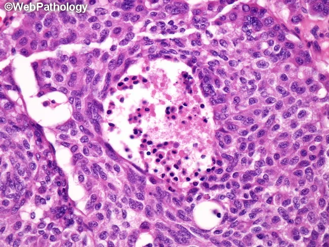
A histologic section of lung tissue stained with Hematoxylin and Eosin, examined under light microscopy at high magnification (approximately 400x). The tumor displays neuroendocrine morphology with organoid nesting and trabecular growth patterns. Tumor cells are uniform, polygonal with moderate cytoplasm and round nuclei with finely dispersed, salt-and-pepper chromatin. Notable mitotic activity is present, ranging from 2 to 10 mitoses per 10 high-power fields, and punctate necrosis is evident within some tumor nodules. The architectural arrangement suggests a well-differentiated neuroendocrine neoplasm consistent with an atypical carcinoid, as opposed to a typical carcinoid which lacks necrosis and shows lower mitotic rates. Immunohistochemical studies (not shown) would typically show neuroendocrine marker positivity (chromogranin A, synaptophysin, CD56) supporting a neuroendocrine neoplasm. The combination of mitotic activity and necrosis distinguishes atypical carcinoid from typical carcinoid, which lacks significant necrosis and has lower mitotic rates. This histology correlates with a higher risk of nodal and distant metastasis and a more aggressive clinical course, with a 5-year survival around 60% in reported series. Smoking history is more commonly associated compared with typical carcinoids but not universal. TP53 and RB1 mutations are seen in a subset (~20%) of atypical carcinoids. This image is representative for educational purposes in pathology, pulmonology, oncology, and medical training to illustrate diagnostic criteria and prognostic implications.

A histologic section of lung tissue stained with Hematoxylin and Eosin, examined under light microscopy at high magnification (approximately 400x). The tumor displays neuroendocrine morphology with organoid nesting and trabecular growth patterns. Tumor cells are uniform, polygonal with moderate cytoplasm and round nuclei with finely dispersed, salt-and-pepper chromatin. Notable mitotic activity is present, ranging from 2 to 10 mitoses per 10 high-power fields, and punctate necrosis is evident within some tumor nodules. The architectural arrangement suggests a well-differentiated neuroendocrine neoplasm consistent with an atypical carcinoid, as opposed to a typical carcinoid which lacks necrosis and shows lower mitotic rates. Immunohistochemical studies (not shown) would typically show neuroendocrine marker positivity (chromogranin A, synaptophysin, CD56) supporting a neuroendocrine neoplasm. The combination of mitotic activity and necrosis distinguishes atypical carcinoid from typical carcinoid, which lacks significant necrosis and has lower mitotic rates. This histology correlates with a higher risk of nodal and distant metastasis and a more aggressive clinical course, with a 5-year survival around 60% in reported series. Smoking history is more commonly associated compared with typical carcinoids but not universal. TP53 and RB1 mutations are seen in a subset (~20%) of atypical carcinoids. This image is representative for educational purposes in pathology, pulmonology, oncology, and medical training to illustrate diagnostic criteria and prognostic implications.

Searching Images

gastric carcinoid pathogenesis sequence atrophic gastritis hypergastrinemia ECL cell hyperplasia diagram

Reading File
Reading File
Finding Sources
Finding Sources
Reading Web Page

https://www.ncbi.nlm.nih.gov/books/NBK279162

Carcinoid tumor and carcinoid syndrome

Modern term: A carcinoid tumor is a well-differentiated neuroendocrine tumor (NET). The older name remains common in MBBS examinations.

Core concept

TermMeaning
Carcinoid tumorA neoplasm arising from neuroendocrine cells, commonly in the gastrointestinal tract or bronchus
Carcinoid syndromeSystemic symptoms caused by excessive release of vasoactive tumor products, especially serotonin, usually after liver metastasis in a midgut tumor
Carcinoid sequenceThe pathogenetic chain from neuroendocrine cell proliferation to tumor formation, and then to syndrome when hormones escape hepatic metabolism

1. Origin and common sites

Carcinoids arise from the diffuse neuroendocrine cell system in mucosa. In the intestine, many arise from enterochromaffin (EC) cells, which can produce serotonin. Gastric carcinoids typically arise from enterochromaffin-like (ECL) cells, which are stimulated by gastrin.
Embryological groupTypical sitesMain secretory products / features
ForegutBronchus, stomach, duodenum, pancreasHistamine, 5-HTP; may give atypical flushing
MidgutIleum, appendix, caecum, ascending colonSerotonin; most typical source of carcinoid syndrome
HindgutDistal colon and rectumUsually non-functional; syndrome uncommon
Most important site for exams: Terminal ileum, followed by appendix and rectum.

2. Carcinoid tumor: morphology

Gross features

FeatureDescription
SizeOften small initially, but may infiltrate deeply
AppearanceFirm, yellow-tan, submucosal nodule
Intestinal effectCauses fibrosis and desmoplastic reaction in mesentery
ConsequenceKinking of bowel, obstruction, ischemia, abdominal pain
SpreadRegional lymph nodes and liver, especially with ileal tumors

Microscopy

Histological featureDescription
Cell typeUniform, polygonal/cuboidal cells
ArrangementNests, trabeculae, ribbons, cords, rosettes, or glands
NucleiRound nuclei with finely granular “salt-and-pepper” chromatin
CytoplasmModerate, pale to eosinophilic cytoplasm
StromaDelicate fibrovascular stroma
Mitoses/necrosisUsually scant in well-differentiated tumors; increased mitoses and necrosis suggest a more aggressive neoplasm
Carcinoid tumor histology showing neuroendocrine nesting and salt-and-pepper chromatin

Immunohistochemistry

MarkerResult in well-differentiated NET/carcinoid
Chromogranin APositive
SynaptophysinPositive
CD56Often positive, but less specific
CytokeratinPositive
Ki-67Used to assess proliferative activity and grade
Synaptophysin and chromogranin are standard markers for confirming neuroendocrine differentiation. - Yamada's Textbook of Gastroenterology, neuroendocrine tumor pathology section

3. Carcinoid sequence: tumor formation

A. General sequence

Normal neuroendocrine cell
        ↓
Persistent trophic / hormonal stimulation
        ↓
Neuroendocrine cell hyperplasia
        ↓
Dysplasia / micronodular proliferation
        ↓
Well-differentiated neuroendocrine tumor
        ↓
Local invasion + lymph node / liver metastasis
        ↓
Hormones reach systemic circulation
        ↓
CARCINOID SYNDROME

B. Important gastric carcinoid sequence

This is a frequently asked sequence in pathology.

Type 1 gastric carcinoid: most common

Autoimmune chronic atrophic gastritis
        ↓
Parietal-cell destruction
        ↓
↓ HCl secretion (achlorhydria)
        ↓
Loss of acid-mediated inhibition on antral G cells
        ↓
↑ Gastrin secretion: hypergastrinemia
        ↓
Persistent ECL-cell stimulation
        ↓
ECL-cell hyperplasia
        ↓
Multiple type 1 gastric NETs / carcinoids
Diagram relating gastric neuroendocrine tumors and carcinoid syndrome

Gastric carcinoid types

TypeAssociationGastrin levelNumberBehavior
Type 1Autoimmune chronic atrophic gastritis, pernicious anemiaHighMultiple, smallUsually indolent
Type 2Zollinger-Ellison syndrome, MEN1-associated gastrinomaHighMultipleUsually low metastatic risk
Type 3SporadicNormalSolitary, largerMore aggressive, higher metastatic risk
Type 1 lesions are linked to autoimmune atrophic gastritis and chronic hypergastrinemia, while type 3 tumors arise independently of hypergastrinemia and are more likely to have metastasized. - Clinical Gastrointestinal Endoscopy, p. 452

4. Carcinoid syndrome

Definition

Carcinoid syndrome is a group of symptoms due to systemic circulation of vasoactive substances released by a functional NET.

Main mediators

MediatorMajor effects
Serotonin (5-HT)Diarrhea, increased bowel motility, fibrosis, valvular heart disease
Bradykinin / kallikreinFlushing, vasodilatation
HistamineFlushing and bronchospasm, especially in foregut tumors
Tachykinins and prostaglandinsMay contribute to flushing, diarrhea, bronchospasm

Why is liver metastasis usually required?

Primary ileal carcinoid
        ↓
Serotonin enters portal venous blood
        ↓
Liver metabolizes serotonin
        ↓
No systemic symptoms initially
But:
Ileal carcinoid + liver metastases
        ↓
Tumor products drain directly into hepatic veins
        ↓
Escape hepatic first-pass metabolism
        ↓
Enter systemic circulation
        ↓
CARCINOID SYNDROME
Thus, midgut carcinoid syndrome generally indicates liver metastasis. The important exception is a bronchial carcinoid, whose secretions enter systemic circulation directly, so syndrome can occur without liver metastases. Endotext explains this portal-metabolism mechanism.

Clinical features: “Flushing, diarrhea, wheezing and right heart disease”

SystemManifestationMechanism
SkinEpisodic flushing of face, neck, upper chestVasodilator mediators
GITWatery diarrhea, abdominal crampsIncreased intestinal secretion and motility due mainly to serotonin
RespiratoryBronchospasm, wheezeVasoactive mediators
CardiovascularHypotension or tachycardia during attacksVasodilatation
HeartRight-sided valvular fibrosisSerotonin-mediated endocardial fibrosis
NutritionPellagra-like dermatitis, diarrhea, dementiaTryptophan is diverted to serotonin synthesis causing relative niacin deficiency

Easy mnemonic: “F-D-W-R”

F = Flushing
D = Diarrhea
W = Wheezing
R = Right-sided valvular lesions

5. Carcinoid heart disease

High circulating serotonin
        ↓
Plaque-like fibrous endocardial deposits
        ↓
Tricuspid valve fibrosis and retraction
        ↓
Tricuspid regurgitation
        ↓
Pulmonary valve fibrosis
        ↓
Pulmonary stenosis / regurgitation
Mostly affectedUsually spared
Tricuspid valveMitral valve
Pulmonary valveAortic valve
Right side of heartLeft side of heart
Why left heart is usually spared: vasoactive substances are inactivated in the lungs.
Exception: left-sided lesions may occur with bronchial carcinoid or a right-to-left cardiac shunt.

6. Diagnosis

InvestigationFinding / usefulness
24-hour urinary 5-HIAABest classical biochemical test for serotonin-secreting carcinoid
Plasma 5-HIAAAlternative biochemical test in appropriate settings
Serum chromogranin AMarker of tumor burden, but not highly specific
Histopathology + IHCConfirms NET: chromogranin and synaptophysin positive
CT/MRIDetects primary lesion and hepatic metastases
Somatostatin receptor imagingDemonstrates receptor-positive NETs
EchocardiographyAssesses tricuspid and pulmonary valvular involvement

Biochemical sequence

Tryptophan
   ↓
5-hydroxytryptophan
   ↓
Serotonin (5-HT)
   ↓ hepatic metabolism
5-hydroxyindoleacetic acid (5-HIAA)
   ↓
Excreted in urine
Exam point: Raised urinary 5-HIAA supports serotonin-secreting carcinoid syndrome.

7. Typical exam comparison

FeatureCarcinoid tumorCarcinoid syndrome
MeaningWell-differentiated NETClinical hormonal syndrome
OccurrenceCan occur at many sitesOccurs in a minority of NETs
SymptomsMay be silent, obstruction, bleeding, mass effectFlushing, diarrhea, bronchospasm
Hormone releaseMay or may not be functionalSystemic vasoactive hormone effect present
Liver metastasisNot mandatoryUsually required for midgut primary
Diagnostic clueHistology + IHCRaised 24-hour urinary 5-HIAA

8. One-minute revision flowchart

NEUROENDOCRINE CELL
        ↓
Well-differentiated NET (carcinoid tumor)
        ↓
Often terminal ileum / appendix / rectum / bronchus
        ↓
May produce serotonin
        ↓
Portal blood → liver metabolism
        ↓
No syndrome while localized
        ↓
Liver metastasis or bronchial primary
        ↓
Systemic serotonin and vasoactive mediators
        ↓
FLUSHING + DIARRHEA + BRONCHOSPASM
        ↓
RIGHT-SIDED VALVULAR FIBROSIS
        ↓
↑ urinary 5-HIAA

High-yield takeaways

  1. Carcinoid = well-differentiated neuroendocrine tumor.
  2. The most typical GI primary associated with syndrome is ileum.
  3. Histology shows organoid nests/trabeculae and salt-and-pepper chromatin.
  4. Confirm with chromogranin A and synaptophysin.
  5. Midgut carcinoid syndrome generally develops after liver metastasis.
  6. The triad is flushing, diarrhea, bronchospasm.
  7. Chronic serotonin exposure causes right-sided carcinoid heart disease.
  8. The classic urine test is 24-hour urinary 5-HIAA.
  9. In the stomach, the key sequence is: atrophic gastritis → achlorhydria → hypergastrinemia → ECL hyperplasia → type 1 gastric carcinoid.
This is a shared conversation. Sign in to Orris to start your own chat.