
antigen recognition in the adaptive immune system
Reading File
Now let me search the library for antigen recognition in adaptive immunity, running parallel searches.
Finding Sources
Finding Sources
Searching the Web
antigen recognition adaptive immune system T cell B cell MHC overview
The best sources are Janeway's Immunobiology and Cellular and Molecular Immunology. Let me read the key sections in parallel.
Reading File
Reading File
Reading File
Good content from both sources. Let me now read more from Janeway's on MHC structure and T cell recognition, plus search for BCR/TCR structure and MHC antigen presentation details.
Reading File
Finding Sources
Finding Sources
Excellent content. Let me now fetch the TCR structure diagram and read more about MHC/antigen presentation and clonal selection, which are central to antigen recognition.
Loading Image
Reading File
Finding Sources
I have rich content. Let me now read about MHC structure and the clonal selection/costimulation aspects from the Cellular and Molecular Immunology text, and also fetch the costimulation diagram.
Loading Image
Reading File
Finding Sources
I now have comprehensive material from Janeway's Immunobiology and Cellular and Molecular Immunology. Let me fetch one of the MHC structure diagrams to include visually.
Loading Image
Reading File
I now have all the core material needed. Let me compose a thorough, well-structured answer.
Antigen Recognition in the Adaptive Immune System
The adaptive immune system achieves its extraordinary specificity through two fundamentally different recognition strategies - one used by B cells and one by T cells. Both rely on clonally distributed receptors generated by somatic recombination, meaning each lymphocyte carries receptors of a single unique specificity.
1. Overview: What Counts as an "Antigen"
An antigen is any molecule that can be specifically recognized by a lymphocyte receptor. The portion of an antigen actually contacted by the receptor is called an epitope (or antigenic determinant). B cells and T cells recognize epitopes in chemically distinct ways.
2. B Cell Antigen Recognition - The B Cell Receptor (BCR)
Structure of the BCR
B cells recognize antigen through membrane-bound immunoglobulin (Ig), which constitutes the BCR. Each BCR consists of two identical heavy chains and two identical light chains, forming a Y-shaped structure. The antigen-binding portion is the Fab fragment, where the variable (V) domains of the heavy (V_H) and light (V_L) chains come together to create the antigen-binding site.
The hypervariable loops within the V regions - called complementarity-determining regions (CDRs) - make direct contact with antigen. X-ray crystallographic analyses confirm that most or all CDRs contribute contacts that determine both specificity and affinity. Binding forces include electrostatic interactions, hydrogen bonds, van der Waals forces, and hydrophobic interactions. - Janeway's Immunobiology 10e
What the BCR Recognizes
Critically, the BCR binds antigen directly in its native, intact form - no processing required. Antibodies (secreted BCR equivalents) typically contact the surface of protein antigens, recognizing residues that are discontinuous in the primary sequence but come together in the folded 3D structure. These are called conformational (discontinuous) epitopes.
Smaller molecules such as haptens and carbohydrates typically bind within the cleft between the heavy and light chain V regions. Antibodies can distinguish between molecules differing by a single conservative amino acid substitution - reflecting their extraordinary specificity. Some antibodies produced against one antigen may cross-react with structurally similar antigens, which can be a basis for autoimmune disease. - Cellular and Molecular Immunology
Avidity vs. Affinity
Each antibody molecule is bivalent (IgG) or higher (IgM has 10 binding sites). This allows simultaneous engagement of multiple epitopes on a target, dramatically increasing overall avidity - the total binding strength - even when individual site affinity is modest.
3. T Cell Antigen Recognition - The T Cell Receptor (TCR)
The Fundamental Difference
T cells do NOT recognize native antigen directly. They can only recognize short peptide fragments of protein antigens bound to MHC molecules on the surface of other cells. This is one of the most distinctive features of the adaptive immune system. - Janeway's Immunobiology 10e
Structure of the TCR
The TCR is a membrane-bound heterodimer - structurally analogous to the Fab fragment of an antibody:
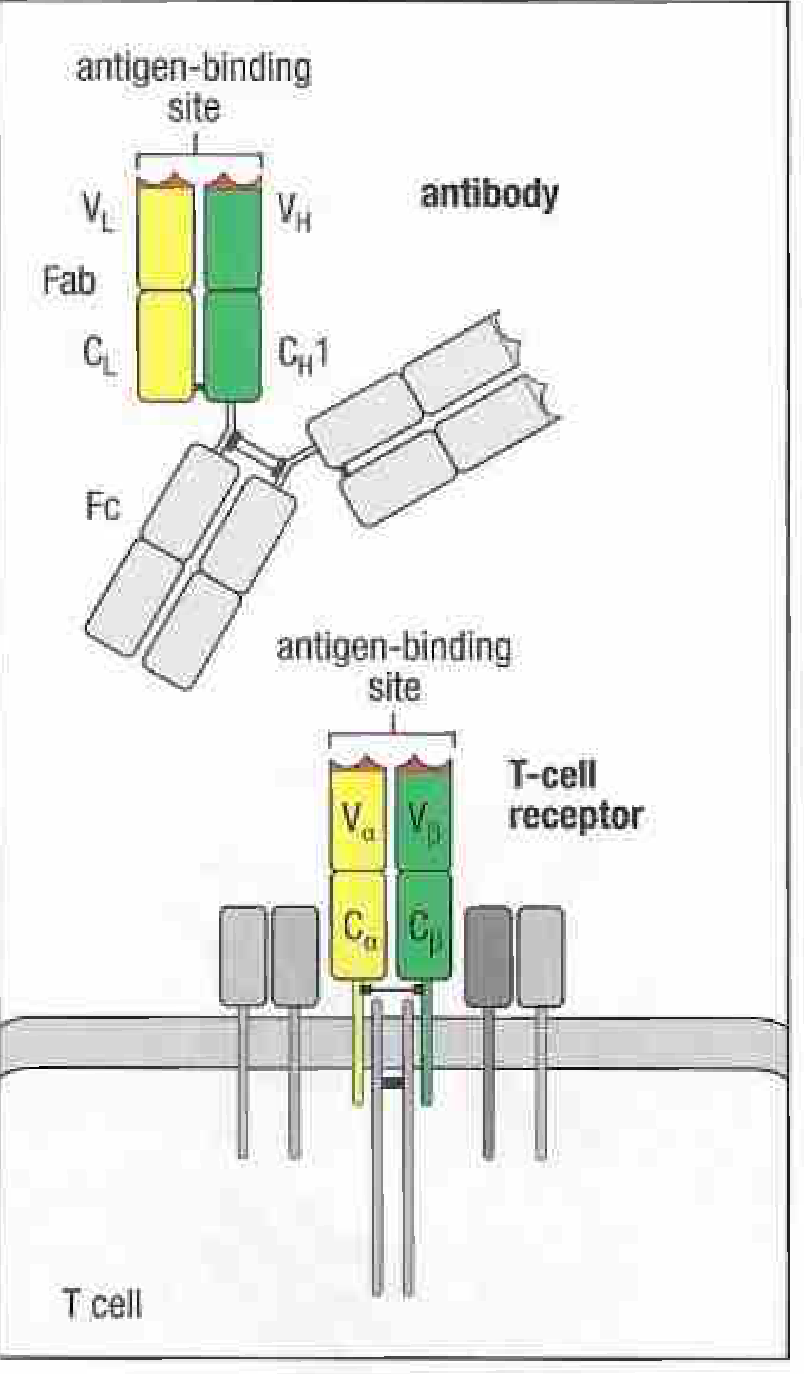
The most common form is the αβ TCR: TCRα and TCRβ chains, each containing an Ig-like variable domain and a constant domain, linked by a disulfide bond. The juxtaposition of the Vα and Vβ domains forms the antigen-recognition site.
Each T cell carries approximately 30,000 identical TCRs on its surface. The αβ heterodimer is always co-expressed with the CD3 complex (γ, δ, ε, and ζ chains), which handles signal transduction rather than antigen binding. A minority of T cells express an alternative γδ TCR with distinct recognition properties and functions still being clarified. - Janeway's Immunobiology 10e
What the TCR Recognizes: The pMHC Complex
T cells recognize a peptide:MHC (pMHC) complex - the TCR makes simultaneous contacts with BOTH the peptide fragment AND the MHC molecule presenting it. This "dual recognition" is called MHC restriction.
Antigen cannot be recognized by the TCR unless the protein is first unfolded, proteolytically processed into peptide fragments, and loaded into MHC molecules - a process handled by antigen-presenting cells (APCs).
4. MHC Molecules - The Presentation Platform
MHC molecules (encoded in the major histocompatibility complex, chromosome 6 in humans) are the essential intermediaries that display peptides to T cells.
MHC Class I vs. Class II
| Feature | MHC Class I | MHC Class II |
|---|---|---|
| Structure | α chain + β2-microglobulin | α chain + β chain (both MHC-encoded) |
| Peptide groove | Closed at both ends (8-10 aa peptides) | Open at both ends (13-25 aa peptides) |
| Source of peptides | Endogenous/cytosolic proteins (viruses, tumor antigens) | Exogenous proteins (endocytosed pathogens) |
| T cell type activated | CD8+ cytotoxic T cells | CD4+ helper T cells |
| Expression | All nucleated cells | Professional APCs (DCs, macrophages, B cells) |
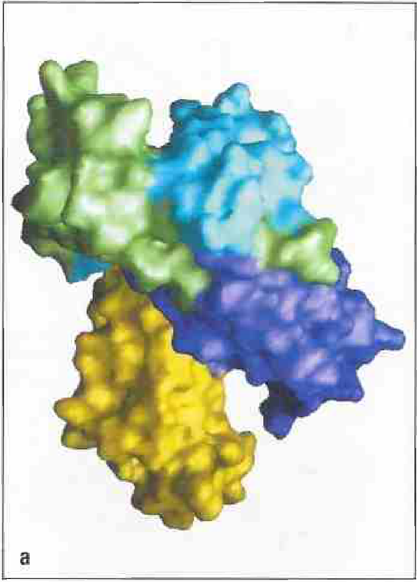
The peptide-binding groove is formed by the α1 and α2 domains of MHC class I, or the α1 and β1 domains of MHC class II. Polymorphic residues lining these pockets determine which peptides each MHC allele can present - explaining why different individuals respond differently to the same pathogen.
5. Antigen Presentation - How Peptides Get Loaded
Endogenous Pathway (MHC Class I)
- Cytosolic proteins (e.g., viral proteins) are degraded by the proteasome
- Peptides are transported into the ER by TAP transporters
- Loaded onto newly synthesized MHC class I molecules
- Complex travels to the cell surface → recognized by CD8+ T cells
Exogenous Pathway (MHC Class II)
- Extracellular antigens are taken up by endocytosis into phagolysosomes
- Proteases (cathepsins) degrade proteins into peptides
- CLIP (class II-associated invariant chain peptide) is displaced by the peptide
- Loaded MHC class II complex travels to the cell surface → recognized by CD4+ T cells
Cross-Presentation
Dendritic cells can also present exogenous antigens via MHC class I (cross-presentation), enabling CD8+ T cell responses against extracellular pathogens.
6. The Role of Antigen-Presenting Cells (APCs) and Costimulation
Naive T cells scan APCs by moving along the fibroblast reticular network in secondary lymphoid organs. When a T cell's TCR recognizes its specific pMHC complex, biochemical signals cause the T cell to arrest, stabilizing contact with the APC and initiating activation.
However, TCR engagement alone is not sufficient for full T cell activation. A second "costimulatory" signal is required:
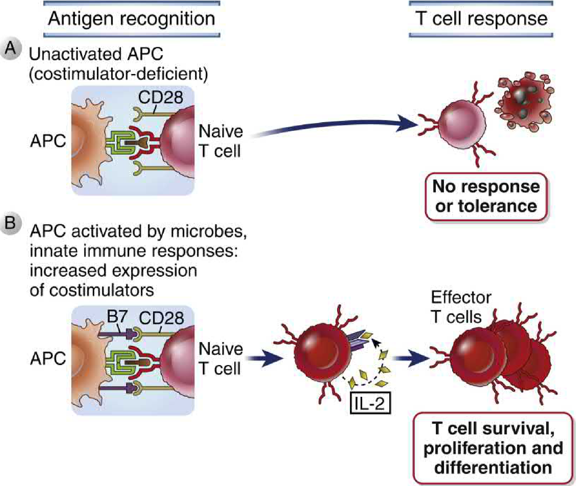
- Signal 1: TCR binds pMHC complex (antigen-specific)
- Signal 2: B7 molecules on APCs (upregulated by microbial stimuli/innate immunity) bind CD28 on T cells
- Without Signal 2: T cells become anergic (unresponsive) or die - a critical mechanism of peripheral tolerance
- With both signals: T cells produce IL-2, proliferate, and differentiate into effector and memory cells
This two-signal requirement ensures T cells are only activated against pathogens (which trigger innate immunity and APC activation), not against harmless self-antigens. - Cellular and Molecular Immunology
7. Clonal Selection and Receptor Diversity
Each B and T lymphocyte expresses receptors of a single unique specificity, generated by V(D)J recombination of inherited germline gene segments during lymphocyte development. This process:
- Generates a receptor repertoire on the order of 10^15-10^18 different specificities
- Concentrates diversity in the CDR loops of the V regions
- Is active only in B and T lymphocytes (same machinery in both)
When a lymphocyte encounters its cognate antigen, it is clonally selected - it proliferates and differentiates into effector cells, all bearing identical receptors. This is the molecular basis of immunological specificity and memory. - Cellular and Molecular Immunology
Summary Table
| Feature | B Cells / BCR | αβ T Cells / TCR |
|---|---|---|
| Ligand recognized | Native antigen (protein, carbohydrate, lipid) | Peptide + MHC molecule (pMHC complex) |
| Epitope type | Conformational or linear | Linear peptide only |
| Processing required? | No | Yes (proteolytic + MHC loading) |
| Where recognition occurs | Extracellular fluid / body surfaces | Cell surface of APCs |
| Effector output | Antibody secretion | Cytotoxicity (CD8+) or cytokine help (CD4+) |
| MHC requirement | None | MHC class I (CD8+) or II (CD4+) |
| Costimulation needed | T cell help (CD40L-CD40) | B7-CD28 interaction |
Sources:
- Janeway's Immunobiology, 10th Edition - Sections 4-14 through 4-16
- Cellular and Molecular Immunology - Chapters 5 and 9
This is a shared conversation. Sign in to Orris to start your own chat.