
Historical aspects of hernia
inguinal hernia repair history
Historical Aspects of Hernia
- Lotheissen introduced the first true Cooper's ligament repair in 1898, fixing the pectineal (Cooper's) ligament to Poupart's (inguinal) ligament, which addressed both inguinal and femoral defects.
- McVay later popularized this Cooper's ligament repair, adding a relaxing incision to reduce wound tension.
- The Shouldice technique, introduced in 1958 as a refinement of Bassini's operation, used meticulous multilayer closure of the transversalis fascia (in two layers) and is associated with excellent long-term recurrence rates, though it has a steeper learning curve.
| Period | Development |
|---|---|
| ~1500 BC | First documented reports of abdominal wall hernias (Egypt/Greece) |
| 1st century AD | First operative repairs (scrotal incision + orchiectomy) |
| ~700 AD | Sac excision with cord/testis en bloc |
| 14th century | First inguinal vs. femoral classification |
| 1559 | Direct vs. indirect inguinal hernia described |
| 1700s | Paré's hernia truss |
| Late 1700s-1800s | Anatomical era (Hesselbach, Cooper, Camper, Scarpa, Gimbernat) |
| 1884-1889 | Bassini's tissue repair |
| 1898 | Lotheissen's Cooper's ligament repair |
| Early 1900s | Silver wire filigree patches; McVay repair |
| 1918 | Handley's silk darn |
| 1958 | Shouldice repair |
| Mid-1900s | Usher's polyethylene mesh |
| 1980s | Lichtenstein tension-free mesh repair |
| Early 1990s | Ger's laparoscopic repair, TAPP/TEP evolution |
| 2000s-present | Robot-assisted inguinal hernia repair |
- Schwartz's Principles of Surgery, 11th Ed. - Inguinal Hernia, History section, p. 1626-1627
- Maingot's Abdominal Operations - Groin Hernia, History section, p. 142-143
- Fischer's Mastery of Surgery, 8th Ed. - Open Preperitoneal Inguinal Hernia Repair, Introduction
Create a presentation on key hernia surgeons and their innovations
Bassini inguinal hernia repair surgical technique diagram
Lichtenstein tension-free mesh inguinal hernia repair diagram
laparoscopic TEP TAPP inguinal hernia repair diagram
inguinal hernia anatomy groin
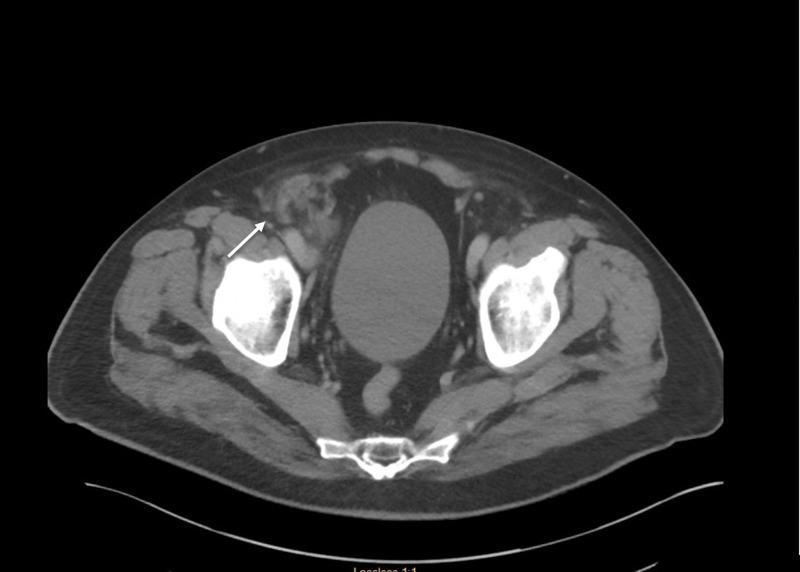
This diagnostic image is an axial computed tomography (CT) scan of the lower abdomen and pelvis. It reveals the bony anatomy of the pelvis, including the bilateral iliac bones, sacrum, and right femoral head, characterized by high-density (bright white) attenuation. In the midline, a fluid-filled urinary bladder is visible. The primary pathological finding, indicated by a white arrow, is a heterogeneous soft tissue mass in the right groin region, consistent with an incarcerated right femoral hernia. The hernia sac contains an inflamed appendix (De Garengeot hernia), evidenced by a tubular structure with surrounding fat stranding and increased attenuation, indicating localized inflammation. The mass is positioned medial to the femoral vessels and posterior to the inguinal ligament, typical for femoral hernia anatomy. This image illustrates a rare clinical presentation where the vermiform appendix becomes trapped within a femoral hernia sac, leading to appendicitis within the groin bulge.
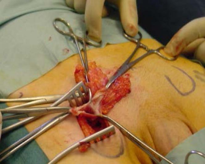
This clinical photograph captures an intraoperative view of a right femoral hernia repair using a low inguinal (Lockwood) approach. The surgical field displays a transverse skin incision in the groin area, with the underlying subcutaneous tissues and fascia dissected to reveal the femoral hernia sac. Retractors are positioned to provide exposure, while surgical clamps and forceps are utilized to manipulate the tissues. A prominent preperitoneal fat pad is visible, appearing as a yellowish, lobulated mass associated with the hernia sac. The sac itself exhibits a thin, fibrous, and membranous texture. The surrounding tissues demonstrate typical surgical hyperemia. This image is an educational example of inguinal anatomy during hernia management, specifically highlighting the identification of the hernia sac and its contents before reduction and mesh placement.
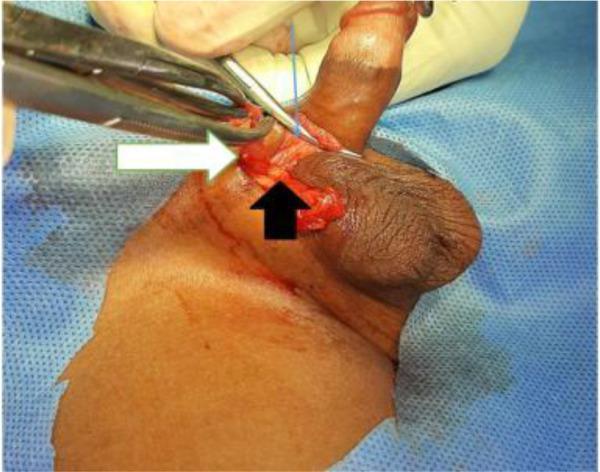
This clinical photograph captures an intraoperative view of a pediatric inguinal hernia repair performed via a high scrotal approach. The surgical field displays the dissected inguinal-scrotal region with key anatomical structures identified by colored arrows. A thick white arrow points to the translucent, reddish hernia sac being held and retracted by surgical forceps. A thin blue arrow indicates the spermatic fascia, which has been opened to expose the internal structures. A short black arrow highlights the cord structures, encompassing the vas deferens and testicular vessels, which are being carefully separated from the hernia sac. The image illustrates the critical surgical step of isolating the sac from the spermatic cord to ensure high ligation while preserving vital reproductive and vascular components. This visual is used to demonstrate surgical anatomy during inguinal herniotomy and to contrast the scrotal incision technique with the traditional groin crease approach.
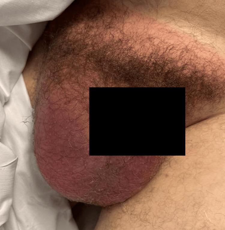
Clinical photograph of the male pelvic and groin region depicting a large, right-sided indirect inguinal hernia with scrotal extension. The image shows a significant, asymmetrical protrusion originating from the lower right inguinal area, descending deeply into the scrotum, causing marked distension and displacement of scrotal anatomy. The overlying skin exhibits diffuse, intense erythema and localized edema, suggesting an acute inflammatory or vascular process. These visual features are consistent with a strangulated or incarcerated inguinal hernia, where compromising of the blood supply to the herniated contents (such as small bowel or colon) and secondary ischemia of the scrotal structures, including the testicle, may occur. The presentation illustrates a surgical emergency requiring immediate diagnostic evaluation and operative intervention. This clinical image is intended for educational use in general surgery, emergency medicine, and urology to demonstrate the physical examination findings of complicated hernias.
Edoardo Bassini surgeon portrait public domain
Ambroise Paré Astley Cooper portrait wikimedia commons public domain
Irving Lichtenstein surgeon hernia photo Edward Shouldice photo
mkdir -p /tmp/workspace/hernia-surgeons-ppt/assets && ls -la /tmp/workspace/
cd /tmp/workspace/hernia-surgeons-ppt && python /tmp/skills/shared/scripts/fetch_images.py \ "https://upload.wikimedia.org/wikipedia/commons/thumb/b/b7/Portrait_of_Edoardo_Bassini_Wellcome_M0017969.jpg/960px-Portrait_of_Edoardo_Bassini_Wellcome_M0017969.jpg" \ "https://upload.wikimedia.org/wikipedia/commons/thumb/c/c9/Portrait_of_Ambroise_Pare_%281510_-_1590%29%2C_French_surgeon_Wellcome_V0004474ER.jpg/330px-Portrait_of_Ambroise_Pare_%281510_-_1590%29%2C_French_surgeon_Wellcome_V0004474ER.jpg" \ "https://michigansurgery.com/wp-content/uploads/2015/04/Lichtenstein.jpg" \ "https://michigansurgery.com/wp-content/uploads/2015/04/shouldice.jpg" \ > fetch_result.json 2>fetch_err.log; echo EXIT:$?; cat fetch_result.json | python3 -c "import json,sys; d=json.load(sys.stdin); [print(x['url'], 'OK' if x['base64'] else 'FAIL: '+str(x['error'])) for x in d]"
~/hernia-surgeons-ppt/build_deck.py
#!/usr/bin/env python3
"""Build 'Key Hernia Surgeons and Their Innovations' presentation."""
import json
import base64
from io import BytesIO

from pptx import Presentation
from pptx.util import Inches, Pt, Emu
from pptx.dml.color import RGBColor
from pptx.enum.text import PP_ALIGN, MSO_ANCHOR
from pptx.enum.shapes import MSO_SHAPE
from pptx.oxml.ns import qn

# ---------- palette ----------
NAVY = RGBColor(0x0B, 0x2E, 0x4E)
TEAL = RGBColor(0x11, 0x6C, 0x7A)
GOLD = RGBColor(0xC9, 0x9A, 0x3A)
LIGHT_BG = RGBColor(0xF4, 0xF1, 0xEA)
WHITE = RGBColor(0xFF, 0xFF, 0xFF)
DARK_TEXT = RGBColor(0x22, 0x22, 0x22)
GREY_TEXT = RGBColor(0x5A, 0x5A, 0x5A)
MAROON = RGBColor(0x7A, 0x22, 0x22)

with open("fetch_result.json") as f:
    imgs = json.load(f)
IMG = {i["url"]: i["base64"] for i in imgs if i.get("base64")}

def img_stream(url):
    b64 = IMG[url]
    data = b64.split(",", 1)[1] if "," in b64 else b64
    return BytesIO(base64.b64decode(data))

BASSINI_URL = "https://upload.wikimedia.org/wikipedia/commons/thumb/b/b7/Portrait_of_Edoardo_Bassini_Wellcome_M0017969.jpg/960px-Portrait_of_Edoardo_Bassini_Wellcome_M0017969.jpg"
PARE_URL = "https://upload.wikimedia.org/wikipedia/commons/thumb/c/c9/Portrait_of_Ambroise_Pare_%281510_-_1590%29%2C_French_surgeon_Wellcome_V0004474ER.jpg/330px-Portrait_of_Ambroise_Pare_%281510_-_1590%29%2C_French_surgeon_Wellcome_V0004474ER.jpg"
LICHTENSTEIN_URL = "https://michigansurgery.com/wp-content/uploads/2015/04/Lichtenstein.jpg"
SHOULDICE_URL = "https://michigansurgery.com/wp-content/uploads/2015/04/shouldice.jpg"

prs = Presentation()
prs.slide_width = Inches(13.333)
prs.slide_height = Inches(7.5)
SW, SH = prs.slide_width, prs.slide_height
BLANK = prs.slide_layouts[6]

def add_slide():
    return prs.slides.add_slide(BLANK)

def set_bg(slide, color=LIGHT_BG):
    bg = slide.background
    bg.fill.solid()
    bg.fill.fore_color.rgb = color

def rect(slide, x, y, w, h, color, line=None):
    sh = slide.shapes.add_shape(MSO_SHAPE.RECTANGLE, x, y, w, h)
    sh.fill.solid()
    sh.fill.fore_color.rgb = color
    if line is None:
        sh.line.fill.background()
    else:
        sh.line.color.rgb = line
        sh.line.width = Pt(1)
    sh.shadow.inherit = False
    return sh

def textbox(slide, x, y, w, h, text, size=18, color=DARK_TEXT, bold=False,
            italic=False, align=PP_ALIGN.LEFT, font="Calibri", anchor=None,
            line_spacing=None, wrap=True):
    tb = slide.shapes.add_textbox(x, y, w, h)
    tf = tb.text_frame
    tf.word_wrap = wrap
    if anchor:
        tf.vertical_anchor = anchor
    tf.margin_left = 0
    tf.margin_right = 0
    tf.margin_top = 0
    tf.margin_bottom = 0
    lines = text.split("\n")
    for i, line in enumerate(lines):
        p = tf.paragraphs[0] if i == 0 else tf.add_paragraph()
        p.text = line
        p.alignment = align
        if line_spacing:
            p.line_spacing = line_spacing
        for r in p.runs:
            r.font.size = Pt(size)
            r.font.color.rgb = color
            r.font.bold = bold
            r.font.italic = italic
            r.font.name = font
    return tb

def bullets(slide, x, y, w, h, items, size=15, color=DARK_TEXT, font="Calibri",
            bullet_color=None, line_spacing=1.15, space_after=8, bold_first=False):
    tb = slide.shapes.add_textbox(x, y, w, h)
    tf = tb.text_frame
    tf.word_wrap = True
    tf.margin_left = 0
    tf.margin_right = 0
    for i, item in enumerate(items):
        p = tf.paragraphs[0] if i == 0 else tf.add_paragraph()
        p.text = f"•  {item}"
        p.line_spacing = line_spacing
        p.space_after = Pt(space_after)
        for r in p.runs:
            r.font.size = Pt(size)
            r.font.color.rgb = color
            r.font.name = font
    return tb

def page_number(slide, n):
    textbox(slide, SW - Inches(0.8), SH - Inches(0.45), Inches(0.6), Inches(0.3),
            str(n), size=11, color=GREY_TEXT, align=PP_ALIGN.RIGHT)

def header_bar(slide, title, subtitle=None):
    rect(slide, 0, 0, SW, Inches(1.15), NAVY)
    rect(slide, 0, Inches(1.15), SW, Pt(3), GOLD)
    textbox(slide, Inches(0.6), Inches(0.18), Inches(11), Inches(0.6), title,
            size=28, color=WHITE, bold=True, font="Georgia")
    if subtitle:
        textbox(slide, Inches(0.6), Inches(0.72), Inches(11.5), Inches(0.4), subtitle,
                size=14, color=RGBColor(0xCF, 0xDD, 0xE3), italic=True, font="Calibri")

def picture_framed(slide, url, x, y, w, h, border=GOLD):
    frame = rect(slide, x - Pt(3), y - Pt(3), w + Pt(6), h + Pt(6), border)
    pic = slide.shapes.add_picture(img_stream(url), x, y, height=h)
    # center crop width if too wide, else keep aspect via height only
    if pic.width > w:
        pic.width = w
        pic.left = x
    else:
        pic.left = int(x + (w - pic.width) / 2)
    return pic

# =====================================================================
# SLIDE 1 — TITLE
# =====================================================================
s = add_slide()
set_bg(s, NAVY)
rect(s, 0, Inches(6.7), SW, Inches(0.8), GOLD)
textbox(s, Inches(0.9), Inches(2.3), Inches(11.5), Inches(1.6),
        "Key Hernia Surgeons\nand Their Innovations", size=44, color=WHITE,
        bold=True, font="Georgia", line_spacing=1.05)
textbox(s, Inches(0.9), Inches(4.0), Inches(11), Inches(0.6),
        "A Historical Journey Through Groin Hernia Surgery: 1500 BC to the Robotic Era",
        size=18, color=RGBColor(0xD8, 0xC9, 0x9A), italic=True, font="Calibri")
textbox(s, Inches(0.9), Inches(6.85), Inches(8), Inches(0.4),
        "Compiled from Schwartz's Principles of Surgery, Maingot's Abdominal Operations & Fischer's Mastery of Surgery",
        size=11, color=NAVY, font="Calibri")

# =====================================================================
# SLIDE 2 — TIMELINE OVERVIEW
# =====================================================================
s = add_slide()
set_bg(s)
header_bar(s, "Overview: A Timeline of Innovation", "From ancient trusses to robotic mesh repair")

events = [
    ("1500s", "Ambroise Paré", "Popularized the hernia truss"),
    ("1700s-1800s", "The Anatomists", "Hesselbach, Cooper, Scarpa, Gimbernat map the groin"),
    ("1884", "Edoardo Bassini", "First tissue-based tension repair"),
    ("1898", "Georg Lotheissen", "Cooper's ligament repair"),
    ("1942-58", "Chester McVay / E.E. Shouldice", "McVay & multilayer Shouldice repairs"),
    ("1950s-60s", "Francis Usher", "Polyethylene synthetic mesh"),
    ("1984", "Irving Lichtenstein", "Tension-free mesh repair"),
    ("1990s", "Ralph Ger & pioneers", "Laparoscopic TAPP / TEP repair"),
    ("2000s→", "Modern Innovators", "Robotic-assisted hernia repair"),
]
top = Inches(1.55)
row_h = Inches(0.62)
x0 = Inches(0.6)
rect(s, Inches(1.85), top + Inches(0.05), Pt(2.5), row_h*len(events)-Inches(0.1), GOLD)
for i, (yr, name, note) in enumerate(events):
    y = top + row_h*i
    circ = s.shapes.add_shape(MSO_SHAPE.OVAL, Inches(1.77), y+Inches(0.09), Inches(0.18), Inches(0.18))
    circ.fill.solid(); circ.fill.fore_color.rgb = TEAL; circ.line.fill.background(); circ.shadow.inherit=False
    textbox(s, x0, y, Inches(1.1), Inches(0.4), yr, size=13, color=MAROON, bold=True, align=PP_ALIGN.RIGHT)
    textbox(s, Inches(2.15), y-Inches(0.02), Inches(3.3), Inches(0.4), name, size=15, color=NAVY, bold=True)
    textbox(s, Inches(5.6), y, Inches(7.1), Inches(0.5), note, size=13, color=GREY_TEXT)
page_number(s, 2)

# =====================================================================
# SLIDE 3 — Ambroise Paré
# =====================================================================
s = add_slide()
set_bg(s)
header_bar(s, "Ambroise Paré (1510–1590)", "The truss era: conservative management")
picture_framed(s, PARE_URL, Inches(0.7), Inches(1.7), Inches(3.0), Inches(4.6))
textbox(s, Inches(0.7), Inches(6.45), Inches(3.0), Inches(0.4), "French army surgeon",
        size=12, color=GREY_TEXT, italic=True, align=PP_ALIGN.CENTER)
bullets(s, Inches(4.2), Inches(1.7), Inches(8.4), Inches(4.5), [
    "Renowned 16th-century French military surgeon, often called the father of modern surgery.",
    "In the 1700s (era associated with his teachings and successors), the hernia truss became the dominant recommended treatment for abdominal wall and inguinal hernias.",
    "Reflected the prevailing philosophy of the time: conservative, non-operative management was preferred, with surgery reserved for strangulation or other complications.",
    "Operative repairs at that time still relied on wide scrotal incisions and often required orchiectomy (removal of the testicle), so avoiding surgery whenever possible was the rational choice.",
    "His legacy set the stage for centuries of truss-based management before anatomical understanding advanced enough to make elective surgical repair safe.",
], size=16)
rect(s, Inches(4.2), Inches(6.55), Inches(8.4), Inches(0.55), TEAL)
textbox(s, Inches(4.4), Inches(6.62), Inches(8.0), Inches(0.4),
        "Innovation: Championed the hernia truss as safe, effective non-operative therapy",
        size=13, color=WHITE, bold=True)
page_number(s, 3)

# =====================================================================
# SLIDE 4 — The Anatomists
# =====================================================================
s = add_slide()
set_bg(s)
header_bar(s, "The Anatomists (Late 1700s–1800s)", "Mapping the groin: the foundation for safe surgery")
names = [
    ("Franz Kaspar Hesselbach", "Described Hesselbach's triangle — the region bounded by the inguinal ligament, inferior epigastric vessels, and rectus sheath, still used to classify direct vs. indirect hernias."),
    ("Sir Astley Cooper", "Detailed the pectineal (Cooper's) ligament along the pubic bone, later the anchor point for the Cooper's ligament and McVay repairs."),
    ("Antonio Scarpa", "Clarified the superficial fascial layers of the abdominal wall and groin (Scarpa's fascia)."),
    ("Antonio de Gimbernat", "Described Gimbernat's ligament, defining the medial boundary of the femoral canal."),
    ("Peter Camper & August Richter", "Further defined fascial planes and named partial (Richter's) hernia of the bowel wall."),
]
y = Inches(1.55)
colw = Inches(6.0)
for i, (name, desc) in enumerate(names):
    col = i % 2
    row = i // 2
    x = Inches(0.6) + col*Inches(6.3)
    yy = y + row*Inches(1.85)
    rect(s, x, yy, Inches(0.08), Inches(1.6), GOLD)
    textbox(s, x+Inches(0.25), yy, colw, Inches(0.4), name, size=15, color=NAVY, bold=True)
    textbox(s, x+Inches(0.25), yy+Inches(0.4), colw-Inches(0.2), Inches(1.3), desc, size=12.5, color=GREY_TEXT, line_spacing=1.1)
textbox(s, Inches(0.6), Inches(6.85), Inches(11.5), Inches(0.5),
        "Innovation: Cadaveric dissection gave surgeons the anatomical vocabulary and landmarks needed for precise, reproducible groin repair.",
        size=13, color=MAROON, bold=True, italic=True)
page_number(s, 4)

# =====================================================================
# SLIDE 5 — Edoardo Bassini
# =====================================================================
s = add_slide()
set_bg(s)
header_bar(s, "Edoardo Bassini (1844–1924)", "Father of modern inguinal hernia surgery")
picture_framed(s, BASSINI_URL, Inches(0.7), Inches(1.7), Inches(3.0), Inches(4.6))
bullets(s, Inches(4.2), Inches(1.7), Inches(8.4), Inches(4.3), [
    "First performed his novel operation in 1884 in Padua, Italy; published outcomes in 1889.",
    "Reported just 5 recurrences among over 250 patients with complete 5-year follow-up — an unheard-of result for the era.",
    "Technique: high ligation of the hernia sac at the internal ring, plus suture reinforcement of the posterior inguinal wall — approximating the transversalis fascia and conjoint tendon medially to the inguinal ligament laterally.",
    "Closed the canal in a deep layer (transversalis fascia repair) and a superficial layer (external oblique aponeurosis).",
    "Transformed hernia repair from a high-morbidity, high-recurrence operation into a safe, reliable procedure — the template for all subsequent tissue repairs (McVay, Shouldice, Desarda).",
], size=15.5)
rect(s, Inches(4.2), Inches(6.4), Inches(8.4), Inches(0.7), MAROON)
textbox(s, Inches(4.4), Inches(6.48), Inches(8.0), Inches(0.55),
        "Innovation: The first successful tissue-based tension repair of the groin\n(the 'Bassini repair') — foundation of modern herniorrhaphy",
        size=13, color=WHITE, bold=True, line_spacing=1.1)
page_number(s, 5)

# =====================================================================
# SLIDE 6 — Lotheissen & McVay
# =====================================================================
s = add_slide()
set_bg(s)
header_bar(s, "Georg Lotheissen & Chester McVay", "The Cooper's ligament repair")
rect(s, Inches(0.6), Inches(1.55), Inches(5.9), Inches(5.0), WHITE, line=RGBColor(0xDD,0xDD,0xDD))
rect(s, Inches(6.8), Inches(1.55), Inches(5.9), Inches(5.0), WHITE, line=RGBColor(0xDD,0xDD,0xDD))
textbox(s, Inches(0.9), Inches(1.75), Inches(5.4), Inches(0.5), "Georg Lotheissen — 1898", size=17, color=NAVY, bold=True)
bullets(s, Inches(0.9), Inches(2.35), Inches(5.3), Inches(3.9), [
    "Introduced the first true Cooper's ligament repair.",
    "Fixed the pectineal (Cooper's) ligament to Poupart's (inguinal) ligament.",
    "Key advance: a single repair that addressed both inguinal AND femoral hernia defects simultaneously — closing the femoral canal as well as the inguinal floor.",
], size=14.5, line_spacing=1.2)
textbox(s, Inches(7.1), Inches(1.75), Inches(5.4), Inches(0.5), "Chester McVay — 1940s", size=17, color=NAVY, bold=True)
bullets(s, Inches(7.1), Inches(2.35), Inches(5.3), Inches(3.9), [
    "Popularized and refined the Cooper's ligament repair for widespread clinical use.",
    "Added a relaxing incision in the anterior rectus sheath to reduce excessive wound tension from pulling tissue down to Cooper's ligament.",
    "The McVay repair became the preferred tissue repair specifically for femoral hernias and large direct defects.",
], size=14.5, line_spacing=1.2)
textbox(s, Inches(0.6), Inches(6.75), Inches(12.1), Inches(0.5),
        "Innovation: Extended tissue repair to reliably treat femoral hernias, not just inguinal defects",
        size=13, color=MAROON, bold=True, italic=True)
page_number(s, 6)

# =====================================================================
# SLIDE 7 — Shouldice
# =====================================================================
s = add_slide()
set_bg(s)
header_bar(s, "Edward Earle Shouldice (1890–1965)", "The multilayer Shouldice repair")
picture_framed(s, SHOULDICE_URL, Inches(0.7), Inches(1.7), Inches(3.0), Inches(4.6))
bullets(s, Inches(4.2), Inches(1.7), Inches(8.4), Inches(4.3), [
    "Canadian surgeon who introduced his technique in 1958 as a refinement of Bassini's original operation.",
    "Involves meticulous dissection of the entire inguinal floor, with the transversalis fascia closed in two overlapping layers (rather than Bassini's single layer), followed by two further layers of the internal oblique/conjoint tendon and external oblique.",
    "Popularized local anesthesia and early post-operative ambulation — patients walked soon after surgery, unusual for the time.",
    "Founded the Shouldice Hospital (Toronto), a dedicated high-volume hernia centre; the technique is technically demanding for beginners but achieves excellent long-term recurrence rates.",
    "Remains one of the best-performing pure tissue (non-mesh) repairs in the world literature.",
], size=15)
rect(s, Inches(4.2), Inches(6.45), Inches(8.4), Inches(0.6), TEAL)
textbox(s, Inches(4.4), Inches(6.53), Inches(8.0), Inches(0.45),
        "Innovation: Multilayer imbrication repair + early ambulation model of hernia care",
        size=13, color=WHITE, bold=True)
page_number(s, 7)

# =====================================================================
# SLIDE 8 — Usher & synthetic mesh
# =====================================================================
s = add_slide()
set_bg(s)
header_bar(s, "Francis Usher & the Birth of Synthetic Mesh", "Early-to-mid 20th century")
bullets(s, Inches(0.7), Inches(1.7), Inches(11.9), Inches(3.2), [
    "Early 1900s: silver wire filigree sheets were the first prosthetic patches used to reinforce the inguinal floor — but they suffered metal fatigue, recurrence, and could erode into adjacent structures or even the peritoneal cavity.",
    "1918: Handley introduced silk as a prosthetic 'darn' material to bridge tissue gaps without tension; nylon followed a few years later, but both lost strength over time and increased infection risk when used in heavier weights.",
    "Francis Usher introduced the modern synthetic patch: a plastic monofilament polymer (polyethylene) mesh placed across the inguinal floor — the direct forerunner of today's polypropylene mesh.",
    "This breakthrough proved that a durable, biologically inert synthetic material could reinforce the abdominal wall indefinitely without degrading — paving the way for the tension-free mesh era.",
], size=16, line_spacing=1.25)
rect(s, Inches(0.7), Inches(5.4), Inches(11.9), Inches(0.7), MAROON)
textbox(s, Inches(0.9), Inches(5.5), Inches(11.5), Inches(0.5),
        "Innovation: First durable synthetic (polyethylene) mesh patch for hernia reinforcement",
        size=14, color=WHITE, bold=True)
textbox(s, Inches(0.7), Inches(6.35), Inches(11.9), Inches(0.7),
        "This set the stage for Irving Lichtenstein's tension-free repair using improved polypropylene mesh in the 1980s.",
        size=13, color=GREY_TEXT, italic=True)
page_number(s, 8)

# =====================================================================
# SLIDE 9 — Lichtenstein
# =====================================================================
s = add_slide()
set_bg(s)
header_bar(s, "Irving Lichtenstein (1920–2000)", "The tension-free mesh repair revolution")
picture_framed(s, LICHTENSTEIN_URL, Inches(0.7), Inches(1.7), Inches(3.0), Inches(4.6))
bullets(s, Inches(4.2), Inches(1.7), Inches(8.4), Inches(4.3), [
    "American surgeon who, in the 1980s, applied a flat sheet of polypropylene mesh over the floor of the inguinal canal — described formally in 1984.",
    "Created a truly tension-free repair: instead of suturing native tissues together under tension (as in Bassini, McVay, Shouldice), the mesh itself restored the strength of the transversalis fascia.",
    "Demonstrated outcomes superior to tissue-based repairs, was reproducible regardless of hernia size or type, and had a markedly short learning curve.",
    "Results were achievable by both expert and non-expert hernia surgeons alike — a critical factor in its rapid, worldwide adoption.",
    "The open Lichtenstein mesh repair remains the most widely performed open inguinal hernia operation in the world today.",
], size=15.5)
rect(s, Inches(4.2), Inches(6.45), Inches(8.4), Inches(0.6), TEAL)
textbox(s, Inches(4.4), Inches(6.53), Inches(8.0), Inches(0.45),
        "Innovation: The open tension-free mesh repair — the modern gold-standard open technique",
        size=13, color=WHITE, bold=True)
page_number(s, 9)

# =====================================================================
# SLIDE 10 — Laparoscopic pioneers
# =====================================================================
s = add_slide()
set_bg(s)
header_bar(s, "Ralph Ger & the Laparoscopic Pioneers", "Minimally invasive hernia repair, 1980s–1990s")
bullets(s, Inches(0.7), Inches(1.7), Inches(11.9), Inches(3.4), [
    "Ralph Ger provided the first description of a laparoscopic approach to inguinal hernia closure in the early 1980s, laying the conceptual groundwork for minimally invasive groin surgery.",
    "As laparoscopic techniques diffused through general surgery in the early 1990s, this evolved into two dominant modern approaches:",
    "     – Transabdominal Preperitoneal repair (TAPP): entry through the peritoneal cavity to place mesh in the preperitoneal space.",
    "     – Totally Extraperitoneal repair (TEP): mesh placed in the preperitoneal space without entering the peritoneal cavity, avoiding intra-abdominal adhesion risk.",
    "Intraperitoneal Onlay Mesh (IPOM) was also developed as a further laparoscopic variant.",
], size=15.5, line_spacing=1.2)
rect(s, Inches(0.7), Inches(5.75), Inches(11.9), Inches(0.7), MAROON)
textbox(s, Inches(0.9), Inches(5.85), Inches(11.5), Inches(0.5),
        "Innovation: Minimally invasive access to the preperitoneal space, reducing pain and speeding recovery",
        size=14, color=WHITE, bold=True)
textbox(s, Inches(0.7), Inches(6.6), Inches(11.9), Inches(0.5),
        "Multicenter data (1998) reported a recurrence rate of just 0.4% across over 10,000 laparoscopic repairs.",
        size=12.5, color=GREY_TEXT, italic=True)
page_number(s, 10)

# =====================================================================
# SLIDE 11 — Robotic era
# =====================================================================
s = add_slide()
set_bg(s)
header_bar(s, "The Robotic Era", "2000s to present: the newest chapter")
bullets(s, Inches(0.7), Inches(1.7), Inches(11.9), Inches(3.6), [
    "Robotic-assisted platforms extended laparoscopic principles (TAPP/TEP) with articulated wrists, 3D visualization, and tremor filtration.",
    "Rapidly adopted by general surgeons for improved ergonomics, dexterity in tight preperitoneal planes, and enhanced visualization compared with straight-stick laparoscopy.",
    "Facilitates precise mesh fixation and complex bilateral or recurrent hernia repairs with a more favorable ergonomic learning curve for surgeons transitioning from open surgery.",
    "Represents the continuation of a centuries-long trend in hernia surgery: progressively less invasive access with equal or improved durability of repair.",
], size=16, line_spacing=1.25)
rect(s, Inches(0.7), Inches(5.35), Inches(11.9), Inches(0.7), TEAL)
textbox(s, Inches(0.9), Inches(5.45), Inches(11.5), Inches(0.5),
        "Innovation: Robot-assisted inguinal hernia repair — better ergonomics and visualization",
        size=14, color=WHITE, bold=True)
textbox(s, Inches(0.7), Inches(6.3), Inches(11.9), Inches(0.6),
        "Outcome metrics today center on three pillars: recurrence rate, chronic pain, and quality of life — the same goals Bassini pursued in 1884.",
        size=13, color=GREY_TEXT, italic=True)
page_number(s, 11)

# =====================================================================
# SLIDE 12 — Summary table
# =====================================================================
s = add_slide()
set_bg(s)
header_bar(s, "Summary: Surgeon → Innovation", "At a glance")
rows = [
    ("Surgeon", "Era", "Key Innovation"),
    ("Ambroise Paré", "1500s", "Hernia truss (conservative therapy)"),
    ("Hesselbach, Cooper, Scarpa, Gimbernat", "1700s–1800s", "Anatomical mapping of the groin"),
    ("Edoardo Bassini", "1884–1889", "First successful tissue tension repair"),
    ("Georg Lotheissen", "1898", "Cooper's ligament repair"),
    ("Chester McVay", "1940s", "Relaxing-incision Cooper's ligament repair"),
    ("Edward Earle Shouldice", "1958", "Multilayer imbrication repair"),
    ("Francis Usher", "1950s–60s", "Polyethylene synthetic mesh"),
    ("Irving Lichtenstein", "1984", "Open tension-free mesh repair"),
    ("Ralph Ger & successors", "1980s–90s", "Laparoscopic TAPP / TEP repair"),
    ("Modern surgeons", "2000s→", "Robotic-assisted repair"),
]
top = Inches(1.55)
rh = Inches(0.51)
col_x = [Inches(0.6), Inches(5.4), Inches(8.3)]
col_w = [Inches(4.7), Inches(2.8), Inches(4.4)]
rect(s, Inches(0.6), top, Inches(12.1), rh, NAVY)
for c, (x, w) in enumerate(zip(col_x, col_w)):
    textbox(s, x+Inches(0.1), top+Inches(0.08), w-Inches(0.2), Inches(0.4), rows[0][c],
            size=13, color=WHITE, bold=True)
for i, row in enumerate(rows[1:]):
    y = top + rh*(i+1)
    bg = WHITE if i % 2 == 0 else RGBColor(0xEA, 0xE6, 0xDA)
    rect(s, Inches(0.6), y, Inches(12.1), rh, bg)
    for c, (x, w) in enumerate(zip(col_x, col_w)):
        textbox(s, x+Inches(0.1), y+Inches(0.07), w-Inches(0.2), Inches(0.4), row[c],
                size=12, color=DARK_TEXT, bold=(c==0))
page_number(s, 12)

# =====================================================================
# SLIDE 13 — Sources
# =====================================================================
s = add_slide()
set_bg(s, NAVY)
textbox(s, Inches(0.9), Inches(0.7), Inches(11), Inches(0.8), "Sources", size=32, color=WHITE, bold=True, font="Georgia")
rect(s, Inches(0.9), Inches(1.55), Inches(3.2), Pt(3), GOLD)
bullets(s, Inches(0.9), Inches(1.9), Inches(11.3), Inches(4.5), [
    "Schwartz's Principles of Surgery, 11th Edition — Inguinal Hernia, History (p. 1626–1627)",
    "Maingot's Abdominal Operations — Groin Hernia, History (p. 142–143)",
    "Fischer's Mastery of Surgery, 8th Edition — Open Preperitoneal Inguinal Hernia Repair, Introduction",
    "Bulyk I, Shkarban V, Vasyliuk S. The history of inguinal hernia surgery. Rozhl Chir. 2023. PMID: 37344194",
    "Grebić D, Havidić K. Historical review of surgical techniques for operative treatment of inguinal hernia. Acta Med Hist Adriat. 2025. PMID: 40213846",
    "Portrait images: Wellcome Collection / Wikimedia Commons (Bassini, Paré, public domain); Michigan Surgery — 'A Brief History of Hernia Surgery' (Lichtenstein, Shouldice)",
], size=15, color=RGBColor(0xE9, 0xE4, 0xD4), line_spacing=1.3)
textbox(s, Inches(0.9), Inches(6.8), Inches(11), Inches(0.4), "Prepared for educational purposes.",
        size=12, color=RGBColor(0xB9, 0xC4, 0xCF), italic=True)

prs.save("Key_Hernia_Surgeons_and_Innovations.pptx")
print("Saved.")
cd /tmp/workspace/hernia-surgeons-ppt && python3 build_deck.py
cd /tmp/workspace/hernia-surgeons-ppt && python3 -c " from pptx import Presentation prs = Presentation('Key_Hernia_Surgeons_and_Innovations.pptx') print('Slides:', len(prs.slides)) for i, slide in enumerate(prs.slides,1): texts = [sh.text_frame.text[:40] for sh in slide.shapes if sh.has_text_frame and sh.text_frame.text.strip()] print(i, '->', texts[:2]) " ls -la Key_Hernia_Surgeons_and_Innovations.pptx
Key Hernia Surgeons and Innovations
Presentation · PPTX
- Title slide
- Timeline overview (visual chronology of all figures)
- Ambroise Paré - the hernia truss era (with portrait)
- The Anatomists - Hesselbach, Cooper, Scarpa, Gimbernat, Camper/Richter
- Edoardo Bassini - first successful tissue tension repair (with portrait)
- Georg Lotheissen & Chester McVay - Cooper's ligament repair
- Edward Earle Shouldice - multilayer repair (with portrait)
- Francis Usher - birth of synthetic (polyethylene) mesh
- Irving Lichtenstein - tension-free mesh repair revolution (with portrait)
- Ralph Ger & laparoscopic pioneers - TAPP/TEP
- The robotic era
- Summary table (surgeon → era → innovation, at a glance)
- Sources/citations