
Ossifying fibroma histopathology
Reading File
Ossifying fibroma is a benign fibro-osseous lesion of the jaw. Histologically it shows a cellular fibrous stroma with mineralized material (woven bone trabeculae and/or psammomatoid calcifications). Let me pull the detailed textbook content now.
Searching sources...
Finding Sources
Finding Sources
Searching PubMed
"ossifying fibroma" AND histopathology
Reading File
Reading File
Reading File
Finding Sources
Reading File
Reading File
Finding Sources
I now have comprehensive information from multiple authoritative sources. Let me also check for any histology images available.
Finding Sources
I now have thorough coverage from multiple textbooks. Here is the detailed answer:
Ossifying Fibroma - Histopathology
Overview
Ossifying fibroma (OF) is a true benign neoplasm (not a developmental anomaly) belonging to the fibro-osseous lesion group. This distinction from fibrous dysplasia is critical and was formally proposed by Reed in 1963. The 2017 WHO classification divides OF into three clinicopathologic entities based on location and calcification pattern.
Core Histopathological Features
All variants share two main tissue components:
1. Fibrous Stroma
- Cellular fibrous connective tissue background
- The stroma is initially highly cellular and may become less cellular as the lesion matures
- The stroma is separated from surrounding bone by a fibrous capsule/pseudocapsule, which distinguishes OF from fibrous dysplasia and allows the lesion to shell out cleanly during surgical enucleation
2. Mineralized Component
The type and pattern of calcification varies by subtype:
| Feature | Classic OF / COF | Juvenile Trabecular OF | Juvenile Psammomatoid OF |
|---|---|---|---|
| Calcification pattern | Woven bone trabeculae + cementicles | Predominantly osteoid/trabecular bone | Psammomatoid bodies |
| Osteoblastic rimming | Peripheral osteoblasts present | Plump, prominent osteoblasts | Less prominent |
| Stroma cellularity | Moderate, becomes less cellular with maturity | Highly cellular | Highly cellular |
| Special structures | Rounded cementicles + dystrophic calcifications | Irregular bony spicules | Small acellular ossicles resembling psammoma bodies |
Detailed Features by Variant
A. Classic Ossifying Fibroma (and Cemento-Ossifying Fibroma)
- Fibrous and cellular stroma early on, then develops:
- Trabeculae of woven bone with peripheral osteoblasts
- Dystrophic calcifications
- Rounded cementicles (cementum-like droplets)
- With maturation, calcifications fuse forming a dense calcified mass
- A peripheral fibrous zone (capsule) remains, enabling clean surgical shelling
(Histological appearances mirror radiographic progression: starts radiolucent, becomes mixed, then radiopaque)
B. Juvenile Trabecular Ossifying Fibroma
- Stroma is highly cellular
- Calcification pattern is trabecular - irregular bony spicules of osteoid rimmed by plump osteoblasts
- Affects the maxillary alveolar process in children/adolescents
- More locally aggressive behavior
C. Juvenile Psammomatoid Ossifying Fibroma (APOF)
- Stroma is cellular and fibrous
- Calcification pattern: numerous small, rounded, lamellated ossicles resembling psammoma bodies (as seen in meningiomas)
- These psammomatoid bodies are acellular and EMA (epithelial membrane antigen)-positive on immunohistochemistry
- Arises mainly in fronto-orbital and ethmoid bones
Key Histological Distinction: OF vs. Fibrous Dysplasia
| Feature | Ossifying Fibroma | Fibrous Dysplasia |
|---|---|---|
| Capsule | Present (fibrous capsule) | Absent (merges with adjacent bone) |
| Bone maturity | More mature (woven → lamellar), osteoblastic rimming | Immature woven bone, no osteoblastic rimming |
| Stroma | Cellular fibrous, demarcated | Fibrous, blends with bone |
| Calcification | Trabeculae + cementicles/psammomatoid bodies | Irregular "Chinese letter" or "alphabet soup" trabeculae |
| Nature | True neoplasm | Genetic developmental anomaly |
Summary of Histopathology (high-yield)
- Fibrous cellular stroma + mineralized component (woven bone / cementum-like material)
- Woven bone trabeculae with peripheral osteoblastic rimming
- Rounded cementicles (in classic/COF)
- Psammomatoid ossicles in APOF (EMA+, acellular)
- Well-defined fibrous capsule enabling enucleation
- Absence of osteoblastic rimming in FD is the key differentiating feature
Sources:
- Scott-Brown's Otorhinolaryngology Head & Neck Surgery, Vol 1, p. 1150
- Scott-Brown's Otorhinolaryngology Head & Neck Surgery (Jaw lesions), p. Neoplastic OF section
- Cummings Otolaryngology Head and Neck Surgery, p. 996 (Ossifying Fibroma & Fibrous Dysplasia)
- K.J. Lee's Essential Otolaryngology, p. 762
A recent 2025 systematic review (PMID 39631529, Gautier et al.) on OF and juvenile OF covers clinical/radiological parameters and recurrence - no contradictions with established histopathological descriptions above.
Images of both
Looking for histopathology images of ossifying fibroma and fibrous dysplasia from the textbooks.
Searching sources...
Finding Sources
Finding Sources
Reading File
Finding Sources
Finding Sources
Finding Sources
Good - I found images. Now let me search specifically for histopathology images of fibrous dysplasia (the "Chinese letter" / whorled pattern) and ossifying fibroma, and fetch them.
Finding Sources
Reading File
I found exactly what's needed. Now let me fetch all the key histopathology images:
Loading Image
Loading Image
Here are both histopathology images from the textbooks:
Ossifying Fibroma - Histopathology
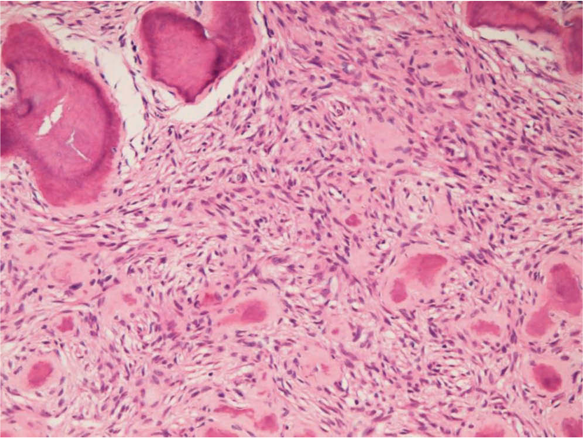
Figure 25.11 - Pathological image of a fibro-osseous lesion (ossifying fibroma / cemento-osseous dysplasia). H&E stain showing:
- Cellular fibrous stroma (spindle-shaped fibroblasts in the background)
- Rounded, well-defined calcified bodies (cementicles / psammomatoid bodies) - the pink globular structures scattered throughout
- Woven bone trabeculae with surrounding fibrous tissue (upper left)
- The mineralized islands have a smooth, rounded outline, consistent with cementum-like material
(Source: Scott-Brown's Otorhinolaryngology Head & Neck Surgery, Fig. 25.11)
Fibrous Dysplasia - Histopathology (for comparison)
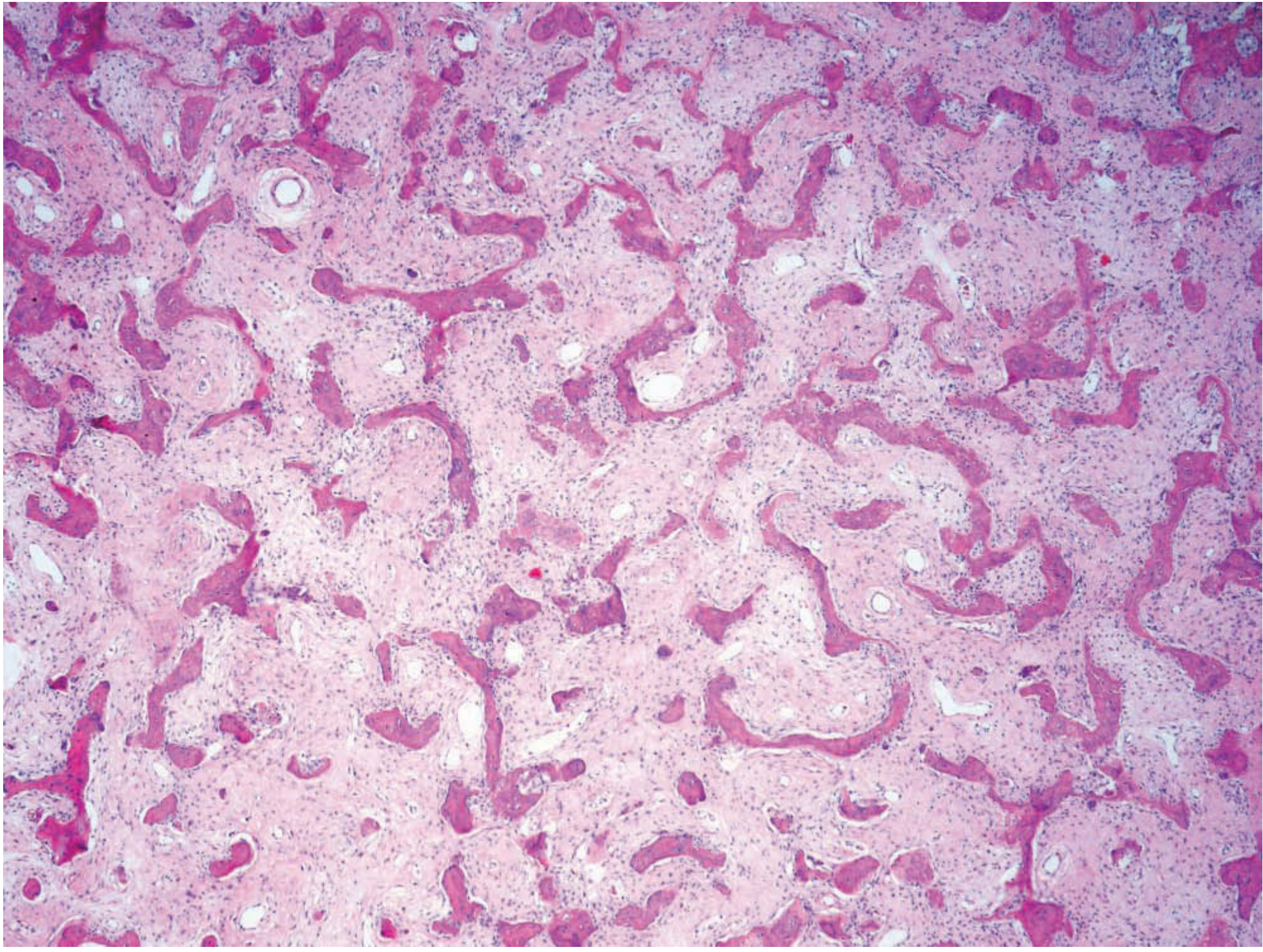
Figure 25.12 - Histological picture of typical fibrous dysplasia. H&E stain showing:
- Cellular fibrous stroma (similar background to OF)
- Curved, irregular "Chinese character" (alphabet soup) trabeculae of woven bone - the highly irregular, curvilinear pink bone islands that resemble Chinese script
- No osteoblastic rimming around the trabeculae (key distinguishing feature from OF)
- Trabeculae blend directly into the surrounding fibrous stroma without a capsule
(Source: Scott-Brown's Otorhinolaryngology Head & Neck Surgery, Fig. 25.12)
Key Visual Distinction
| Ossifying Fibroma | Fibrous Dysplasia | |
|---|---|---|
| Bone shape | Rounded / curved trabeculae with osteoblastic rim | Curvilinear "Chinese character" trabeculae, no rim |
| Boundary | Well-defined capsule | Merges with adjacent bone |
| Calcifications | Rounded cementicles / psammomatoid bodies | Irregular woven bone spicules only |
This is a shared conversation. Sign in to Orris to start your own chat.