
Blow in and blow out fractures in orbital fractures
Reading File
Blow-in and blow-out fractures are two distinct types of orbital wall fractures caused by opposing mechanisms. In a blow-out, a blunt force to the orbit increases intraorbital pressure and herniates the orbital floor (or medial wall) downward/inward into the maxillary sinus, trapping orbital fat and sometimes the inferior rectus. In a blow-in, orbital wall fragments are pushed inward into the orbit itself, reducing the orbital volume.
Searching sources...
Finding Sources
Finding Sources
Reading File
Reading File
Reading File
Finding Sources
Reading File
Reading File
Reading File
Reading File
Reading File
Reading File
Reading File
Reading File
Loading Image
Loading Image
Here is a comprehensive overview of blow-out and blow-in orbital fractures, drawn from multiple authoritative sources.
Blow-Out and Blow-In Orbital Fractures
Overview
Isolated orbital wall fractures are classified as either blow-out or blow-in based on the direction of fragment displacement relative to the orbit. These two fracture types are almost mirror images of each other mechanically.
Blow-Out Fracture
Definition
A fracture of the orbital floor (or medial wall) with an intact orbital rim. Bony fragments are displaced outward - away from the orbit, into the adjacent paranasal sinuses (most commonly the maxillary antrum, or into the ethmoid air cells for medial wall fractures).
Most Common Site
The posteromedial orbital floor (maxillary bone, medial to the infraorbital neurovascular bundle) - this is the weakest point of the orbital floor.
Mechanism (Two Theories)
-
Hydraulic Theory (Smith and Converse, 1956 cadaveric study): A blunt object smaller than the orbital diameter strikes the globe/orbital rim, pushing orbital contents posteriorly. The resulting sudden increase in intraorbital pressure causes fracture at the weakest point - the posteromedial orbital floor.
-
Buckling Theory: A direct blow to the orbital rim causes a buckling stress wave that fractures the thinnest part of the floor, without necessarily compressing orbital contents.
Both mechanisms can operate simultaneously and are not mutually exclusive. - K.J. Lee's Essential Otolaryngology, p. 1119
Key Clinical Features
| Feature | Detail |
|---|---|
| Globe position | Enophthalmos (globe sinks back due to increased orbital volume) |
| Diplopia | Binocular vertical/oblique - especially on upgaze |
| Sensory loss | Ipsilateral cheek, upper lip, and teeth (infraorbital nerve hypesthesia) |
| Emphysema | Subcutaneous/conjunctival crepitus after nose blowing |
| Other | Epistaxis, eyelid edema, ecchymosis |
Important note: Enophthalmos may be masked initially by orbital edema and hemorrhage; the true extent becomes apparent 2-4 weeks after injury.
"White-Eye" Blow-Out Fracture (Trapdoor Fracture)
A pediatric variant involving a trapdoor mechanism:
- A small floor fracture opens under orbital pressure, letting orbital soft tissue (and sometimes inferior rectus) herniate through
- The elastic pediatric bone recoils quickly, trapping the tissue before it can retract
- Children often show no subconjunctival hemorrhage (hence "white eye")
- Presents with oculocardiac reflex: bradycardia, nausea, vomiting, syncope - a surgical emergency, as muscle ischemia and fibrosis can occur within hours
- More common in children due to more flexible (greenstick) bones - Wills Eye Manual, p. 107; Bailey and Love's Surgery, p. 434
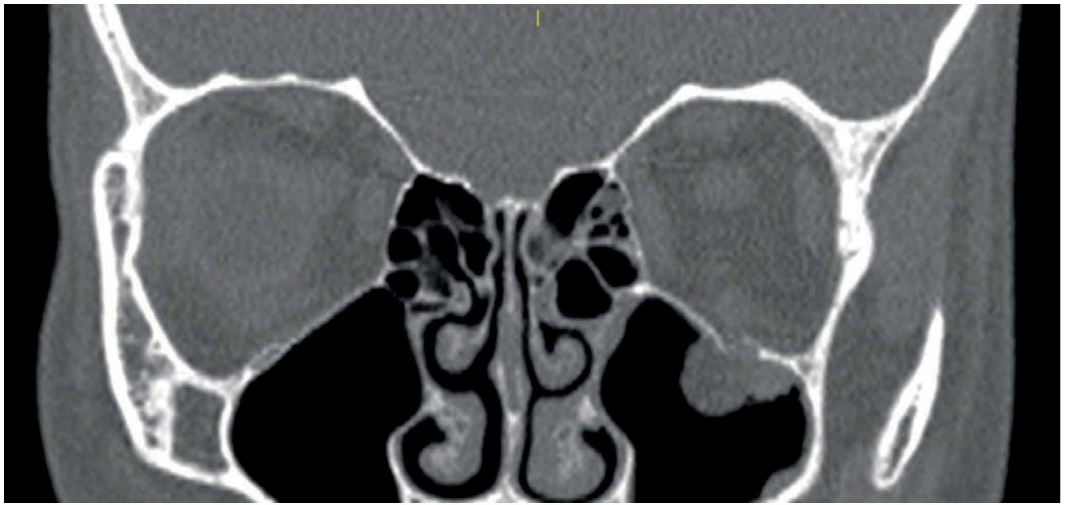
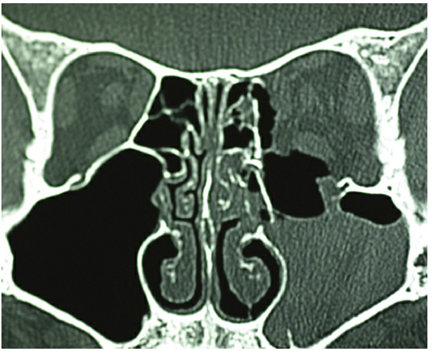
Blow-In Fracture
Definition
The opposite of blow-out: direct trauma to the orbital rim causes a bone fragment to be displaced into the orbit (rather than into an adjacent sinus). This reduces the orbital volume.
Key Clinical Feature
- Exophthalmos (proptosis), not enophthalmos - because orbital volume is reduced by the inward-displaced fragment
- Orbital roof blow-in ("impure blow-in fracture") is the most commonly encountered type of orbital roof fracture - K.J. Lee's Essential Otolaryngology, p. 1128
Orbital Roof Blow-In
- Most often an "impure" blow-in - involves both the orbital roof and the orbital rim
- More common in children (thinner frontal bone; pediatric facial fractures are 18-35% orbital roof involvement)
- Features may include: upper eyelid hematoma, forehead hypoesthesia, contour deformity, CSF rhinorrhea, neurologic deficits, and proptosis from prolapsed intracranial contents
- Management always requires interdisciplinary input (ENT, neurosurgery, ophthalmology); intracranial injuries are addressed first
Direct Comparison
| Feature | Blow-Out | Blow-In |
|---|---|---|
| Fragment direction | Away from orbit (into sinus) | Into the orbit |
| Orbital volume | Increased | Decreased |
| Globe position | Enophthalmos | Exophthalmos/proptosis |
| Rim involvement | Rim intact ("pure") | Rim typically involved ("impure") |
| Common site | Orbital floor (floor or medial wall) | Orbital roof |
| Mechanism | Hydraulic pressure or buckling | Direct trauma to orbital rim |
| Common finding | Inferior rectus entrapment, diplopia on upgaze | Possible proptosis, vision compromise from fragment |
Surgical Indications
For Blow-Out Fractures (Dutton criteria):
- Persistent symptomatic diplopia with positive forced ductions and CT evidence of entrapment, not improving in 1-2 weeks
- Early enophthalmos ≥3 mm
- Significant globe ptosis (hypoglobus)
- Floor defect >50% (or >2 cm²) likely to cause late enophthalmos
- Associated rim/facial fractures
For Blow-In Fractures:
- Position of bone fragments may compromise vision - surgical indication in itself
- Displaced orbital roof fractures with pulsatile proptosis or CSF leak unresolved after 1 week require repair
- K.J. Lee's Essential Otolaryngology, pp. 1119-1128; Wills Eye Manual, pp. 104-111; Schwartz's Principles of Surgery, p. 2852; Bailey and Love's Surgery, p. 434
Evaluation
- CT scan (thin-section axial and coronal, non-contrast): gold standard - evaluates fracture extent, size, configuration, soft tissue entrapment, orbital volume, and hemorrhage
- Forced duction test: grasping the conjunctival tissue at the limbus to passively move the globe - distinguishes muscle entrapment (restricted) from cranial nerve palsy (no restriction)
- Rule out intraocular injury and optic neuropathy in every orbital fracture patient
This is a shared conversation. Sign in to Orris to start your own chat.