
Flow chart for pathophysiology for asthma
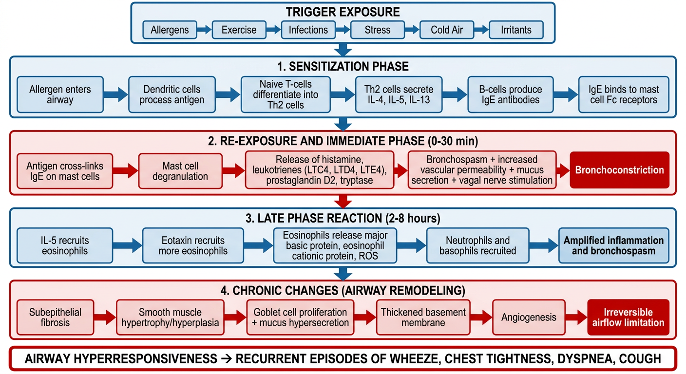
Detailed medical flowchart showing the pathophysiology of asthma. The flowchart starts at the top with "Trigger Exposure" (allergens, exercise, infections, stress, cold air, irritants) and flows downward through these sequential steps with connecting arrows: 1. Sensitization Phase: Allergen enters airway → Dendritic cells process antigen → Naive T-cells differentiate into Th2 cells → Th2 cells secrete IL-4, IL-5, IL-13 → B-cells produce IgE antibodies → IgE binds to mast cell Fc receptors. 2. Re-exposure and Immediate Phase (0-30 min): Antigen cross-links IgE on mast cells → Mast cell degranulation → Release of histamine, leukotrienes (LTC4, LTD4, LTE4), prostaglandin D2, tryptase → Bronchospasm + increased vascular permeability + mucus secretion + vagal nerve stimulation → Bronchoconstriction. 3. Late Phase Reaction (2-8 hours): IL-5 recruits eosinophils → Eotaxin recruits more eosinophils → Eosinophils release major basic protein, eosinophil cationic protein, ROS → Neutrophils and basophils recruited → Amplified inflammation and bronchospasm. 4. Chronic Changes (Airway Remodeling): Subepithelial fibrosis → Smooth muscle hypertrophy/hyperplasia → Goblet cell proliferation + mucus hypersecretion → Thickened basement membrane → Angiogenesis → Irreversible airflow limitation. Final outcome box: "Airway Hyperresponsiveness → Recurrent Episodes of Wheeze, Chest Tightness, Dyspnea, Cough". Use a clean clinical white background with blue and red color coding for immune cells vs structural changes, clear box shapes, and professional medical education style.
Asthma Pathophysiology — Summary
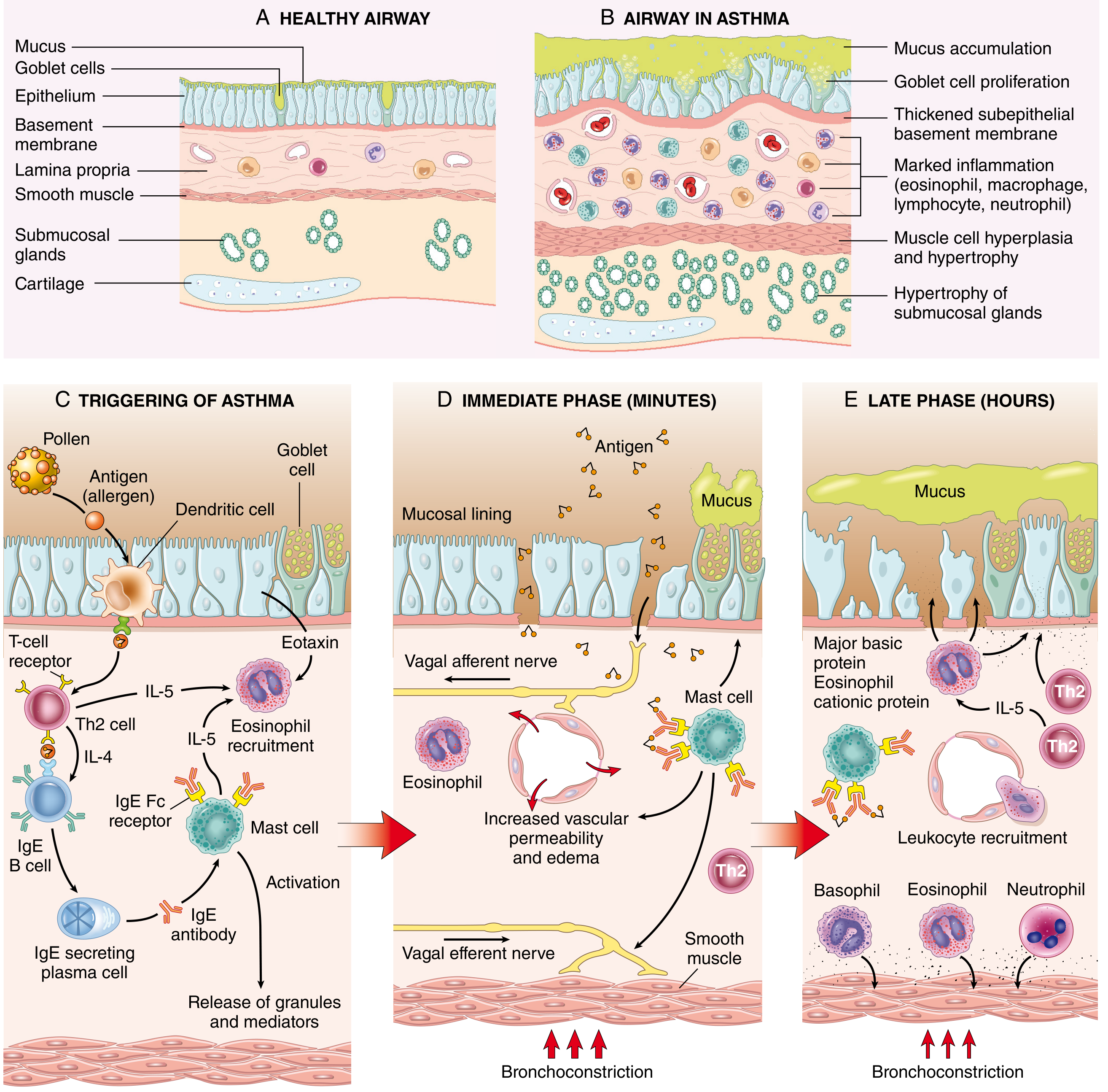
1. Sensitization Phase (First Exposure)
- Trigger (allergen, e.g., pollen, dust mite) enters the airway
- Dendritic cells process and present antigen to naïve T-cells
- T-cells differentiate into Th2 cells, secreting:
- IL-4 → stimulates B-cells to produce IgE
- IL-5 → activates eosinophils
- IL-13 → mucus secretion + IgE production
- IgE binds to Fc receptors on mast cells (sensitization complete)
2. Immediate Phase (0–30 min on re-exposure)
- Antigen cross-links IgE on mast cells → mast cell degranulation
- Released mediators:
- Leukotrienes (LTC4, LTD4, LTE4) — prolonged bronchoconstriction + mucus
- Histamine — bronchospasm + vasodilation
- Prostaglandin D2 — bronchoconstriction
- Acetylcholine (via vagal reflex) — smooth muscle contraction
- Result: bronchospasm, increased vascular permeability, mucus hypersecretion
3. Late-Phase Reaction (2–8 hours)
- IL-5 + eotaxin recruit eosinophils, neutrophils, basophils, and more T-cells
- Eosinophils release:
- Major basic protein (MBP) and eosinophil cationic protein (ECP) → epithelial damage
- ROS → oxidative injury
- Galectin-10 → forms Charcot-Leyden crystals → amplifies inflammation
- Sustained bronchoconstriction and airway wall inflammation
4. Chronic Changes — Airway Remodeling
- Subepithelial fibrosis (thickened basement membrane)
- Smooth muscle hypertrophy and hyperplasia
- Goblet cell proliferation → excess mucus
- Angiogenesis and submucosal gland enlargement
- Result: irreversible airflow limitation and increased exacerbation severity
Final Outcome
Airway hyperresponsiveness → Recurrent episodes of wheeze, chest tightness, dyspnea, and cough
- Robbins, Cotran & Kumar — Pathologic Basis of Disease, Ch. 15 (Pathogenesis of Asthma, Fig. 15.10)
- Lippincott Illustrated Reviews: Pharmacology, Ch. 41 (Pathophysiology of Asthma)
- Fishman's Pulmonary Diseases and Disorders (Lymphocytes and T-cell subsets in asthma)