

What’s the impression No itching
purpura petechiae feet ankles non-blanching bilateral
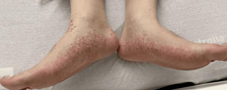
Type: Clinical Photograph. Anatomical Region: Bilateral feet and ankles (medial and lateral aspects). Findings: The image shows a symmetric, dense distribution of numerous small, non-blanching, erythematous to violaceous macules. These lesions are morphologically consistent with petechiae. The distribution is most concentrated around the ankles and heels, extending across the dorsal and lateral surfaces of the feet. The surrounding skin appears otherwise intact with no obvious ulceration, scaling, or crusting, though there is a subtle erythematous background suggesting underlying inflammation. Clinical Significance: These visual findings are characteristic manifestations of small-vessel vasculitis, such as leukocytoclastic vasculitis or Henoch-Schönlein purpura (IgA vasculitis), where cutaneous hemorrhage occurs due to vessel wall inflammation. This image is an educational example of dependent petechiae and purpura, often found in systemic inflammatory or autoimmune conditions.
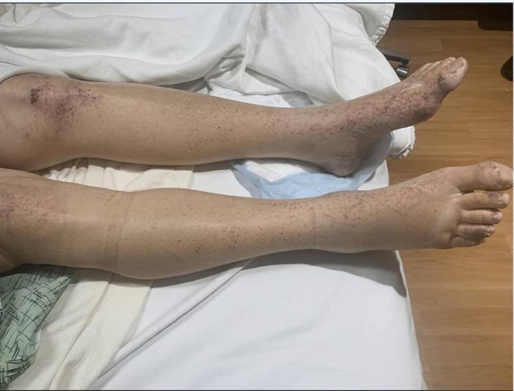
Clinical photograph of bilateral lower extremities from the knees to the feet, illustrating a widespread purpuric rash. The lesions consist of numerous reddish-purple, non-blanching petechiae and palpable purpura. The distribution is diffuse but demonstrates significant clustering and confluence over the patellar regions (knees) and distal extremities, including the ankles and dorsum of the feet. On the knees, the lesions form dark, dense ecchymotic-like patches. Distally, the feet and ankles show mild soft tissue swelling suggestive of dependent edema. The calves exhibit a more sparse, punctate distribution of petechiae. This visual presentation is characteristic of small-vessel vasculitis, such as IgA vasculitis (Henoch-Schönlein purpura) or leukocytoclastic vasculitis, where inflammation leads to the extravasation of red blood cells into the dermis. The density of lesions in dependent areas like the feet and friction-prone areas like the knees is a key clinical finding for medical students and clinicians.
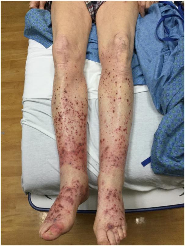
This clinical photograph displays a bilateral dermatological presentation on the lower extremities of a patient. The primary finding is widespread palpable purpura, appearing as non-blanching, reddish-purple to violaceous papules and macules. The distribution is symmetric, extending from the dorsal and plantar surfaces of the feet and ankles proximally toward the knees, involving both the anterior and posterior surfaces of the legs. The lesions vary in size and morphology, ranging from small petechiae to larger, confluent purpuric patches. The density of the eruption is highest in the distal regions (ankles and shins), gradually thinning out near the infrapatellar area. This visual presentation is characteristic of small-vessel vasculitis or cutaneous manifestations of systemic infections. In this specific clinical context, the findings represent a rare cutaneous vasculopathy associated with Varicella-zoster virus (VZV) infection. The image serves as a clinical example for students and practitioners to recognize the morphology and dependent distribution of palpable purpura.
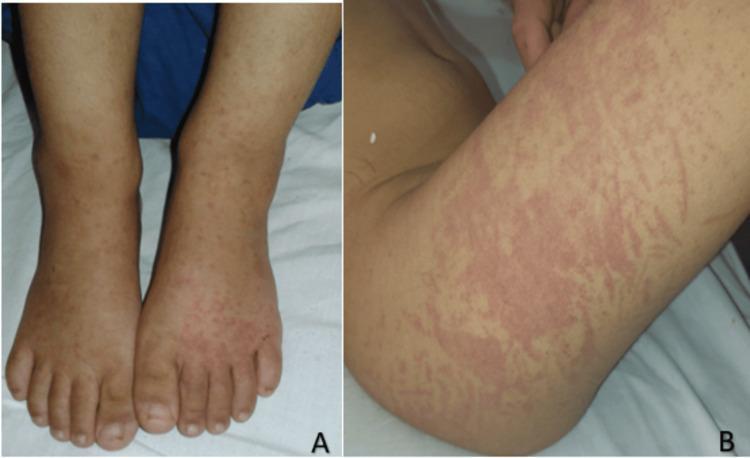
Two-panel clinical photograph illustrating the progression of vascular purpura in a pediatric patient. Panel A shows the dorsal aspect of both feet and lower legs featuring scattered, non-blanching, reddish-purple petechiae and small purpuric macules. Mild bilateral dependent edema is visible around the ankles. Panel B demonstrates the extension of the rash on the third day of admission, located on the thigh. This panel shows more confluent and larger purpuric patches with a distinctive reticular or livedoid pattern, characterized by net-like erythematous streaks and mottled discoloration. The lesions are typical of Henoch-Schönlein purpura (IgA vasculitis), demonstrating orthostatic distribution and evolution from discrete spots to confluent plaques. No secondary changes like ulceration or scaling are observed. The images serve as a teaching tool for recognizing small-vessel vasculitis morphology and its clinical evolution in pediatric dermatology and rheumatology.
Clinical Impression
Primary Diagnosis: Pigmented Purpuric Dermatosis (PPD) / Capillaritis
Morphological Analysis from the Image
- Non-blanching macules and petechiae - ranging from 1-2 mm pinpoint lesions to 1.5-2 cm confluent patches
- Multi-stage coloring: active dark-red/violaceous lesions (fresh extravasation) alongside reddish-brown and golden-brown areas (hemosiderin deposition from older, resolving lesions)
- Bilateral and roughly symmetrical distribution over the dorsum of both feet and ankles
- No ulceration, no scale, no vesicles
- No itching (as reported by the patient)
Why Schamberg Disease / PPD Fits Best
"The typical lesions are thumbprint-sized and composed of aggregates of pinhead-sized petechiae resembling grains of cayenne pepper on a background of golden-brown hemosiderin staining... The lesions usually begin on the lower legs... These lesions seldom itch. The favored sites are on the lower shins and ankles."
- No itching - hallmark of PPD (vs. eczematous PPD variant which does itch)
- Bilateral symmetric lower extremity distribution
- Mixed colors - fresh red/violaceous + brownish hemosiderin staining = lesions of varying ages
- Confined to feet and ankles (dependent/gravity-dependent area)
- No ulceration or necrosis (against vasculitis)
- No palpable purpura apparent (against leukocytoclastic vasculitis)
Differential Diagnoses to Consider
| Diagnosis | For | Against |
|---|---|---|
| Schamberg disease (PPD) | Non-itchy, bilateral feet/ankles, mixed red-brown hemosiderin, no necrosis | - |
| Leukocytoclastic vasculitis | Non-blanching lower limb distribution | Usually burning/painful, often palpable purpura, may ulcerate |
| IgA vasculitis (HSP) | Lower limb purpura | Typically in children, associated arthritis/renal involvement, palpable |
| Lichen aureus | Unilateral golden-brown patch, PPD type | Typically unilateral, single-segment |
| Thrombocytopenic purpura | Petechiae | Usually widespread, not limited to feet |
| Stasis purpura (gravitational) | Dependent areas, bilateral | Usually with edema, varicosities, lipodermatosclerosis |
Recommended Workup
- Baseline bloods: CBC (rule out thrombocytopenia), PT/INR, LFTs, renal function, ESR/CRP
- Urinalysis - to rule out IgA vasculitis (hematuria/proteinuria)
- Skin biopsy (punch biopsy) - confirmatory: shows lymphocytic capillaritis with extravasated RBCs and hemosiderin; Prussian Blue stain highlights hemosiderin
- Dermoscopy - may show coppery-red pigmentation, linear vessels, red dots
Management (if PPD confirmed)
- Reassurance
- Compression stockings (reduce hydrostatic pressure)
- Topical corticosteroids (limited evidence, mild cases)
- Colchicine or pentoxifylline (for persistent/widespread disease)
- Address any contributing drugs (NSAIDs, furosemide, amlodipine are known triggers)
Give me differentials with pictures
Schamberg disease pigmented purpuric dermatosis cayenne pepper legs
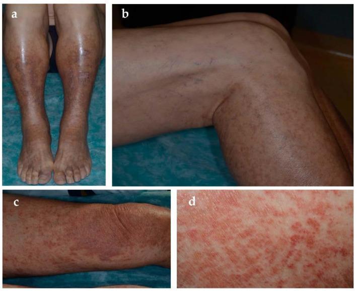
This composite of four clinical photographs (a–d) illustrates Schamberg disease, the most common form of pigmented purpuric dermatosis (PPD). Images (a) and (b) display a bilateral, diffuse distribution of skin lesions across the lower legs and feet. The primary visible feature is a persistent yellow-orange-brown discoloration caused by hemosiderin deposition, frequently associated with underlying venous hypertension. Overlaid on these hyperpigmented patches are numerous pinpoint, non-blanching petechiae, often described as having a 'cayenne pepper' appearance. Images (c) and (d) provide closer views of discrete yellow-red patches with dense, superimposed petechial spots, demonstrating the active inflammatory phase of the condition. The educational focus is on the characteristic morphology and color progression of PPD, which typically presents as chronic, asymptomatic macules and patches on the lower extremities. This visual material is relevant for dermatology training, emphasizing the clinical diagnosis of capillaritis and its differentiation from other purpuric or vasculitic conditions.
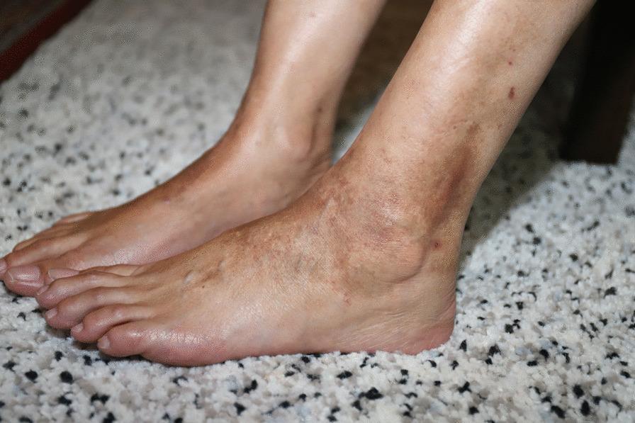
This clinical photograph displays the bilateral lower limbs and feet of a patient presenting with Pigmented Purpuric Dermatosis (PPD), likely Schamberg disease. The image shows a symmetrical distribution of non-blanching, reddish-brown to orange-brown macules and patches concentrated around the ankles and shins. Key visual features include a characteristic 'cayenne pepper' appearance caused by petechial hemorrhage within the lesions. The affected areas exhibit a combination of fresh purpura and chronic hyperpigmented patches resulting from hemosiderin deposition. The skin texture appears largely intact with fine wrinkling, without prominent edema, ulceration, or signs of major venous insufficiency. This image is educational for identifying the morphology of pigmented purpuric eruptions, which are clinically distinguished from vasculitis by their typical color and indolent appearance. Relevant medical concepts include red blood cell extravasation, capillary fragility, and medication-induced dermatoses.
leukocytoclastic vasculitis palpable purpura lower legs
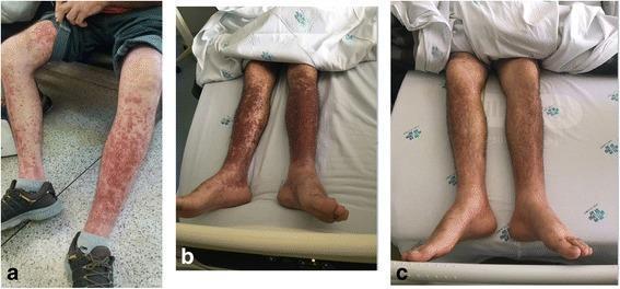
This series of clinical photographs (panels a, b, and c) documents the temporal progression and resolution of leukocytoclastic vasculitis manifesting as palpable purpura on the lower limbs of a patient following chemotherapy. Panel 'a' shows the initial presentation with discrete, erythematous to violaceous macules and papules concentrated on the shins and calves. Panel 'b' demonstrates the peak intensity of the condition, where the lesions have become confluent, forming dense, dark-purple purpuric plaques covering a significant surface area of the bilaterally affected legs. Panel 'c' captures the resolution phase, showing a marked decrease in the number and intensity of lesions, with the skin returning to near-normal pigmentation, though residual faint hyperpigmentation persists. The images illustrate a classic dermatological drug reaction or systemic response, characterized by the symmetrical distribution of purpuric lesions in gravity-dependent areas, typical of small-vessel vasculitis.
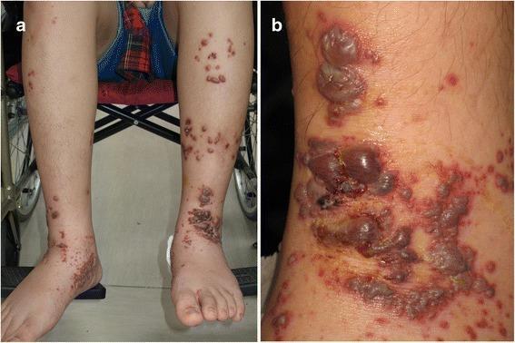
Clinical photograph of the lower extremities of a pediatric patient presenting with severe cutaneous manifestations of leukocytoclastic vasculitis, consistent with Henoch-Schönlein purpura (HSP). Panel (a) provides a bilateral view of both legs, showing extensive, palpable purpuric macules and papules distributed from the dorsal feet to the mid-shins. Interspersed among the purpura are multiple elevated vesicles and bullae. Panel (b) shows a close-up of the left ankle and dorsal foot, highlighting the progression of lesions. Visible features include large, tense hemorrhagic bullae (ranging from 2-20 mm), several of which have ruptured to form dark, adherent crusts and superficial erosions. The surrounding skin exhibits confluent erythematous and violaceous purpuric patches. These findings illustrate a rare, bullous variant of IgA vasculitis, demonstrating the characteristic progression from maculopapular purpura to hemorrhagic vesiculobullous lesions on gravity-dependent areas.
IgA vasculitis Henoch-Schonlein purpura lower extremity rash
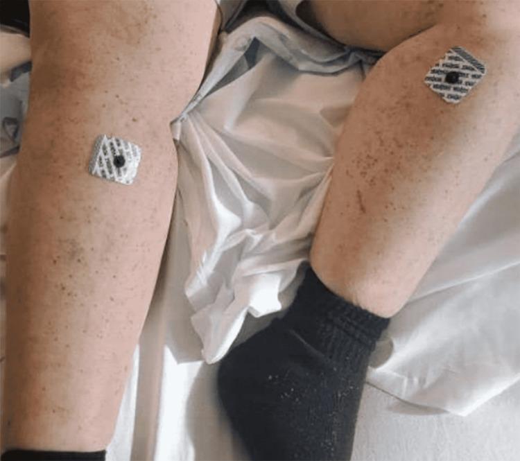
Clinical photograph of the bilateral lower extremities of a 47-year-old female, illustrating the resolving phase of a maculopapular rash, diagnosed as adult-onset IgA vasculitis (Henoch-Schönlein purpura). The skin shows a diffuse distribution of residual erythematous macules and small papules, primarily located on the shins and lower thighs. Some lesions exhibit post-inflammatory hyperpigmentation, indicating healing. Two square ECG/monitoring adhesive electrode patches are visible: one on the right lower thigh and one on the left shin. The patient is wearing a black compression-style sock on the left foot and ankle. This image serves as an educational example of the clinical progression and partial resolution of systemic vasculitis following corticosteroid treatment, highlighting the characteristic predilection for lower extremity involvement.
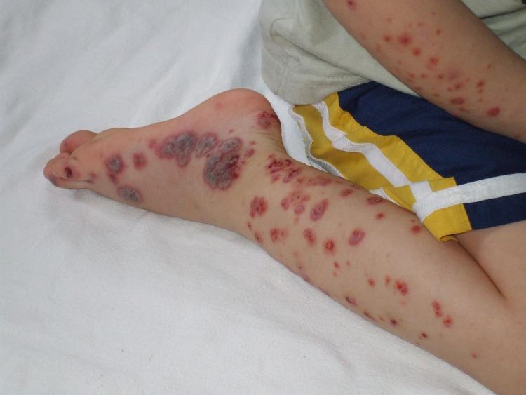
This clinical photograph displays characteristic dermatological manifestations of Henoch-Schönlein purpura (HSP), also known as IgA vasculitis, on the right lower extremity and forearm of a pediatric patient. The image shows a classic distribution of palpable purpura, presenting as numerous erythematous to violaceous macules, papules, and larger confluent plaques. On the lateral aspect and dorsal surface of the foot, the lesions are more severe, appearing as dark, purplish-brown nodular plaques with evidence of central crusting or possible necrosis. The pretibial region and lower leg exhibit a denser concentration of discrete, smaller purpuric spots compared to the proximal thigh. Morphologically, the lesions range from petechiae to larger ecchymotic patches, which are non-blanching and typically found in gravity-dependent areas such as the lower limbs. This visual is an essential educational reference for distinguishing vasculitic rashes from accidental bruising or non-accidental trauma in pediatric medicine, emphasizing the symmetrical and dependent distribution typical of HSP.
lichen aureus golden brown patch leg ankle
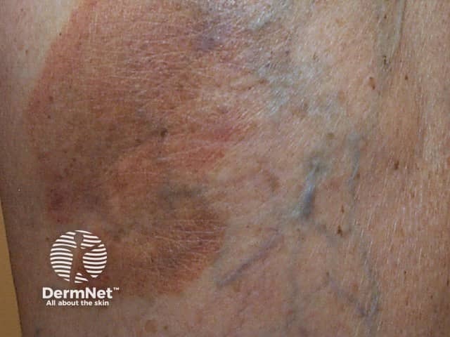
This is a high-resolution clinical photograph of a skin fragment consistent with lichen aureus, a localized variant of the pigmented purpuric dermatoses spectrum. Modality: clinical photography of the skin; view: close‑up, surface appearance of the lower leg region (pretibial area) with oblique perspective. The image demonstrates a well‑defined, rust‑orange to golden‑brown patch, often described as lichenoid or patch‑like, with subtle violaceous remodeling at the periphery. Surface texture is largely flat with minimal scale; there are no erosions or vesicles. Within the patch, faint peppering or punctate purpura may be suggested, compatible with superficial capillaritis and red blood cell extravasation. The lesion appears non-tender and asymptomatic or mildly pruritic, and there is no evidence of systemic illness in this image. These features support a clinical diagnosis of lichen aureus, typically a solitary or few lesions confined to the lower extremities and persisting for weeks to months. Pathophysiology involves capillary inflammation with hemosiderin deposition, producing the characteristic color. Diagnostic significance lies in distinguishing pigmented purpuric dermatoses from vasculitis or inflammatory dermatitis; biopsy is reserved for atypical cases. Clinical utility includes education, differential diagnosis practice, and guiding conservative management with topical steroids or observation, and vascular risk assessment if indicated.
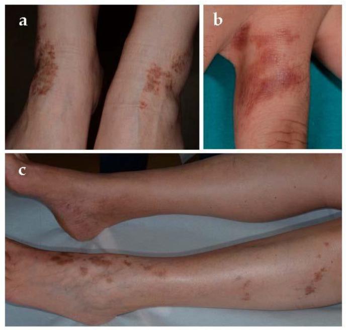
This clinical photograph displays characteristic skin lesions of Lichen aureus, a localized variant of pigmented purpuric dermatosis. Panel (a) illustrates bilateral, symmetrical yellow-brown to golden patches situated over the medial and lateral malleoli of the lower legs. These patches are formed by the coalescence of individual purpuric papules, creating a 'cayenne pepper' appearance. Panel (b) shows a focal, rust-colored to purple-brown patch on the first phalanx of the third finger, demonstrating a linear and well-circumscribed morphology following the digit's contour. Panel (c) provides a wider view of the lower extremities, highlighting the chronic distribution of these copper-toned macules and patches along the ankles and distal shins. The images demonstrate the hallmark 'golden' (aureus) discoloration caused by hemosiderin deposition within the dermis. This material is primarily intended for dermatology education regarding the clinical recognition of localized purpuric eruptions and their distinct morphological features compared to other chronic pigmented purpuras.
stasis dermatitis gravitational purpura hemosiderin ankles varicose
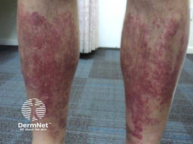
Clinical photography of bilateral lower legs demonstrating pigmented, non-blanching purpuric patches consistent with golfer's capillaritis (pigmented purpuric dermatosis). The image shows the pretibial surfaces of the shins with diffuse reddish-brown to purple macules and patches, irregular borders, and variable intensity, extending from the distal thirds to above the ankles. Superficial pigment deposition (hemosiderin) produces a mottled appearance, with faint residual erythema and mild edema in places. There is no obvious ulceration or vesiculation. The distribution is symmetric and predominantly on exposed areas subjacent to prolonged standing or exercise, a classic clinical pattern for capillaritis. The color progression may range from red to brick-red to brown over weeks, reflecting resolving inflammation and pigmentary changes. Clinically, these findings are characteristic of pigmented purpuric dermatoses, in particular Schamberg-like patterns, and can be exacerbated by venous insufficiency or venous hypertension without systemic involvement. Important differentials include vasculitic purpura, stasis dermatitis, drug-induced purpura, and other pigmented rash disorders. Correlating with history, venous examination, and laboratory studies helps distinguish benign pigmented purpura from inflammatory vasculitides. Management focuses on skin protection, compression therapy, leg elevation, and monitoring for progression; prognosis is generally favorable with recurrence possible. This image serves educational purposes for dermatology and primary care training, teledermatology triage, and dermatosis differential education.
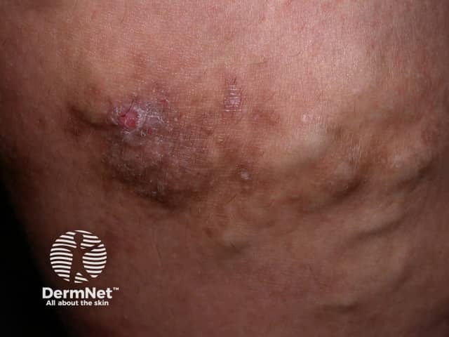
This is a color clinical photograph of the lower leg illustrating chronic venous insufficiency manifesting as varicose dermatitis or stasis dermatitis. The image highlights typical stasis changes including brown hemosiderin pigmentation, edema with thickened, leathery skin (lipodermatosclerosis), and eczematous plaques with crusting and fissures; hyperpigmented patches extend around the medial malleolus and distal lower leg. A shallow ulceration or crusted erosion may be present in the affected zone, accompanied by surrounding erythema. The appearance is consistent with chronic venous hypertension and venous stasis, rather than acute infection. Clinically relevant features include chronic edema, skin induration, and pigmentation, with potential secondary infection risk. This image can aid in diagnosing stasis dermatitis, differentiating from cellulitis or contact dermatitis, and evaluating response to therapy such as compression stockings, leg elevation, emollients, and anti-inflammatory topical agents. Educational use cases include dermatology exams, vascular medicine training, and wound care planning; also useful for illustrating differential diagnoses of leg edema-related dermatitis and for teaching recognition of lipodermatosclerosis. High-quality imaging helps learners connect clinical signs with pathophysiology of venous insufficiency, including microvascular hemosiderin deposition, edema-driven skin changes, and chronic inflammatory processes that predispose to ulcer formation. This image is suitable for bedside teaching and exam review purposes.
thrombocytopenic purpura petechiae skin
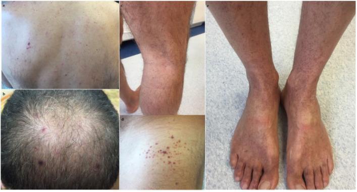
A series of clinical photographs documenting the dermatological manifestations of immune thrombocytopenic purpura (ITP). The images show widespread petechiae—small (1-3 mm), non-blanching, reddish-purple macules caused by capillary hemorrhage—distributed across several anatomical regions. On the back and scalp, the petechiae are relatively sparse and discrete. In contrast, the lower extremities, including the shins and the dorsal surfaces of the feet, demonstrate a higher density of lesions, appearing more confluent and clustered. The morphology is consistent with thrombocytopenia-induced microhemorrhages under the skin. The color variation from bright red to deep purple suggests varying stages of extravasated blood. This visual evidence serves as a clinical marker for evaluating systemic coagulation disorders and platelet dysfunction in a medical education context.
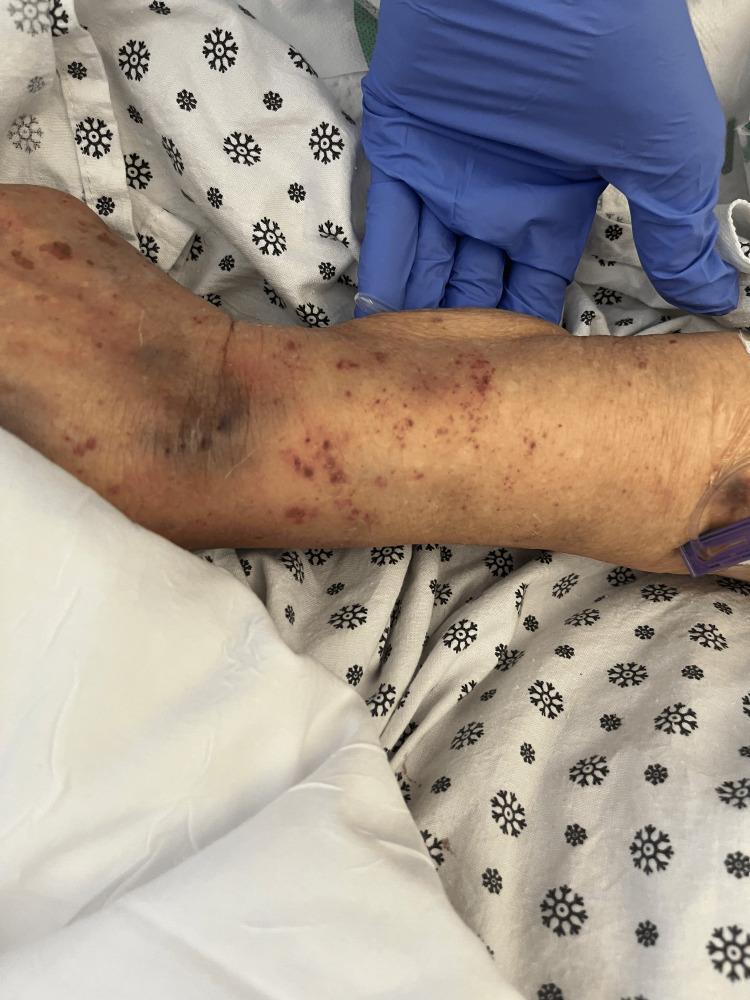
Clinical photograph of a patient's left forearm showing dermatologic manifestations of thrombocytopenia. The skin displays a diffuse distribution of petechiae—non-blanching, small, red-to-purple macules—measuring 1-2 mm in diameter. These are particularly concentrated on the volar aspect of the forearm and near the antecubital fossa. Larger, confluent areas of ecchymosis (bruising) are visible around the elbow and proximal forearm, indicating subcutaneous extravasation of blood. The skin appears thin and xerotic, with an overall uneven tone. A gloved hand is visible for clinical context. This visual presentation is characteristic of severe platelet deficiency, consistent with clinical entities such as Thrombotic Thrombocytopenic Purpura (TTP), and serves as a teaching aid for identifying bleeding diathesis in a hospital setting.
Differentials: Non-Itchy Bilateral Purpura of the Feet & Ankles
1. ⭐ Schamberg Disease (Pigmented Purpuric Dermatosis) — Most Likely
2. Leukocytoclastic Vasculitis (Small Vessel Vasculitis)
3. IgA Vasculitis (Henoch-Schonlein Purpura)
4. Lichen Aureus (Localized PPD variant)
5. Stasis Purpura (Chronic Venous Insufficiency)

6. Immune Thrombocytopenic Purpura (ITP)
Quick Comparison Table
| Feature | Schamberg (PPD) | LCV Vasculitis | IgA Vasculitis | Stasis Purpura | ITP |
|---|---|---|---|---|---|
| Itch | None | Burning/pain | Pain | Variable | None |
| Palpable | No | Yes | Yes | No | No |
| Bilateral symmetric | Yes | Yes | Yes | Medial > bilateral | Widespread |
| Hemosiderin staining | Prominent | Mild | Mild | Prominent | No |
| Systemic features | No | Possible | Arthritis/renal | No | Bleeding elsewhere |
| Biopsy | Capillaritis | Nuclear dust | IgA deposits | Venous changes | Normal vessels |
| Platelets | Normal | Normal | Normal | Normal | Low |

What’s the impression
phytophotodermatitis linear streak dripping hyperpigmentation skin

Clinical photography of the back demonstrating phytophotodermatitis. The dorsal trunk displays multiple erythematous macules and patchy plaques with variable edema and mild superficial desquamation; some areas show postinflammatory hyperpigmentation. Lesions are distributed unevenly, with occasional linear or streak-like patterns suggesting prior contact with plant sap, fruit juice, or essential oils and subsequent ultraviolet A (UVA) irradiation. The overall appearance is nonpurpuric and nonbullous in this image, though phototoxic reactions can blister in acute stages. The skin shows a pale in-texture; no obvious vesicles are present here. The patterning is consistent with a phototoxic interface reaction rather than an immune-mediated contact dermatitis. The patient’s exposure history, outdoor activity, and potential contact with citrus fruits, celery, fennel, bergamot, or other furocoumarin-containing organisms should be evaluated. Postinflammatory hyperpigmentation may persist for weeks to months. Clinically, phytophotodermatitis is diagnosed by history and pattern recognition; biopsy is rarely required. This image is useful for education about sun-exposed, plant-associated phototoxic reactions, differential diagnoses such as other photosensitivities, and for illustrating the classical streaking distribution. It supports clinical decision-making regarding avoidance of triggering plants, photoprotection, and topical anti-inflammatory therapy when indicated. Educational value includes recognizing pattern recognition, teaching exposure history, and documenting phototoxic risk factors for clinicians.
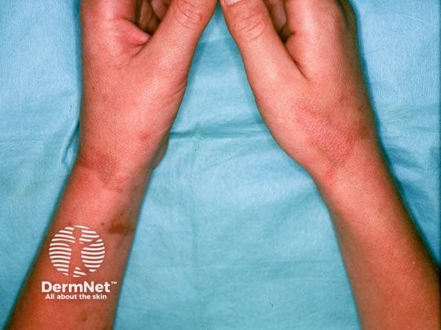
Clinical photography demonstrates bilateral forearm skin with residual post-inflammatory hyperpigmentation following phytophotodermatitis. The image, captured in a standard clinical setting with even room lighting, shows multiple irregular, brown to reddish-brown macules and patches distributed along sun-exposed forearm surfaces. There is minimal surrounding erythema, consistent with a quiescent or resolving phase after an acute phototoxic eruption. The pigmentation is mottled and can appear reticulated in places, reflecting prior inflammatory injury to the epidermis and superficial dermis. The pattern often tracks lines or contact with plant juice (furocoumarins) and sunlight, producing linear or streak-like hyperpigmented bands that gradually fade over months. No vesiculation or active edema is evident in this image; there may be faint residual accentuation on the wrists and dorsal forearms. Clinically, this appearance is typical of post-inflammatory hyperpigmentation after phytophotodermatitis and has relevance for prognosis and counseling. Differential considerations include other causes of PIH such as melasma, solar lentigines, or pigmentary contact dermatitis; however, the distribution and history support a phototoxic etiology. Management focuses on photoprotection, barrier care, and gradual pigment clearance; dermatologic follow-up may be necessary to document improvement and address cosmetic concerns. This image serves educational and comparative purposes for dermatology training and patient counseling and documentation.

This dermatologic image shows a dorsal trunk clinical photograph of phytophotodermatitis produced by contact with lime juice and subsequent sun exposure. Modality: clinical photography; No contrast. The focal points are linear, pigmented streaks and erythematous patches corresponding to areas of juice exposure on the left upper back and scapular region, extending medially toward the midline. Early findings include superficial erythema and slight edema; later findings show brown tan hyperpigmentation along the same linear distribution. The pattern is distinctive: streaks rather than irregular patch; margins are relatively well-defined and asymmetrical. The pathophysiology: photosensitizing furocoumarins (psoralens) in citrus juice transfer to the skin and, upon UVA exposure, generate reactive oxygen species causing epidermal damage and melanin deposition, resulting in post-inflammatory hyperpigmentation. Clinically, this condition mimics contact dermatitis but follows a photodistributed, streak-like pattern. Diagnostic significance: history of lime juice exposure, sun exposure, and a linear pattern; differential includes contact dermatitis from other citrus components, berloque dermatitis, photodermatoses, and pigmented contact dermatitis. Potential clinical use: educate patients on phototoxic contact reactions; differentiate from sunburn; monitor for persistent hyperpigmentation. This image aids dermatology education and phototoxicity recognition in emergency and primary care settings.