
Types of collagen
Collagen is the most abundant protein in the body, making up about 30% of total protein. There are at least 28 types, classified by structure and function. The major ones include Type I (skin, bone, tendons), Type II (cartilage), Type III (blood vessels, fetal skin), Type IV (basement membranes), and Type V (hair, placenta).
Searching sources...
Reading File
Finding Sources
Finding Sources
The best sources are Junqueira's Basic Histology, the Histology A Text and Atlas, Biochemistry Lippincott, and Andrews' Diseases of the Skin. Let me pull the detailed content from the top sources simultaneously.
Reading File
Reading File
Reading File
Reading File
I have rich content from the textbooks. Let me also fetch the detailed collagen table from Junqueira's and the Lippincott figure, and get the full table from Histology A Text and Atlas.
Reading File
Reading File
Loading Image
I now have comprehensive data from multiple authoritative textbooks. Here is the full synthesized answer:
Types of Collagen
Collagen is the most abundant protein in the human body (~30% of dry weight). The family includes at least 28 types (named with Roman numerals in order of discovery), each differing in α-chain composition, structure, location, and function. They are grouped into functional categories.
Classification by Functional Group
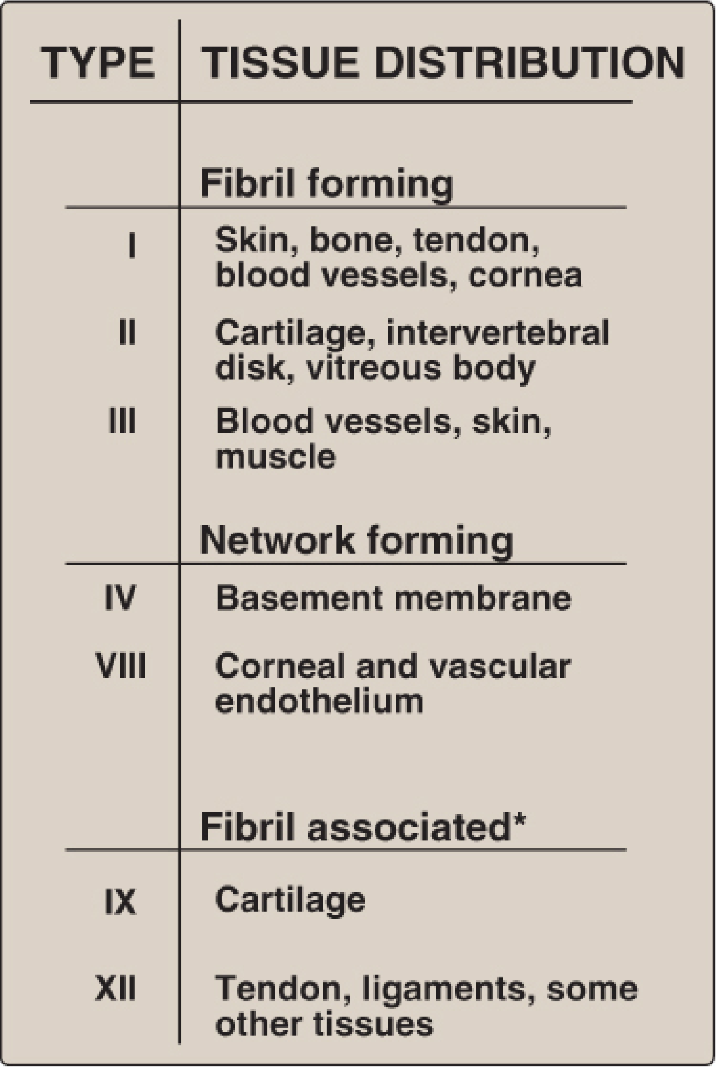
(From Lippincott's Biochemistry, 8th ed.)
1. Fibril-Forming Collagens (Types I, II, III, V, XI)
These have uninterrupted Gly-Pro-Hydroxyproline repeats and form 67-nm-banded fibrils visible by electron microscopy. They are the most abundant collagens in the body.
| Type | α-Chain Composition | Major Location | Function / Notes |
|---|---|---|---|
| I | [α1(I)]₂α2(I) | Skin, tendon, bone, dentin, cornea | Resistance to tension; most abundant collagen (60-90% dry weight of skin/ligaments/bone); forms thick, eosinophilic bundles |
| II | [α1(II)]₃ | Cartilage, vitreous body, intervertebral disc | Resistance to pressure; target antigen in relapsing polychondritis |
| III | [α1(III)]₃ | Blood vessels, fetal skin, muscle, gut; often co-distributed with Type I | Structural maintenance in distensible organs; 35% of adult skin collagen, up to 40% in inflamed skin |
| V | [α1(V)]₃ | Fetal tissues, skin, bone, placenta, most interstitial tissues | Participates in type I collagen function; N-terminal globular domain retained |
| XI | [α1(XI)][α2(XI)][α3(XI)] | Cartilage | Participates in type II collagen function; small fibers |
- Type I fibrils often contain small amounts of Types III, V, and XI co-assembled into heterotypic fibrils.
- Type II fibrils co-assemble with Type XI.
2. Network-Forming Collagens (Types IV, VIII, X)
Instead of fibrils, these form 2D sheets or hexagonal meshworks.
| Type | Location | Function |
|---|---|---|
| IV | All basement membranes (basal and external laminae) | Forms a 2D cross-linked network; major structural component of basement membranes; supports epithelial cells and acts as a filtration barrier |
| VIII | Corneal and vascular endothelium | Forms hexagonal lattices |
| X | Hypertrophic cartilage (zone of endochondral ossification) | Increases matrix density |
3. Fibril-Associated Collagens with Interrupted Triple Helices (FACITs)
These do not form fibrils themselves but bind to the surface of existing fibrils. Their interrupted triple helices provide molecular flexibility. Includes: Types IX, XII, XIV, XVI, XIX, XX, XXI, XXII, XXVI, XXVII, XXIX.
| Type | Location | Function |
|---|---|---|
| IX | Cartilage, vitreous body | Binds Type II collagen fibrils to proteoglycans of ECM; stabilizes cartilage matrix |
| XII | Tendon, ligaments, skin, placenta | Interacts with Type I collagen fibrils |
| XIV | Placenta, bone | Binds Type I fibrils; works with Types V and XI to strengthen fiber formation |
4. Linking / Anchoring Collagens
| Type | Location | Function |
|---|---|---|
| VI | Widespread connective tissue | Forms beaded microfilaments; links large collagen fibers to surrounding matrix |
| VII | Epithelial basement membranes (dermal-epidermal junction) | Forms anchoring fibrils attaching basal lamina to reticular lamina; target antigen in bullous lupus and epidermolysis bullosa acquisita |
5. Transmembrane Collagens
These are type II transmembrane proteins with collagen-like ectodomains.
| Type | Location | Notes |
|---|---|---|
| XIII | Focal adhesions | Cell-surface collagen |
| XVII | Hemidesmosomes (skin) | Also known as BP150/BP180; target antigen in bullous pemphigoid and pemphigoid gestationis |
| XXIII | Metastatic cancer cells | Upregulated in cancer |
| XXV | Brain neurons | Brain-specific |
6. Multiplexins (Multiple Triple-Helix Domains with Interruptions)
| Type | Location | Notes |
|---|---|---|
| XV | Basement membrane zones (especially vascular endothelium) | May have angiogenic roles |
| XVIII | Basement membranes | Endostatin (anti-angiogenic fragment) is derived from Type XVIII collagen |
7. Basement Membrane-Forming Collagens (additional)
| Type | Location | Notes |
|---|---|---|
| XXVIII | Peripheral nerve basement membranes | Plays a role in basement membrane assembly of supporting cells (Schwann cells) |
Key Clinical Correlations
| Collagen Type | Disease |
|---|---|
| Type I | Osteogenesis imperfecta (OI Types I-IV), Ehlers-Danlos syndrome (classical type) |
| Type II | Relapsing polychondritis (autoantibodies to Type II) |
| Type III | Ehlers-Danlos syndrome (vascular type - EDS IV); deficiency leads to vessel/gut rupture |
| Type IV | Alport syndrome (mutations in COL4A3/4/5 genes) |
| Type V | Ehlers-Danlos classical type (COL5A mutations) |
| Type VII | Epidermolysis bullosa (dystrophic type), bullous lupus |
| Type XVII | Bullous pemphigoid, pemphigoid gestationis |
Biosynthesis Highlights
Fibrillar collagen biosynthesis proceeds: pre-procollagen (ribosome) → hydroxylation of Pro/Lys (Vit C-dependent) → triple helix assembly → procollagen secretion → propeptide cleavage by procollagen peptidases → fibril assembly → cross-linking by lysyl oxidase (copper-dependent). Deficiency of Vitamin C impairs hydroxylation (scurvy); deficiency of copper impairs lysyl oxidase (Menkes disease), leading to fragile collagen and elastin.
Sources:
- Junqueira's Basic Histology, 17th ed., Table 5-3
- Histology: A Text and Atlas (Pawlina), Table 6.2
- Lippincott's Illustrated Reviews: Biochemistry, 8th ed., Ch. 4
- Andrews' Diseases of the Skin, Table 25.1
This is a shared conversation. Sign in to Orris to start your own chat.