
drunkometer in forensic medicine
I'll look this up in the medical library first.
Searching sources...
Reading File
Finding Sources
Finding Sources
Finding Sources
Good - found relevant content in three forensic medicine textbooks. Let me read all three sections in parallel.
Reading File
Reading File
Reading File
I now have comprehensive content from three authoritative forensic medicine textbooks. Let me also fetch the breath analyzer image and read a bit more from the PC Dikshit text.
Loading Image
Reading File
I now have all the information needed for a thorough answer. Here is a complete, well-organized summary:
Drunkometer in Forensic Medicine
What It Is
The drunkometer (also called breath analyser, alcometer, or intoximeter) is a device used for detecting alcohol in expired (exhaled) air. It is one of the most common preliminary screening tests used by law enforcement agencies for roadside detection of drunken driving.
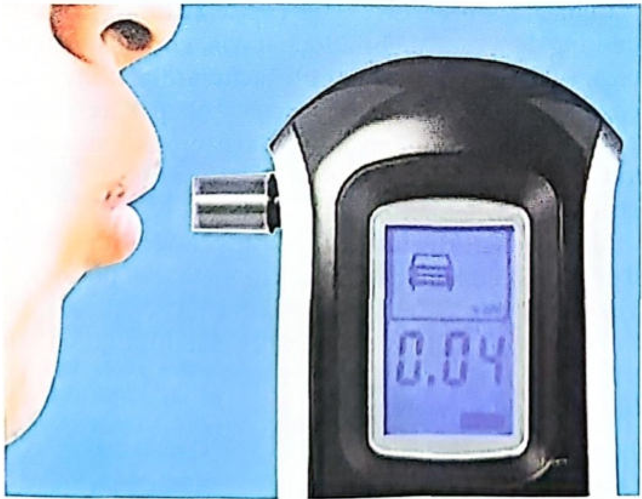
Fig: Modern digital breath analyzer (alcometer/drunkometer)
Basic Principle
The test is based on the Breath-Blood Alcohol Ratio:
2100 mL of alveolar air = 1 mL of blood in terms of alcohol content
This ratio is constant at a given temperature (reference temperature of exhaled air ~34°C). The alcohol concentration in deep lung (alveolar) air is dependent on arterial blood. So, the breath alcohol content closely mirrors the blood alcohol concentration (BAC). - The Essentials of Forensic Medicine and Toxicology, 36th ed. and PC Dikshit Textbook of Forensic Medicine and Toxicology
Chemical Reaction (Classic Drunkometer)
The subject blows 60-100 mL of breath into:
- A dry balloon or plastic bag, OR
- Directly into a glass tube containing the reagent mixture
The reagent is a crystalline bichromate-sulfuric acid (dichromate-H₂SO₄) mixture.
Reaction:
When exhaled air containing ethyl alcohol is passed over concentrated sulfuric acid + dichromate crystals:
Ethyl alcohol + Dichromate (orange/yellow) → Greenish colour change
- The intensity of the green colour is proportional to the amount of alcohol present
- A BAC >80 mg% is considered a positive result (green colour) - PC Dikshit Textbook
Procedure
- The person is asked to blow into a special container or directly into the breath analyser
- 60-100 mL of exhaled (deep lung) breath is collected
- The breath contacts the bichromate-sulfuric acid reagent
- Colour change (yellow/orange → green) indicates the presence and amount of alcohol
- This is a qualitative/semi-quantitative screening test
Modern (electronic) breathalyzers do NOT necessarily rely on chemical oxidation - they use electrochemical or infrared sensors and give quantitative results, with some producing printed output accepted for court work. - Parikh's Textbook of Medical Jurisprudence
Causes of False Positive Results
| Cause | Explanation |
|---|---|
| Residual mouth alcohol | Alcohol may remain in the mouth up to 20-30 minutes after even a small peg - test must be repeated after 20 minutes |
| Hyperventilation | Lowers alveolar alcohol concentration (actually gives false low) |
| Physical exercise | Can affect readings |
| Emesis / Regurgitation | Stomach contents containing alcohol can contaminate breath sample |
| Acetone, ether, paraldehyde | May be estimated as alcohol by some devices |
To preserve breath samples, plastic, aluminium, and other flexible bags are available, allowing samples to be kept for several hours for re-analysis. - PC Dikshit Textbook
Comparison with Other Samples for Alcohol Analysis
| Sample | Notes |
|---|---|
| Blood | Best sample; gives direct evidence of BAC in brain; peripheral vein preferred |
| Urine | 20% higher than blood; two samples needed (1st to empty bladder, 2nd after 25-30 min); urine alcohol lags blood by 20-25 min |
| Breath | Rapid, non-invasive; good screening tool; 2100 mL air = 1 mL blood |
| Saliva | ~same as blood alcohol |
| Vitreous humour | Useful postmortem; resistant to putrefaction; used when blood unavailable |
Legal / Medicolegal Significance
- The drunkometer/breathalyser is the primary roadside screening test used by police
- In India, the statutory blood alcohol limit is 30 mg% under Section 185, Motor Vehicles Act 1988
- First offence: fine up to ₹5,000 or 6 months imprisonment or both
- Subsequent offences: fine up to ₹3,000 or 2 years imprisonment or both
- Electronic breath analysers with printed output have been accepted for court work in some countries abroad
- A limitation: if alcohol absorption is not yet complete at the time of testing, BAC may be overestimated - Parikh's Textbook
Widmark Formula (Supplementary)
When direct BAC is unavailable, the Widmark formula estimates total body alcohol:
a = p × r × c
Where:
- a = weight of alcohol in the body (grams)
- p = body weight (kg)
- r = distribution constant (0.68 for males; 0.55 for females)
- c = concentration of alcohol in blood (mg/kg)
This is used in forensic reconstruction of BAC at the time of an incident. - PC Dikshit Textbook of Forensic Medicine and Toxicology
Sources:
SLE
Asking for Preferences
SLE is a broad topic. What aspect are you most interested in?
I'll do a comprehensive search across the medical library covering all major aspects of SLE.
Searching sources...
Finding Sources
Finding Sources
Excellent. Let me now pull the main SLE sections from Harrison's (most comprehensive) and Goldman-Cecil, plus the dermatology/skin manifestation section.
Finding Sources
Finding Sources
Reading File
Reading File
I have very rich content from Harrison's. Let me read the classification criteria table from Goldman-Cecil and then more of Harrison's for pathogenesis, manifestations, and treatment.
Reading File
Reading File
I have very comprehensive content. Let me fetch the key treatment flowchart image and read the clinical manifestations section.
Reading File
Loading Image
Excellent - I have a very thorough dataset from Harrison's 22e (2025) and Goldman-Cecil. I now have everything needed for a comprehensive answer.
Systemic Lupus Erythematosus (SLE)
Definition
SLE is a multisystem autoimmune disease in which overactive innate and adaptive immune systems cause tissue damage through the effects of autoantibodies and immune complexes. It targets multiple organ systems - cutaneous, musculoskeletal, renal, pulmonary, cardiovascular, hematologic, serosal, and CNS. The course is typically waxing and waning. - Harrison's 22e (2025)
Epidemiology
| Feature | Detail |
|---|---|
| Sex ratio | Female : Male = 9:1 in adults |
| Peak age | 15-44 years (childbearing age) |
| US prevalence | ~72.8 per 100,000 |
| Incidence | ~5.1 per 100,000/year |
| Racial disparity | Black women > Hispanic > White; Black men have highest prevalence among men |
| Children / >55 yrs | F:M ratio closer to 2:1 |
~90% of affected individuals are women. Prognosis varies by geographic region, ethnicity, and access to care. - Harrison's 22e; Goldman-Cecil Medicine
Pathogenesis
Genetic Factors
- Multiple susceptibility genes (polygenic, cumulative-hit model)
- Impaired inactivation of the X-chromosome may explain the female predominance
- Strong HLA associations (HLA-DR2, HLA-DR3)
- Complement deficiencies (C1q, C2, C4) predispose to SLE
Environmental Triggers
- UV light - alters DNA methylation, generates stimulatory nucleic acids, activates keratinocyte immune responses
- Epstein-Barr virus - induces IFN response, expresses viral proteins activating SLE susceptibility genes
- Low socioeconomic status, mercury, pesticides, diet
- Gut microbiome - increased gut permeability promotes translocation of gut microbes, contributing to autoantibody development
Innate Immunity Defects
- Neutrophils have higher turnover in SLE, delivering large loads of stimulatory nucleic acids
- Defective macrophage/dendritic cell clearance of cellular debris and immune complexes
- Excess uncleared debris triggers nucleic acid sensors and TLRs in plasmacytoid dendritic cells
- Result: massive upregulation of type I interferon (IFN) - the "IFN signature" central to SLE
Adaptive Immunity Defects
- B cells lose tolerance - expand as "age-associated B cells (ABCs)" that mature into autoantibody-secreting plasma cells
- T follicular and T peripheral helper cells promote B-cell differentiation into high-affinity autoantibody plasma cells
- T regulatory cells are defective - cannot maintain tolerance
- Autoantibodies form immune complexes that deposit in tissues causing inflammation
Autoantibodies in SLE
| Autoantibody | Sensitivity | Specificity | Clinical Association |
|---|---|---|---|
| ANA | >95% | Low | Screening test; almost all SLE patients positive |
| Anti-dsDNA | 70% | 97% | Highly specific; correlates with disease activity and nephritis |
| Anti-Sm (Smith) | 25-30% | 55-100% | Highly specific for SLE |
| Anti-histone | 70% | Low | Drug-induced lupus |
| Antiphospholipid (aPL) | 30-40% | Moderate | Antiphospholipid syndrome (thrombosis, fetal loss) |
| Anti-Ro/SSA | 30-40% | Low | Neonatal lupus, subacute cutaneous lupus, Sjogren overlap |
| Anti-La/SSB | 15-20% | Low | Associated with anti-Ro; neonatal lupus |
| Low C3/C4 | Variable | Moderate | Complement consumption; active disease |
Key rule: ANA sensitivity is >95% (excellent screening), but Anti-dsDNA and Anti-Sm are the most specific antibodies for SLE. - Harrison's 22e; Harriet Lane Handbook
Classification Criteria: 2019 EULAR/ACR
Step 1 - Entry criterion: ANA titer ≥1:80. If absent, do NOT classify as SLE.
Step 2 - Additional weighted criteria (count only the highest criterion per domain):
| Domain | Criterion | Points |
|---|---|---|
| Constitutional | Fever | 2 |
| Neuropsychiatric | Delirium | 2 |
| Psychosis | 3 | |
| Seizure | 5 | |
| Mucocutaneous | Non-scarring alopecia | 2 |
| Oral ulcers | 2 | |
| Subacute cutaneous / discoid lupus | 4 | |
| Acute cutaneous lupus (malar rash) | 6 | |
| Musculoskeletal | Joint involvement | 6 |
| Serosal | Pleural or pericardial effusion | 5 |
| Acute pericarditis | 6 | |
| Hematologic | Leukopenia | 3 |
| Thrombocytopenia | 4 | |
| Autoimmune hemolysis | 4 | |
| Renal | Proteinuria >0.5 g/24h | 4 |
| Renal biopsy class II or V LN | 8 | |
| Renal biopsy class III or IV LN | 10 | |
| Antiphospholipid Ab | Anticardiolipin OR anti-β2GPI OR lupus anticoagulant | 2 |
| Complement | Low C3 OR low C4 | 3 |
| Low C3 AND low C4 | 4 | |
| SLE-specific Ab | Anti-dsDNA | 6 |
| Anti-Sm | 6 |
Classify as SLE if total score ≥ 10 points - Goldman-Cecil Medicine
Note: A renal biopsy showing class III or IV lupus nephritis alone scores 10 points and is sufficient.
Clinical Manifestations
Cutaneous (70-80%)
- Malar (butterfly) rash - erythema over cheeks and nose bridge, sparing nasolabial folds; triggered/worsened by UV
- Discoid lupus - chronic scarring plaques, may cause permanent alopecia
- Subacute cutaneous lupus (SCLE) - annular or papulosquamous lesions, associated with anti-Ro
- Photosensitivity
- Non-scarring alopecia - hair loss/thinning during flares
- Oral/nasal ulcers - usually painless
- Vasculitic lesions - Raynaud's phenomenon, livedo reticularis
Musculoskeletal (90%)
- Arthralgia and non-erosive arthritis - symmetrical, involving small joints (hands, wrists, knees)
- Jaccoud's arthropathy - reversible deformity due to tendon/ligament laxity (not erosive)
- Myalgia, myositis (less common)
- Osteonecrosis (avascular necrosis) - especially with steroid use
Renal (40-75%) - Lupus Nephritis
- One of the most serious manifestations
- WHO/ISN-RPS Classes:
- Class I: Minimal mesangial
- Class II: Mesangial proliferative
- Class III: Focal proliferative (< 50% glomeruli)
- Class IV: Diffuse proliferative (≥ 50% glomeruli) - most severe, worst prognosis
- Class V: Membranous
- Class VI: Advanced sclerosis
- Presents with haematuria, proteinuria, hypertension, nephrotic/nephritic syndrome
Neuropsychiatric (50%)
- Headache, cognitive dysfunction, "brain fog"
- Seizures, psychosis, stroke (often aPL-mediated)
- Peripheral neuropathy, myelitis
Cardiovascular
- Pericarditis (most common cardiac manifestation)
- Myocarditis, endocarditis (Libman-Sacks endocarditis - sterile verrucous vegetations on valve surfaces)
- Accelerated atherosclerosis - major cause of morbidity/mortality in long-term disease
- Raynaud's phenomenon
Pulmonary
- Pleuritis/pleural effusion (most common pulmonary manifestation)
- Pneumonitis, pulmonary hypertension
- "Shrinking lung syndrome" - diaphragmatic dysfunction
Hematologic
- Anemia - often autoimmune hemolytic; also anemia of chronic disease
- Leukopenia (lymphopenia is most characteristic)
- Thrombocytopenia
Antiphospholipid Syndrome (aPL)
- Present in ~30-40% of SLE patients
- Causes: venous/arterial thrombosis, recurrent miscarriage, thrombocytopenia
- Lab: lupus anticoagulant, anticardiolipin Ab, anti-β2GPI Ab
Treatment
Overview: Treat-to-Target Approach
- Goal: remission or lowest possible disease activity to prevent organ damage
- Hydroxychloroquine (HCQ) 5 mg/kg/day is the backbone of therapy for ALL patients unless contraindicated - reduces flares, damage, and mortality
- Minimize glucocorticoid exposure; taper as quickly as possible
Treatment by Severity
Mild disease (cutaneous, arthritis, constitutional):
- NSAIDs (with caution - risk of aseptic meningitis, renal dysfunction)
- HCQ +/- low-dose glucocorticoids
- Methotrexate or azathioprine for steroid-sparing
Moderate-Severe disease (nephritis, CNS, haematologic):
- Glucocorticoids - IV methylprednisolone pulse for severe flares, then oral taper
- Mycophenolate mofetil (MMF) - first-line for lupus nephritis induction and maintenance
- Cyclophosphamide (CYC) - IV; used for severe nephritis (NIH regimen or Euro-lupus low-dose)
- Azathioprine (AZA) - maintenance therapy, safe in pregnancy
- Rituximab (RTX) - anti-CD20; for refractory disease
Biologics
- Belimumab (anti-BLyS/BAFF) - approved for active SLE and lupus nephritis; reduces flares; given as add-on to standard therapy with MMF or low-dose CYC
- Anifrolumab (anti-IFN receptor) - approved for moderate-to-severe SLE; particularly effective for skin disease; NOT recommended for severe neuropsychiatric disease
- Voclosporin (VOC) + MMF - calcineurin inhibitor combination for lupus nephritis
Lupus Nephritis Treatment Algorithm (EULAR 2024):
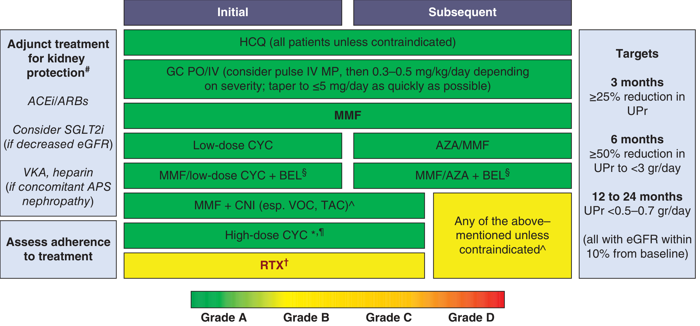
Treatment targets: ≥25% reduction in proteinuria at 3 months; ≥50% reduction to <3 g/day at 6 months; <0.5-0.7 g/day at 12-24 months
Type 1 vs Type 2 Lupus (Harrison's 22e Classification)
| Type 1 SLE | Type 2 SLE | |
|---|---|---|
| Features | Nephritis, arthritis, vasculitis, immune-mediated | Fatigue, diffuse pain, cognitive dysfunction, brain fog, depression |
| Mechanism | Autoimmune inflammation / organ damage | Less clearly autoimmune |
| Response to immunosuppression | Good | Poor |
Prognosis & Monitoring
- Disease activity tracked with SLEDAI (SLE Disease Activity Index) or BILAG scores
- Anti-dsDNA titres and complement (C3/C4) levels correlate with disease activity
- Main causes of death: Infection (especially early), cardiovascular disease (atherosclerosis, especially late), renal failure, CNS disease
- Accelerated atherosclerosis is a major late complication; annual cardiovascular risk assessment recommended
- Blood for alcohol: serum fluoride/oxalate preserved; avoid spirit for skin cleaning (to prevent false-positive alcohol results - from forensic context this time, about SLE, not alcohol!)
Sources:
- Harrison's Principles of Internal Medicine, 22nd ed. (2025) - primary reference
- Goldman-Cecil Medicine, International Edition - classification criteria, pathobiology
- Harriet Lane Handbook, 23rd ed. - autoantibody specificity/sensitivity
Envenoming
Searching sources...
Finding Sources
Finding Sources
Good - found content in Goldman-Cecil, Rosen's EM, Tintinalli's, Harrison's, and Mulholland Surgery. Let me read the key sections in parallel.
Reading File
Reading File
Reading File
Reading File
Excellent data. Let me now read the spider/scorpion/marine sections and the antivenom/treatment section from Goldman-Cecil.
Reading File
Reading File
I now have comprehensive content. Let me compile the full answer.
Envenoming (Envenomation)
Definition
Envenomation occurs when a venomous animal - usually via a bite or sting - injects a physiologically impactful quantity of venom into a target. A bite without sufficient venom injection is called a "dry bite" and produces no signs of envenomation. - Goldman-Cecil Medicine
Epidemiology
| Envenomation type | Annual cases | Annual deaths | Case fatality rate |
|---|---|---|---|
| Venomous snakebite | >2.5 million | >100,000 | ~1:25 |
| Jellyfish stings | >1 million | Hundreds | ~1:10,000 |
| Spider bites (US) | 5,000-10,000 | Rare | Very low |
| Scorpion stings (US) | ~20,000 | Rare | Very low |
- Arthropods overall account for more deaths from envenomation than snakes globally
- Regional variation is marked: in Australia, spider bites cause nearly twice as many hospital visits as snakebites
- In India alone, at least 45,000 people die annually from snakebite
- Asia has the highest snakebite burden globally, predominantly an occupational disease of rural workers
Venom: Nature and Sources
Venom is a complex mixture of toxins, enzymes, peptides, and other bioactive molecules. Sources:
- Specialized glands: snakes, scorpions, spiders, cone snails, stinging fish, stingrays
- Cell structures: jellyfish nematocysts
- Acquired/concentrated: venomous octopuses (acquire tetrodotoxin from environment)
Dry bite rates vary: viperid snakes ~20%, elapids ~50%. Some snakes have no venom apparatus and are entirely harmless.
I. SNAKE ENVENOMATION
Classification of Snakes
- Viperids (Viperidae): vipers, rattlesnakes, pit vipers (Crotalids), Russell's viper, Bothrops
- Elapids (Elapidae): cobras, kraits, mambas, coral snakes, sea snakes
- Colubrids: mostly harmless, a few rear-fanged and mildly venomous
Clinical Syndromes of Snake Envenomation
1. Neurotoxic Paralysis
- Pre- and post-synaptic neurotoxins target the neuromuscular junction
- Causes progressive descending flaccid paralysis
- Presynaptic neurotoxins (e.g., β-bungarotoxin from kraits): destroy the axon terminal - antivenom may not reverse established paralysis; the axon must regrow (days to weeks)
- Postsynaptic neurotoxins (e.g., α-cobratoxin from cobras): bind acetylcholine receptor externally - antivenom CAN reverse this type
- Elapids (coral snakes, cobras, kraits, mambas) are the primary neurotoxic snakes
- Some African/Asian cobras can spit venom up to 2-3 metres, targeting the eyes → instant severe pain, blepharospasm, temporary blindness (venom spit ophthalmia) - requires copious irrigation; does NOT cause systemic envenomation; does NOT need antivenom
2. Coagulopathy (Venom-Induced Consumptive Coagulopathy - VICC)
- Consumptive coagulopathy with reduced fibrinogen → bleeding
- Hemorrhagins damage blood vessels → extravasation, swelling, local necrosis, shock
- Some Bothrops species (Martinique viper) have procoagulant toxins → thrombosis → embolic infarction (brain, heart, lungs)
- Russell's viper: classic cause of VICC in Asia
3. Myotoxicity
- Systemic myotoxins (mainly sea snakes, some Australian elapids) → rhabdomyolysis
- CK can exceed >100,000 IU/L; myoglobinuria → acute kidney injury
- Local myotoxicity at the bite site
4. Local Tissue Injury / Cytotoxicity
- Many viper venoms → tissue necrosis, fluid shifts, secondary shock
- Secondary infection: mainly Streptococcus and Staphylococcus; gram-negatives in South Africa
- Prophylactic amoxicillin-clavulanate is NOT effective in preventing secondary infection
- Can result in compartment syndrome requiring fasciotomy
5. Cardiotoxicity
- Hyperkalemia from rhabdomyolysis → cardiac arrhythmia
- Direct myocardial depression (some species)
Pit Viper Envenomation Grading (Crotalid - North America)
| Grade | Features | Antivenom? | Disposition |
|---|---|---|---|
| 0 (None) | Fang wound only; minimal pain; <1 inch edema; no systemic signs in 12h | No | Observe 8-12h; discharge if labs normal |
| I (Minimal) | Moderate throbbing pain; 1-5 inch edema; no systemic signs after 12h | No | Admit 12-24h; labs q6h |
| II (Moderate) | Severe widespread pain; edema toward trunk; petechiae/ecchymosis; nausea, vomiting, mild fever | Yes | Admit; antivenom |
| III (Severe) | Severe systemic symptoms; significant lab abnormalities; extensive swelling | Yes | ICU; antivenom |
| IV (Very Severe) | Marked systemic toxicity; shock; life-threatening coagulopathy | Yes | ICU; aggressive antivenom |
Antivenoms for Snake Envenomation
- CroFab (USA) - Fab fragments from sheep immunized with 4 pit viper species; FDA-approved for North American pit viper envenomation
- Anavip (USA, 2015) - F(ab')₂ fragments; longer half-life; may reduce need for follow-up lab testing
- Antivenom is indicated for moderate or severe envenomation
- Presynaptic neurotoxin damage cannot be reversed by antivenom once established - early administration before progression is critical
- WHO antivenom database: https://apps.who.int/bloodproducts/snakeantivenoms/database/
Regional Considerations
| Region | Key snakes | Dominant syndrome |
|---|---|---|
| India/South Asia | Russell's viper, krait, spectacled cobra, saw-scaled viper (the "Big Four") | VICC, neurotoxicity, local necrosis |
| Sub-Saharan Africa | Puff adder, mamba, boomslang | Local necrosis, VICC, neurotoxicity |
| Australia | Brown snake, tiger snake, taipan | Neurotoxicity, VICC, myotoxicity |
| North America | Rattlesnakes, copperhead, cottonmouth, coral snake | Local tissue injury, VICC, neurotoxicity (coral) |
| South America | Bothrops, Lachesis, Micrurus, Crotalus | VICC, neurotoxicity (S. American rattlesnake), embolism |
| Europe | Vipera spp. (adders) | Local swelling, VICC, occasional neurotoxicity |
II. ARTHROPOD ENVENOMATION
A. Scorpions
- Medically important species: Almost exclusively in the family Buthidae
- Distributed worldwide, especially Middle East, North Africa, India, Mexico, USA (Southwest), South America
- Most scorpion stings cause only local pain; truly dangerous species are limited
- Dangerous genera: Androctonus, Leiurus (Middle East/Africa), Centruroides (USA/Mexico), Tityus (South America), Mesobuthus (India)
- Mechanism: Neurotoxic - voltage-gated sodium/potassium channel modulators
- Clinical features:
- Local: intense burning pain (often without much swelling - unlike spider bites)
- Systemic: autonomic storm (tachycardia, hypertension, diaphoresis, hypersalivation, priapism)
- Severe: respiratory failure, pulmonary edema, cardiovascular collapse, pancreatitis (Tityus)
- Treatment: Supportive; antivenom available for some species
B. Spiders
- Only ~50 of ~35,000 species are medically significant
- Two main clinical syndromes:
| Syndrome | Spider | Venom | Features |
|---|---|---|---|
| Necrotic arachnidism | Loxosceles (brown recluse) | Sphingomyelinase D | Painless bite → expanding necrotic ulcer; systemic hemolysis in severe cases |
| Latrodectism | Latrodectus (black widow) | α-Latrotoxin | Muscle pain and cramping; autonomic instability; hypertension; diaphoresis; sweating; no necrosis |
- Black widow: α-latrotoxin causes massive release of neurotransmitters at nerve terminals → severe muscle cramps (classic: abdominal rigidity mimicking peritonitis), hypertension, diaphoresis; antivenom available
- Brown recluse: necrotic skin lesion; treat with wound care; no proven specific antidote
- Sydney funnel-web (Atrax robustus, Australia): highly dangerous; atracotoxin causes autonomic excitation, cardiovascular collapse; antivenom available
C. Hymenoptera (Bees, Wasps, Ants)
- Most stings: local pain and swelling; self-limited
- Mass stings (hundreds to thousands): toxic envenomation → hemolysis, rhabdomyolysis, renal failure
- IgE-mediated anaphylaxis: most dangerous consequence; epinephrine is mainstay of treatment
- Solenopsis invicta (fire ant): unique pustule-forming venom; risk of anaphylaxis
III. MARINE ENVENOMATION
Jellyfish / Box Jellyfish
- Nematocysts inject venom on contact
- Range: local urticaria and pain to anaphylaxis
- Chironex fleckeri (box jellyfish, Australia): potentially lethal; cardiotoxic → cardiac arrest within minutes
- Treatment: Remove tentacles; vinegar to inactivate unfired nematocysts (for Chironex); specific antivenom available in Australia
- Irukandji syndrome (Carukia barnesi): delayed severe pain, hypertension, catecholamine surge - small, nearly invisible jellyfish
Stingrays
- Envenomation via barbed tail spines
- Venom: protein toxins causing intense pain, tissue necrosis
- Treatment: immerse in hot water (45°C, non-scalding) to denature heat-labile venom; wound exploration to remove spine fragments
Venomous Fish (Stonefish, Lionfish, Scorpionfish)
- Stonefish (Synanceia): world's most venomous fish; excruciating pain; antivenom available
- Hot water immersion effective for pain
- Lionfish: increasingly encountered in Atlantic/Caribbean (invasive species)
Cone Snails
- Inject conotoxins (potent neurotoxins) via a harpoon-like tooth
- Can cause rapid flaccid paralysis and respiratory arrest
- No antivenom; purely supportive care
IV. GENERAL MANAGEMENT PRINCIPLES
First Aid (Do's and Don'ts)
| Do | Do NOT |
|---|---|
| Immobilize the affected limb | Cut and suck the wound |
| Apply pressure-immobilization bandage for neurotoxic elapid bites | Apply tourniquet (worsens local tissue injury for vipers) |
| Keep patient calm; reduce movement | Apply ice (worsens necrosis) |
| Transport rapidly to medical facility | Apply electric shock |
| Mark and time leading edge of swelling | Delay seeking care |
Note: Pressure-immobilization bandaging is recommended for elapid (neurotoxic) bites but is contraindicated for cytotoxic viper bites as it concentrates tissue-damaging venom locally.
Hospital Management
- ABC: Airway, breathing, circulation - snakebite with neurotoxicity can cause rapid respiratory failure
- IV access: in an unaffected limb; normal saline
- Identify the snake/animal if possible (do not handle it)
- Labs for snakebite:
- Platelet count, fibrinogen, INR, PTT (baseline and serial)
- CK (if myotoxicity suspected)
- D-dimer, urinalysis, renal function
- Repeat at 4 and 8 days post-envenomation for delayed coagulopathy
- Antivenom - only specific effective treatment for most serious envenomations; administer early; monitor for anaphylaxis and serum sickness
- Tetanus prophylaxis
- Analgesia (IV opioids for severe pain)
- Wound care; fasciotomy if compartment syndrome develops
Antivenom Complications
- Early: anaphylaxis/anaphylactoid reactions (pretreatment with antihistamine +/- epinephrine considered in high-risk patients)
- Delayed: serum sickness (7-14 days post-antivenom) - fever, rash, arthralgia, urticaria; treat with corticosteroids
Sources:
- Goldman-Cecil Medicine, International Edition - Ch. 98: Envenomation, Bites, and Stings
- Rosen's Emergency Medicine, 9th ed. - Pit viper classification and management
- Harrison's Principles of Internal Medicine, 22e (2025)
Carboluria in forensic medicine
Searching sources...
Finding Sources
Finding Sources
Excellent - found rich content in all three forensic medicine textbooks. Let me read the full carbolic acid sections in parallel.
Reading File
Reading File
Reading File
Great content from all three books. Now let me read the specific carboluria and postmortem/treatment sections.
Reading File
Reading File
Reading File
Now let me read the postmortem findings and chemical tests section from Parikh's and the Essentials book.
Reading File
Reading File
I now have comprehensive, detailed content from all three authoritative forensic medicine textbooks. Here is a thorough answer:
Carboluria in Forensic Medicine
Context: Carbolic Acid (Phenol) Poisoning
Carboluria is a pathognomonic urinary finding seen in poisoning by carbolic acid (phenol; C₆H₅OH) - a corrosive, neurotoxic, and nephrotoxic substance used as an antiseptic, disinfectant, and germicide. Poisoning by carbolic acid is called carbolism.
Definition of Carboluria
Carboluria refers to the characteristic urinary findings in carbolic acid poisoning, where the patient's urine is initially clear (or has a normal or faintly greenish hue) but on standing or exposure to air turns dark green, smoky, or olive green/black in colour.
- "Urine is scanty and suppressed; when passed there is a normal or greenish hue that becomes dark or olive green on exposure to air." - PC Dikshit Textbook of Forensic Medicine
- "Urinary findings of carbolic acid go by the name of carboluria. In this, the patient's urine is clear in start but later becomes smoky in colour." - Parikh's Textbook of Medical Jurisprudence
Mechanism / Biochemical Basis
The colour change is entirely due to the metabolic products of phenol:
-
In the body, phenol is partly oxidized in the liver to two major metabolites:
- Hydroquinone (para-dihydroxybenzene)
- Pyrocatechol (ortho-dihydroxybenzene / catechol)
-
These metabolites are excreted in the urine in three forms:
- Partly free (unconjugated)
- Partly in unstable combination with sulfuric acid (sulfate conjugates)
- Partly in combination with glucuronic acid (glucuronide conjugates)
-
On standing or on exposure to air (oxygen), the unstable conjugates break down and hydroquinone and pyrocatechol undergo further oxidation to coloured quinone compounds - producing the characteristic dark green → olive green → black colour change
"The further oxidation of hydroquinone and pyrocatechol in the urine is the cause of green coloration. This is known as carboluria." - Essentials of Forensic Medicine & Toxicology, 36th ed.
Complete excretion of phenol from the body takes approximately 36-48 hours.
Carboluria as a Diagnostic Clue
| Feature | Detail |
|---|---|
| Initial urine colour | Clear / normal / faint green |
| Colour on standing / air exposure | Dark green → olive green → black / smoky |
| Cause | Oxidation of hydroquinone and pyrocatechol |
| Other urinary findings | Albumin, blood casts, free haemoglobin, oliguria/suppression |
| Significance | Pathognomonic for carbolic acid poisoning; diagnostic even when other symptoms mimic opium poisoning |
Historical significance: Carboluria served as a warning sign of the toxic action of carbolic acid when it was used as an antiseptic wound dressing in the past (e.g., Lister's antiseptic wound dressings). - Parikh's
Chemical Tests for Carbolic Acid in Urine
| Test | Reagent | Result |
|---|---|---|
| Ferric chloride test | Add a few drops of 10% ferric chloride to 1 mL urine | Blue/violet colour develops (Note: salicylates also give positive result) |
| Bromine water test | Add bromine water to urine | White precipitate of tribromophenol |
| Benedict's / Fehling's test | Add reducing sugar reagent | Urine reduces the solution (positive result) |
Full Clinical Picture of Carbolic Acid Poisoning
Physical Properties
- Pure phenol: colourless, prismatic needle-like crystals; burning sweetish taste; characteristic carbolic/phenolic smell
- Turns pink and liquefies on exposure to air
- Slightly soluble in water; freely soluble in glycerine, ether, alcohol
- Commercial grade: dark brown liquid containing cresol impurities
Key Preparations
| Preparation | Composition |
|---|---|
| Lysol | 50% cresol in saponified vegetable oil (less toxic than pure phenol) |
| Dettol | Chlorinated phenol + terpineol |
| Pure phenol | ~8 times more toxic than Lysol |
| Other derivatives | Cresol, creosote, thymol, menthol, tannic acid |
Fatal Dose & Period
- Fatal dose: 10-15 g (pure phenol); 25-50 mL of household phenol; 20 drops of pure phenol can be fatal
- Fatal period: Usually 3-4 hours; range from 3 minutes to 60 hours
Signs and Symptoms
Local Effects:
- Skin: Burning and numbness (due to nerve ending damage); protein coagulation → white opaque eschar (painless, falls off in days, leaving brown stain); deep burns turn black; necrosis and gangrene; green-white or brown-white dead tissue
- Mucous membranes: Lips, mouth, tongue coroded and hardened; white, bleached, hardened burns → become brown; slough rapidly
- GIT: Hot burning pain mouth to stomach → tingling → anaesthesia; vomiting may NOT occur (anaesthetic action on stomach)
Systemic (Remote) Effects:
| System | Features |
|---|---|
| CNS | Giddiness → insensibility → coma (rapid); convulsions; lockjaw |
| CVS | Shock; pulse weak, feeble and thready |
| Skin | Cold and clammy; pale and cyanosed |
| Eyes | Pupils contracted/pinpoint (can mimic opium poisoning) |
| Temperature | Subnormal |
| Respiration | Slow, laboured, stertorous; strong smell of carbolic acid in breath |
| Blood | Haemolysis; methemoglobinaemia (due to hydroquinone) |
| Kidneys | Nephrotoxic; oliguria/suppression; albuminuria, blood casts → carboluria |
⚠️ Important differential: The combination of contracted pupils, cold clammy skin, and coma can mimic opium poisoning. Carboluria + phenolic smell of breath + white patches in mouth distinguishes carbolic acid poisoning.
Chronic Poisoning: Phenol Marasmus / Ochronosis
Seen in workers (surgeons, nurses) with chronic exposure:
- Anorexia, weight loss, headache, vertigo
- Dark urine chronically
- Pigmentation of skin, sclera, cornea, and cartilages → called Ochronosis
- Hydroquinone and pyrocatechol deposit in connective tissues
- Note: Ochronosis also occurs in alkaptonuria (inborn error of metabolism - homogentisic acid deposits)
Postmortem Findings
- Smell of carbolic acid from body/stomach
- Lips and mouth: white, corroded, hardened burns → brownish
- Stomach: mucosa shows grey/brown opaque, leathery thickening with swollen folds; reddish fluid with mucus and epithelial shreds smelling of phenol
- Duodenum and upper small intestine: similar but milder corrosion
- Liver and spleen: whitish, hardened patches (from transudation of phenol from stomach)
- Kidneys: haemorrhagic nephritis (in delayed death); parenchymal degeneration
- Lungs and brain: congested; brain may be oedematous
- Blood: dark, semifluid, partially coagulated
- Glottis: swelling and congestion
Preservation of viscera: Must be preserved in saturated sodium chloride solution - NOT in rectified spirit (alcohol dissolves phenol and may interfere with analysis)
Treatment
- Emetics often fail - phenol's anaesthetic effect prevents vomiting
- Gastric lavage: Pass soft stomach tube carefully (tissues are hardened); wash with warm water + activated charcoal, olive/castor oil, magnesium or sodium sulphate (form harmless compounds); 10% glycerine or soap solution; continue until washings are clear and odourless
- Leave magnesium sulphate (forms insoluble sulphocarbolate) or liquid paraffin in stomach
- Demulcents: Milk, white of an egg
- IV saline + sodium bicarbonate (7g/L) - combats shock, dilutes phenol in blood, promotes diuresis/excretion
- Oxygen and artificial respiration
- Methylene blue IV for severe methemoglobinaemia
- Haemodialysis for renal failure
- Skin contamination: remove clothing, wash skin with polyethylene glycol (PEG) or copious water
Medicolegal Significance
| Manner | Details |
|---|---|
| Suicide | Most common manner of poisoning |
| Accidental | Indiscriminate/excessive medicinal use; occupational exposure |
| Homicide | Rare - characteristic smell and taste of phenol makes it difficult to administer covertly |
| Abortion | Sometimes injected into vagina/uterus to procure criminal abortion |
Sources:
Vitriolage
Searching sources...
Finding Sources
Finding Sources
Excellent - rich content in all three forensic medicine books plus an image. Let me read all sections in parallel.
Reading File
Reading File
Reading File
Loading Image
Now let me read the acid attack Q&A from Parikh's and the sulphuric acid section from Dikshit for more detail on agents and legal aspects.
Reading File
Reading File
I now have comprehensive content from all three authoritative forensic medicine textbooks. Here is the complete answer:
Vitriolage (Acid Attack / Vitriol Throwing)
Definition
Vitriolage (also called vitriol throwing or acid attack) is the act of deliberately throwing or administering a corrosive substance - usually a strong acid or alkali - on the body or face of a victim with malicious intent to disfigure, destroy vision, cause grievous hurt, or damage clothing.
"Vitriolage is the throwing of strong sulphuric acid, concentrated mineral acid, corrosive alkalis, carbolic acid, or the juice of marking nut over the face or body of the victim for the purpose of disfiguring the face, destroying vision, causing injury on the body, or even destroying the clothing of the victim." - PC Dikshit Textbook of Forensic Medicine
The name "vitriolage" is derived from "oil of vitriol" - the old name for sulphuric acid (H₂SO₄), which is the most commonly used agent.
Agents Used in Vitriolage
| Agent | Features on Skin/Clothing |
|---|---|
| Sulphuric acid (oil of vitriol) - MOST COMMON | Brownish-black discolouration; chars and blackens; penetrating burns |
| Nitric acid (aqua fortis) | Yellow discolouration (xanthoproteic reaction) |
| Carbolic acid (phenol) | White hardened burns → brown staining |
| Hydrochloric acid | Corrosive burns |
| Caustic soda (NaOH) / Caustic potash (KOH) | Alkaline burns; saponification of fat |
| Iodine (strong solution) | Less common; has been recorded |
| Juice of marking nut (Semecarpus anacardium) | Vesicating plant juice |
| Juice of calotropis | Irritant/corrosive plant sap |
Sulphuric acid is the most commonly used because it is widely available and its high corrosive potency and viscosity make it cling to skin and penetrate deeply.
Motive / Circumstances
- Jealousy - romantic rejection, love affairs, marital disputes
- Hatred or enmity - personal vendettas, property disputes
- Revenge - out of malice or rage
- Attack on private parts has been recorded out of sexual jealousy in extramarital situations
- Perpetrators sometimes use old electric bulbs filled with acid as a delivery mechanism to surprise the victim
- Attacks are directed primarily at the face to destroy vision and cause permanent facial disfigurement
Characteristic Features of Vitriolage Wounds
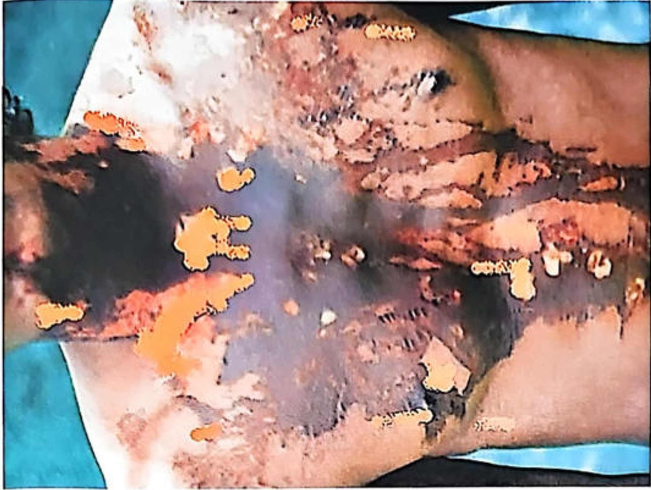
Fig: Vitriolage - showing penetrating chemical burns with brownish-black corrosive staining and trickle marks (Essentials of Forensic Medicine & Toxicology, 36th ed.)
| Feature | Description |
|---|---|
| Pain | Burns are initially painless (acid destroys nerve endings) |
| Type of burn | Penetrating burns - acid devitalizes tissues and predisposes to infection |
| Stain colour | Brown/black (sulphuric acid); yellow (nitric acid) |
| Trickle marks | Characteristic linear runs of acid down the skin from the point of impact |
| Vesication | Absent (unlike thermal burns which form blisters - important distinguishing feature) |
| Red line of demarcation | Absent (unlike thermal burns) |
| Repair | Slow; scar tissue formation causes contracture |
| Clothing | Holes/discolouration in clothing at corresponding sites |
| Eyes | Blindness if eyes involved |
Local and Systemic Effects
Local Effects
- Skin: Severe corrosion with formation of brownish-black burned areas → permanent scarring; necrosis; gangrene; contracture
- Eyes: Intense pain, chemosis, corneal ulceration → blindness (permanent)
- Airways/Respiratory: Volatile acids (nitric acid, HCl) → irritation and oedema of respiratory tract; asphyxia
- Clothing: Distinctive acid holes and staining
Systemic Effects (if absorbed)
- Shock (from extensive burns and pain)
- Toxaemia from systemic absorption
- Death may result from shock, toxaemia, or secondary infection from extensive burns
Treatment of Vitriolage
Immediate First Aid
- Copious water washing of affected parts immediately - the single most important step; dilutes and removes the acid
- Wash with soap solution or dilute sodium or potassium bicarbonate/carbonate solution (neutralises acid)
- Apply a thick paste of magnesium oxide or magnesium carbonate over the burns (acts as a neutralising buffer)
- Cover the raw surface with tannic acid jelly, soframycin, or penicillin tulle gauze dressing
Eye Care
- Wash eyes immediately with large amounts of water
- Irrigate with 1-5% sodium bicarbonate solution
- Instil a few drops of olive oil or castor oil (soothes, forms a protective layer)
- Treat corneal ulcers with:
- Atropine ointment
- Hydrocortisone eye drops
- Antibiotic eye drops/ointment
General/Supportive
- Treat shock with IV fluids
- Analgesics
- Antibiotics for secondary infection
- Surgical: debridement; skin grafting for extensive burns
Medicolegal Aspects
Legal Classification (Indian Law)
| Provision | Description |
|---|---|
| BNS Section 124(1) (formerly IPC 326A) | Causing permanent or partial damage by throwing/administering acid with intent/knowledge = imprisonment not less than 10 years to life + fine (fine paid to victim as compensation) |
| BNS Section 124(2) (formerly IPC 326B) | Attempt to throw acid (even without causing injury) = 5-7 years imprisonment + fine |
| IPC Section 326A (older reference) | Voluntarily causing grievous hurt by acid attack = 10 years to life + fine |
| IPC Section 326B | Attempt to throw acid = 5-7 years imprisonment |
Legal Classification of Injuries
| Injury | Legal Category |
|---|---|
| Blindness resulting from vitriolage | Grievous injury (under law) |
| Scar tissue formation (permanent disfigurement) | Grievous injury |
| Vitriolage as a whole | Amounts to dangerous injury |
Definition (Legal - Acid Attack)
"Acid attack is throwing or attempting to throw acid or any acid substance or a corrosive or burning substance which can cause bodily injury leading to scars or disfigurement or temporary or permanent disability or grievous hurt." - Parikh's Textbook
Other Medicolegal Points
- Scar disfigurement on the face from vitriolage is a permanent disfigurement and provides grounds for compensation in civil law
- Acid attacks can be distinguished from thermal burns forensically by:
- Absence of vesicles/blisters
- Absence of red line of demarcation
- Presence of trickle marks
- Characteristic stain colour (brown-black or yellow)
- Chemical detection of the acid in wound washings/clothing
- Acid attacks have been increasing in incidence, leading to specific dedicated legal provisions
Sources:
Civil responsibility of mentally ill persons
Searching sources...
Finding Sources
Finding Sources
Finding Sources
Finding Sources
Found the dedicated section in Parikh's. Let me also search for related content in other forensic medicine textbooks.
Reading File
Finding Sources
Good content found. Let me read the remaining sections (testamentary capacity and more) plus search for related content in other forensic textbooks.
Reading File
Finding Sources
Now let me search for the same topic in the Essentials and PC Dikshit textbooks.
Finding Sources
Finding Sources
Finding Sources
Finding Sources
Found the Essentials of Forensic Medicine section too. Let me read it.
Reading File
I now have comprehensive content from both Parikh's and the Essentials of Forensic Medicine. Here is the complete answer:
Civil Responsibility of Mentally Ill Persons
Introduction
Civil responsibility of a mentally ill person refers to their legal capacity to exercise civil rights and incur civil obligations. The law does not treat mental illness as a blanket disqualifier - it examines the degree of incapacity at the relevant time and the nature of the specific act in question.
The subject is generally considered under six main heads:
- Management of property
- Contracts
- Marriage
- Competency as a witness
- Validity of consent
- Testamentary capacity
1. Management of Property
The Mental Health Act, 1987 provides the legal framework for protection of both the person and property of mentally ill individuals.
Key provisions:
- The Court may appoint a guardian to take care of the person
- The Court may appoint a manager to manage the property, if the person is unable to look after himself or his affairs
- The Court may order the sale or disposal of property to pay the mentally ill person's debts and expenses
- If it is reported that the unsoundness of mind has ceased, the Court may order a second inquiry and terminate all proceedings
Standard of proof: "The inquiry is not whether the person is insane, but whether he is so insane as to make him incapable of managing his property and affairs." In case of doubt, it is safer to give an opinion in favour of sanity. - Essentials of Forensic Medicine & Toxicology, 36th ed.
2. Contracts
Legal basis: Section 12 of the Indian Contract Act, 1872 (Act IX of 1872)
| Situation | Legal Position |
|---|---|
| Contract made when person was mentally ill and unable to understand it | Invalid / Void |
| Contract made during a lucid interval | Valid and binding |
| Mental illness developing after the contract | Does not make it invalid, unless performance of services becomes impossible |
| Other party unaware of mental illness and contract is fair | May be binding |
| Contract for simple necessities (food, shelter, clothing, medical care) | Mentally ill person IS responsible |
| Order grossly excessive or seller took undue advantage of illness | Mentally ill person is NOT responsible |
Additional rules:
- A person who is usually of unsound mind but occasionally sound may make a contract when of sound mind
- A person usually of sound mind but occasionally unsound may NOT make a contract during unsound periods
- Mental disorder of a partner does not dissolve a partnership unless dissolution steps are taken
3. Marriage and Divorce
Legal basis: Divorce Act, 1869; Hindu Marriage Act; Special Marriage Act
Marriage is regarded as a contract in law. A marriage is considered null and void if at the time of the ceremony either party:
- Was incapable of giving valid consent due to mental illness
- Though capable of giving valid consent, was suffering from such a kind or degree of mental disorder as to be unfit for marriage and procreation of children
- Has been suffering from recurrent attacks of unsoundness of mind or epilepsy
Key rule: Mental illness occurring subsequent to marriage is NOT a ground for divorce, except under exceptional circumstances. - Parikh's Textbook
4. Competency as a Witness
Legal basis: Section 118 of the Indian Evidence Act (now BSA-2023, Section 124)
"All persons shall be competent to testify unless the court considers that they are prevented from understanding the questions put to them, or from giving rational answers to those questions, by tender years, extreme old age, disease whether of body or mind, or any other cause of the same kind."
| Situation | Competency |
|---|---|
| Mentally ill, unable to understand questions or give rational answers | NOT competent to testify |
| Mentally ill but testifying during a lucid interval | Competent |
| Person suffering from delusions but able to relate what they saw and understands the obligation of an oath | Competent |
The court evaluates each witness individually - mental illness is not an automatic bar to testimony.
5. Validity of Consent
Legal basis: Section 90 of the Indian Penal Code (IPC)
"Consent is not valid if given by a person who, from unsoundness of mind or intoxication, is unable to understand the nature and consequence of that to which he gives consent."
Situations where this arises:
- Rape - if a woman gives "consent" while of unsound mind, it is not valid consent and the act constitutes rape
- Causing grievous hurt or death - consent from an insane person cannot legally authorise the act
- Abetment of suicide - if a person incites a mentally ill person to commit suicide, the invalidity of consent is relevant
6. Testamentary Capacity
Definition: The capacity of a person to make a valid will. The legal standard requires that the testator possess a "sound disposing mind" (compos mentis) at the time of making the will, certified by a doctor.
Conditions for a Valid Will
A will is valid only if all of the following are fulfilled:
| # | Condition |
|---|---|
| 1 | The testator must be a major (adult) |
| 2 | Must have a sound disposing mind at the time of making the will |
| 3 | Must understand the nature and consequences of the act |
| 4 | Must know what property he has, to whom he is giving it, and has a rational reason |
| 5 | Executing it voluntarily, without undue influence |
| 6 | Must be signed in the presence of two witnesses, one of whom should be a medical man; both witnesses must sign in the presence of each other and of the testator |
| 7 | None of the witnesses should be beneficiaries under the will |
"Sound Disposing Mind" Defined
A sound disposing mind is a mind which has the capacity of recollecting what property he has, understanding to whom he is giving it, and appreciating the consequences of his act. Force, undue influence, or dishonest misrepresentation invalidates a will even in a sane person.
Special Rules for Testamentary Capacity
| Situation | Validity of Will |
|---|---|
| Mentally ill person during a lucid interval | Valid will can be made |
| Person suffering from delusions not related to the property | Can make a valid will |
| Delusions directly related to the property being bequeathed | Will may be invalid |
| Will made during drunkenness - but person knew what he was doing | Valid, unless repudiated in sober moments |
| Will made when drunk and person did not understand the act | Invalid |
| Extreme age, feeble health, mental sluggishness alone | Do NOT invalidate a will |
| Mind so impaired as to be unable to understand the nature of the act | Invalid |
| Aphasic person who communicates by nods | Can make a valid will |
| Eccentric person (not legally insane) | Will may be contested but cannot be declared invalid on ground of eccentricity alone |
Summary Table
| Civil Matter | Key Legal Principle | Relevant Law |
|---|---|---|
| Property management | Court appoints guardian/manager | Mental Health Act, 1987 |
| Contracts | Invalid if unable to understand; valid in lucid interval | Indian Contract Act, 1872 (S.12) |
| Marriage | Null and void if incapable of consent at time of ceremony | Divorce Act 1869; Hindu Marriage Act |
| Witness competency | Competent if able to understand questions and give rational answers | Evidence Act S.118 / BSA S.124 |
| Validity of consent | Consent of insane person is not valid | IPC S.90 |
| Testamentary capacity | Requires sound disposing mind; valid in lucid interval | Indian Succession Act |
Important Concept: Lucid Interval
A lucid interval is a period during which a mentally ill person temporarily regains their mental faculties to a sufficient degree to perform legal acts. During a genuine lucid interval:
- Contracts entered are valid
- A will made is valid
- Testimony given is admissible
- Consent given is valid
The lucid interval must be genuine (not merely a quiet period) - the person must be capable of full understanding of the nature and consequence of the act.
Sources:
- Parikh's Textbook of Medical Jurisprudence, Forensic Medicine and Toxicology - Chapter 33, Civil Responsibilities of the Mentally Ill
- The Essentials of Forensic Medicine and Toxicology, 36th ed. (2026) - Civil Responsibility section
Kleptomania in forensic medicine
Searching sources...
Finding Sources
Finding Sources
Excellent content in Kaplan & Sadock (both Synopsis and Comprehensive), plus PC Dikshit forensic. Let me read these sections in parallel.
Reading File
Reading File
Finding Sources
Excellent content. Let me now read the etiology, treatment and forensic/medicolegal sections plus the PC Dikshit forensic reference and the DSM criteria.
Reading File
Reading File
Reading File
Now let me read the treatment section and the forensic/differential diagnosis content from Kaplan Synopsis.
Reading File
Reading File
I now have comprehensive, authoritative content from both Kaplan & Sadock's Synopsis and Comprehensive Textbook of Psychiatry. Here is the complete answer:
Kleptomania in Forensic Medicine
Definition
Kleptomania (from Greek kleptēs, "thief" + mania, "madness") is a recurrent failure to resist the impulse to steal objects that are not needed for personal use and have no significant monetary value to the individual - who could easily have afforded them. The act of stealing itself is the goal, not the object stolen.
It is classified under Disruptive, Impulse-Control, and Conduct Disorders in DSM-5-TR, and is a recognised form of monomania (an irresistible impulse in an otherwise rational mind) in forensic psychiatry.
DSM-5-TR Diagnostic Criteria
| Criterion | Description |
|---|---|
| A | Recurrent failure to resist impulses to steal objects not needed for personal use or monetary value |
| B | Increasing sense of tension immediately before committing the theft |
| C | Pleasure, gratification, or relief at the time of committing the theft |
| D | The stealing is NOT to express anger or vengeance, and NOT in response to a delusion or hallucination |
| E | NOT better accounted for by Conduct Disorder, a Manic Episode, or Antisocial Personality Disorder |
Source: DSM-5-TR (APA, 2022) as reproduced in Kaplan & Sadock's Comprehensive Textbook of Psychiatry
Core Characteristics
| Feature | Description |
|---|---|
| Object stolen | Not needed; often of little or no value; could have been purchased |
| What happens after stealing | Objects are given away, returned secretly, hidden, or hoarded |
| Planning | Theft is not preplanned; occurs impulsively |
| Others involved? | No - theft does not involve accomplices |
| Motive | No anger, revenge, financial gain, or delusional drive |
| Awareness of risk | May take some precautions (e.g., not stealing in front of a guard) but does NOT rationally weigh consequences despite repeated arrests |
| Emotions after theft | Guilt, shame, anxiety, depression - but NOT anger or vengeance |
| Ego-syntonic vs dystonic | Ego-dystonic - the urge is distressing and unwanted |
The classic triad: tension before → gratification during → guilt/remorse after
Epidemiology
| Parameter | Data |
|---|---|
| Prevalence in general population | 0.3% to 0.6% (DSM-5-TR) |
| Prevalence among shoplifters arrested | 3.8% to 24% |
| Prevalence in psychiatric inpatients | Current: 7.8%; Lifetime: 9.3% |
| Sex ratio | Female predominance - about two-thirds of patients are women; M:F ratio = 1:3 in clinical samples |
| Age of onset | Commonly adolescence; average onset ~18.75 years; may begin in childhood or adulthood |
| Course | Chronic, waxing and waning |
| Underdiagnosis | Common - patients are embarrassed and ashamed to report symptoms |
Etiology and Neurobiology
The etiology is poorly understood. Several theories exist:
Neurological Associations
- Case reports of kleptomania associated with:
- Head trauma
- Frontal lobe lesions
- Cortical atrophy / dementia
- Hypoglycaemia (secondary to insulinoma)
- Diffusion tensor imaging: Reduced fractional anisotropy in inferior frontal white matter → subtle white matter pathology
- Neuroimaging: Hyperactivity in the right ventral striatum and orbitofrontal white matter damage reported
- Greater severity correlates with impaired executive functioning on neuropsychological testing
Neurochemical Theories
- Serotonin dysregulation - kleptomania as an obsessive-compulsive spectrum disorder
- Opioid system - naltrexone (µ-opioid receptor antagonist) reduces stealing urges, suggesting reward/drive pathway involvement
- Dopamine system - role in reward and impulse regulation
- Glutamate (NMDA) pathway - memantine (NMDA antagonist) reduces urges
Genetic Factors
- Limited data; patients with kleptomania have more first-degree relatives with alcohol use disorders than controls
Comorbidities
Kleptomania has high comorbidity with:
| Disorder | Notes |
|---|---|
| Major depressive disorder | Most common comorbidity |
| Anxiety disorders | Common |
| OCD | Rate 6.5-60%; may be a variant of OCD |
| Eating disorders | Especially bulimia nervosa |
| Substance use disorders | Especially alcohol use disorder |
| Pathological gambling | |
| Compulsive shopping / buying | |
| Personality disorders | |
| Other impulse-control disorders | |
| Suicide | Individuals with kleptomania have higher rates of suicidal ideation and attempts |
Forensic / Medicolegal Significance
1. Kleptomania as a Defence in Criminal Law
Kleptomania is relevant to the criminal responsibility of the accused in theft cases. Under Indian law (IPC Section 84/BNS Section 22):
- A person is not criminally responsible for an act if, at the time of doing it, by reason of unsoundness of mind, they were incapable of knowing the nature of the act or that it was wrong
- However, kleptomania alone is NOT accepted as a complete defence to theft in most jurisdictions - the act is typically considered wilful, even if driven by an uncontrollable impulse
- Courts look at whether the accused knew the act was wrong (they typically do - they avoid guards, conceal the object), which is evidence of the knowledge of wrongdoing
- In practice, kleptomania may be used to mitigate punishment rather than to completely excuse the offence
2. Kleptomania vs. Malingering
"Some individuals with an antisocial personality disorder or no disorder may steal and then claim to suffer from kleptomania. This would be an example of malingering, not kleptomania, although with sufficiently intelligent perpetrators it may be challenging to tell the difference." - Kaplan & Sadock's Synopsis
How to distinguish:
- True kleptomania: objects have no value to the person; guilt and distress are genuine; no financial motive; no planning; ego-dystonic
- Malingering: claims made only after arrest; objects have value or are sold; lack of genuine guilt; history of deliberate theft; antisocial traits
3. Differential Diagnosis of Shoplifting in Forensic Context
| Type of Shoplifting | Features |
|---|---|
| True kleptomania | Rare; objects not needed; genuine tension-relief cycle; guilt; ego-dystonic |
| Ordinary theft (antisocial) | Premeditated; for personal gain; no remorse; may involve others; possible violence |
| Teenage shoplifting | Most common; for "kicks"/goods; in pairs/groups; no psychiatric disorder |
| Psychotic stealing | During mania, psychotic depression, schizophrenia; driven by delusions/hallucinations |
| Substance intoxication | Theft during acute intoxication; reduced inhibition |
| Dementia/Alzheimer's | Forgetfulness - leaves without paying; no larcenous intent |
| Malingering | Claims kleptomania after arrest to escape punishment |
4. Type of Mental Disorder (Forensic Classification)
In forensic psychiatry, kleptomania is classified as a type of monomania - an irresistible impulse directed at a specific act in an otherwise sane mind. It is an impulse control disorder, not a psychosis.
(PC Dikshit Forensic Medicine MCQ: "Kleptomania is a type of: D. Impulse")
5. Legal and Social Consequences
- Repeated arrests and criminal prosecution despite the psychiatric diagnosis
- Jail time and probation
- Marital and relationship conflict from shoplifting episodes
- Decreased quality of life
- Suicidal gestures may occur following arrests (as in the case vignette)
- Courts may order psychiatric evaluation and treatment as a condition of probation
Treatment
Pharmacotherapy
| Drug | Evidence |
|---|---|
| Naltrexone (µ-opioid antagonist) | Best evidence; controlled trial: 66% remission vs 7.7% placebo; reduces urge to steal and stealing behaviour |
| Memantine (NMDA antagonist) | Open-label; reduces urges; improves impulsivity and mood |
| SSRIs (fluoxetine, fluvoxamine, citalopram) | Mixed results; based on OCD-spectrum hypothesis; some benefit, some no effect; paradoxically some developed kleptomania ON SSRIs |
| Lithium | Case reports of benefit |
| Topiramate | Case reports: reduces stealing urges |
| Escitalopram | Controlled trial found NO benefit |
| Tricyclics, trazodone, valproate | Case reports |
| ECT | Case reports |
Psychotherapy
| Modality | Evidence |
|---|---|
| Cognitive Behavioural Therapy (CBT) | Most promising; case reports and series |
| Exposure and Response Prevention (ERP) + CBT | Feasibility study: reduces symptoms and autonomic arousal |
| Systematic desensitisation | Reported successful |
| Aversive conditioning | Reported successful even when motivation was low |
| Insight-oriented psychotherapy / Psychoanalysis | Successful when patient is motivated by guilt and shame |
Most patients seek treatment only after being arrested, due to shame. Combination of pharmacotherapy (naltrexone) and CBT is the most logical approach given current evidence.
Assessment Scales
| Scale | Use |
|---|---|
| K-SAS (Kleptomania Symptom Assessment Scale) | Self-report; validated; monitors urges, behaviour, and cognition |
| Y-BOCS (Yale-Brown OC Scale) | Not validated for kleptomania but used in practice to monitor treatment |
| Barratt Impulsiveness Scale (BIS) | Assesses impulsivity severity |
Sources:
- Kaplan & Sadock's Comprehensive Textbook of Psychiatry - Chapter 24.3: Kleptomania
- Kaplan and Sadock's Synopsis of Psychiatry - Chapter 18: Impulse Control Disorders
- PC Dikshit Textbook of Forensic Medicine and Toxicology
Hallucination in forensic medicine
Searching sources...
Finding Sources
Finding Sources
Finding Sources
Finding Sources
Good - found a dedicated forensic medicine section in PC Dikshit. Let me read that plus search Parikh's and the Essentials for hallucination content.
Reading File
Finding Sources
Good. Now let me search Parikh's and the Essentials for hallucination, and also get the Kaplan & Sadock psychiatry definition for a richer clinical picture.
Finding Sources
Finding Sources
Reading File
Finding Sources
Now let me read the Parikh's hallucination section and the Essentials of Forensic Medicine.
Finding Sources
Reading File
Now let me read the Essentials of Forensic Medicine section on hallucination types.
Reading File
I now have comprehensive content from all key sources. Let me compile the full answer.
Hallucination in Forensic Medicine
Definition
Hallucination is a false sense perception without any external object or stimulus to produce it - a perception that seems entirely genuine to the patient yet occurs in the complete absence of any real external stimulus.
"Hallucination is the apparent perception of an external object when no corresponding real object exists." - PC Dikshit, Textbook of Forensic Medicine and Toxicology
"The experience of a perception that seems genuine yet occurs without an actual external stimulus. The perception may occur in any of the five senses: auditory, gustatory, olfactory, tactile/somatosensory, or visual." - Kaplan & Sadock's Comprehensive Textbook of Psychiatry
A dream is cited as the simplest example of hallucination in normal experience.
Hallucination vs. Illusion vs. Delusion (Key Forensic Distinctions)
| Feature | Hallucination | Illusion | Delusion |
|---|---|---|---|
| Definition | Perception WITHOUT any external stimulus | False INTERPRETATION of a real external stimulus | False, fixed belief not correctable by evidence |
| External stimulus | Absent | Present (but misinterpreted) | Not applicable |
| Example | Hearing voices when alone | Mistaking a rope for a snake | Believing one is being poisoned |
| Domain | Perception | Perception | Thought/belief |
| Correction | Cannot correct | Sane person CAN correct; insane cannot | Cannot be corrected by reasoning |
| Forensic significance | May lead to violence/suicide | May lead to violence; person not responsible | Basis for insanity defence |
Types of Hallucinations
1. Auditory Hallucination
- Person hears voices or sounds when no one is present
- Most common type in functional psychiatric disorders (especially schizophrenia)
- Also occurs in delirium, psychotic mood disorders, toxic and metabolic encephalopathies
- Forensically most significant due to command hallucinations
2. Visual Hallucination
- Person sees objects or persons that do not exist (e.g., imagines being attacked by a lion)
- Most common type in organic mental disorders
- Occurs in: delirium tremens, focal CNS lesions, toxic disturbances, drug withdrawal syndromes, schizophrenia
- In non-Western cultures, may be the most common form in schizophrenia
3. Olfactory Hallucination
- Person smells pleasant or unpleasant odours when none are present
- Occurs in: organic brain disease, major depression, temporal lobe epilepsy
- The smell of burning or rotting is a classic feature
4. Gustatory Hallucination
- Person perceives taste (sweet, sour, bitter, good, or bad) in the mouth without any food present
- Occurs in: organic brain diseases, temporal lobe epilepsy (uncinate fits)
5. Tactile (Haptic) Hallucination
- Person imagines physical sensations - e.g., rats or mice crawling over or into the bed (formication)
- Classic example: "cocaine bugs" - the sensation of insects crawling under the skin in cocaine/stimulant poisoning
- Common in: alcohol withdrawal syndrome (delirium tremens), chronic cocaine poisoning
6. Kinaesthetic / Psychomotor Hallucination
- Feeling of movement of some part of the body when it is stationary
- Sensation of bodily parts moving, floating, or being twisted without actual movement
7. Command Hallucination (Forensically Most Important)
- The patient is ordered by hallucinatory voices to do things which may be frightening or dangerous
- More often unpleasant and alarming
- A person suffering from command hallucinations may be incited to commit suicide or homicide
- Forensically critical - directly links hallucination to violent or self-destructive acts
8. Lilliputian / Microptic / Macroptic Hallucination
- Objects appear abnormally small (microptic/Lilliputian) or abnormally large (macroptic)
- Seen in certain toxic states, alcohol intoxication, and some organic disorders
9. Hypnagogic Hallucination
- False sensory perception occurring midway between wakefulness and falling asleep (drowsy, pre-sleep state)
- A normal phenomenon in many people; not indicative of psychosis
- Associated with narcolepsy and sleep disorders
10. Hypnopompic Hallucination
- Occurs during the drowsy state after deep sleep and before full awakening
- Also normal in healthy individuals; should not be mistaken for psychosis
11. Sexual Hallucination
- Person feels sexually satisfied from unfounded, self-imagined objects or sensations
Conditions Causing Hallucinations
| Category | Examples |
|---|---|
| Psychiatric disorders | Schizophrenia (auditory most common), psychotic depression, acute mania |
| Drugs / Substance abuse | LSD, mescaline, cannabis, cocaine (tactile), alcohol (small doses - tactile/visual) |
| Alcohol withdrawal | Delirium tremens - vivid visual and tactile hallucinations |
| CNS lesions | Temporal lobe lesions, brain tumour, frontal lobe lesions |
| Organic brain disease | Generalised organic brain disease, cortical atrophy, dementia |
| Metabolic / Toxic | Uraemia, subarachnoid haemorrhage, hypoglycaemia, metabolic encephalopathy |
| Febrile states | High fever (febrile delirium) |
| Sleep-wake disorders | Hypnagogic/hypnopompic hallucinations (narcolepsy) |
Medicolegal Importance of Hallucination
1. Criminal Responsibility
"Delusions arising from unpleasant hallucinations may cause the person to commit suicide or homicide. He is not responsible for the deeds." - PC Dikshit
- Under IPC Section 84 / BNS Section 22: A person is not criminally responsible for an act if, by reason of unsoundness of mind, they did not know the nature of the act or that it was wrong or contrary to law
- A person acting under command hallucinations - hearing voices ordering them to kill - may be held to lack the mens rea (guilty mind) for the crime
- Such individuals are typically found not guilty by reason of insanity and are detained in a psychiatric facility rather than prison
- The key forensic question: Were the hallucinations so overwhelming that the person could not understand the nature or wrongfulness of their act?
2. Command Hallucinations and Homicide
- Auditory command hallucinations in schizophrenia are directly linked to acts of violence against others and suicide
- The complexity of the hallucinations is relevant: more complex, commanding, and persistent voices → greater risk of compliance
- Whether the patient believed the voices were real and had no control over their response is central to the insanity defence
3. Hallucinations and Suicide
- Unpleasant hallucinations - especially command hallucinations ordering self-harm - can incite suicide
- A person who kills themselves while experiencing hallucinations is not considered to have had valid consent to their own death in the context of coercion by psychotic symptoms
4. Malingered Hallucinations
- Persons may feign hallucinations to escape criminal responsibility (malingering)
- Features of genuine hallucinations vs. malingering:
| Feature | Genuine Hallucination | Malingered Hallucination |
|---|---|---|
| Onset | Consistent with clinical history | Often appears suddenly on arrest |
| Consistency | Consistent over time | Inconsistent, changes with questioning |
| Behaviour concordant | Actions match the reported experience | Often no concordant behaviour |
| Response to medication | Responds to antipsychotics | No response (or dramatic claims of cure) |
| Affect | Appropriate distress, fear, response | Often exaggerated or theatrical |
| Other psychotic features | Usually present (delusions, disorganised thinking) | Usually absent |
5. Hallucinations and Testamentary Capacity
- A person suffering from hallucinations at the time of making a will lacks a sound disposing mind
- However, if hallucinations are present but do not relate to the property being bequeathed, the will may still be valid (analogous to the rule for delusions)
6. Hallucinations and Consent
- Under IPC Section 90: Consent given by a person experiencing hallucinations or delusions that prevent them from understanding the nature and consequences of an act is not valid consent
7. Hallucinations as Evidence of Insanity
- Persistent, systematised hallucinations - especially auditory command voices in schizophrenia - are strong evidence of legal insanity
- The forensic psychiatrist is called upon to:
- Determine whether hallucinations were genuinely present at the time of the offence
- Assess whether the hallucinations were severe enough to negate knowledge of wrongdoing
- Distinguish true hallucinations from illusions, pseudohallucinations, or malingering
Hallucination in Relation to Specific Disorders (Forensic Context)
| Disorder | Hallucination Type | Forensic Relevance |
|---|---|---|
| Schizophrenia | Auditory (most common); command voices | Homicide, self-harm; insanity defence |
| Delirium tremens (alcohol withdrawal) | Visual (pink elephants, insects) + tactile | Acts during DT; reduced responsibility |
| LSD / Mescaline intoxication | Visual, auditory | Drug-induced psychosis; diminished responsibility |
| Cocaine poisoning | Tactile (cocaine bugs), visual | Drug offences; violence during intoxication |
| Temporal lobe epilepsy | Olfactory, gustatory (uncinate fits) | Automatism defence; offences during ictal period |
| Dementia | Visual | Wandering, aggression; reduced criminal responsibility |
| Febrile delirium | Visual, auditory | Acts in delirium; no criminal responsibility |
Summary of Forensic Points
- Hallucination is a false perception without external stimulus - purely imaginary
- Auditory hallucinations are commonest in functional disorders; visual in organic disorders
- Command hallucinations are the most forensically dangerous type - may drive the patient to commit homicide or suicide
- A person acting on the basis of hallucinations is generally not criminally responsible
- Courts must be alert to malingered hallucinations - careful psychiatric assessment is essential
- Hallucinations affect testamentary capacity, validity of consent, and competency as a witness
- They must be distinguished from illusions (misinterpretation of real stimuli) and delusions (false fixed beliefs)
Sources:
Delusion in forensic medicine
Searching sources...
Finding Sources
Finding Sources
Finding Sources
Finding Sources
Found the dedicated forensic section in PC Dikshit. Let me read it and simultaneously search for delusion content in Parikh's, the Essentials, and Kaplan & Sadock.
Reading File
Finding Sources
Reading File
Finding Sources
Now let me get Parikh's and the Essentials content on delusion.
Finding Sources
Finding Sources
Finding Sources
Finding Sources
Let me now read the Essentials and Parikh's sections on delusion directly and also read the Kaplan & Sadock delusion content.
Reading File
Reading File
Now let me read the rest of the Essentials section on types of delusion and their medicolegal importance.
Reading File
I now have comprehensive content from all three forensic medicine textbooks and Kaplan & Sadock. Here is the complete answer:
Delusion in Forensic Medicine
Definition
Delusion is a false and firm belief in something which is not a fact, and which persists even after its falsity has been clearly demonstrated. The belief is not ordinarily shared by other members of the patient's sociocultural and educational group, and it cannot be corrected by reasoning, argument, or evidence.
"A delusion is a firmly held, false belief based on an incorrect inference that is unshakeable despite evidence to contradict it. It cannot simply be a cultural belief." - Kaplan & Sadock's Comprehensive Textbook of Psychiatry
"Delusion is a false and firm belief in something which is not a fact, and which persists even after its falsity has been clearly demonstrated. A normal person can have a delusion, but is capable of correcting it by his reasoning power, by his past experience and by being convinced by others." - Essentials of Forensic Medicine & Toxicology, 36th ed.
Key Characteristics
| Feature | Description |
|---|---|
| False | Not based on reality |
| Fixed / Firm | Unshakeable; cannot be corrected by logic, evidence, or persuasion |
| Incorrigible | Persists even when falsity is clearly demonstrated |
| Not culturally shared | Not a belief held by the person's sociocultural group |
| Pathognomonic | Delusions are pathognomonic of the psychoses |
| Domain | Disorder of thought content (not perception) |
| Normal person | A normal person CAN have a momentary false belief, but corrects it by reasoning |
| Insane person | Cannot correct - belief is under control of emotional, not rational forces |
Delusion vs. Hallucination vs. Illusion (Forensic Comparison)
| Feature | Delusion | Hallucination | Illusion |
|---|---|---|---|
| Domain | Thought (belief) | Perception | Perception |
| External stimulus | Absent | Absent | Present (misinterpreted) |
| Correctability (sane) | Can correct | Can correct | Can correct |
| Correctability (insane) | Cannot correct | Cannot correct | Cannot correct |
| Example | "My wife is poisoning me" | Hearing voices when alone | Rope mistaken for a snake |
Types of Delusion
1. Delusion of Grandeur / Exaltation (Megalomania)
- Person imagines himself to be very rich, powerful, or a famous personality (e.g., a king, a prophet, a deity) when in reality he is an ordinary person or a pauper
- Seen in: delirium tremens, mania, general paresis of the insane (GPI)
- Often co-exists with delusions of persecution in the same individual
2. Delusion of Persecution (Paranoid Delusion)
- Person believes that attempts are being made to harm, poison, kill, or conspire against him - often by nearest relatives (wife, sons, parents)
- Example: "My wife is putting poison in my food"
- Seen in: paranoid schizophrenia, dementia, depression, paranoid disorder
- Forensically most dangerous type - most frequently leads to homicide
3. Delusion of Reference
- Person believes that people, events, things, strangers, newspapers, radio refer to or are talking about him in a special way
- Example: "The newsreader on TV is sending me messages"; "Strangers in the street are talking about me"
- Seen in: schizophrenia, paranoid states
4. Delusion of Influence / Control
- Person believes his thoughts, feelings, and actions are being influenced or controlled by some outside agency - radio waves, hypnotism, telepathy, aliens, etc.
- Also called "passivity experiences" or "made phenomena"
- Seen in: schizophrenia (first-rank symptom of Schneider)
5. Delusion of Infidelity (Othello Syndrome / Morbid Jealousy)
- Person imagines his spouse or partner to be unfaithful while in fact she/he is completely faithful
- Also called jealous delusion or conjugal paranoia
- Othello Syndrome: The person has delusions of infidelity about wife/mistress and assaults her; if she confesses under severe pressure, he may explode into violent acts and attempt or commit murder
- Forensically very significant - common cause of domestic violence and intimate partner homicide
6. Delusion of Self-Reproach (Self-Accusation)
- Person blames himself excessively for past failures and misdeeds which are often trivial or non-existent
- Seen in: severe depression, melancholia
- Forensic relevance: strong association with suicide
7. Nihilistic Delusion
- Person declares that he does not exist, or that the world does not exist, or that his organs have rotted away
- Seen in: severe depressive psychosis (Cotard's syndrome)
- Associated with self-neglect and suicide
8. Hypochondriacal Delusion
- Person has an unshakeable belief that there is something seriously wrong with his body, despite being medically healthy
- Distinct from ordinary hypochondria (where insight is partially preserved)
- Seen in: severe depression, schizophrenia, delusional disorder (somatic type)
9. Paranoid Delusion
- Characterised by over-suspiciousness leading to complex persecutory delusions
- Person may believe in elaborate conspiracies against him
- May overlap significantly with delusion of persecution
10. Delusion of Jealousy (Morbid Jealousy)
- Related to, but broader than, Othello Syndrome
- Can apply to any relationship, not just marital
11. Religious Delusion
- Person has false fixed beliefs of a religious nature - that he is a divine messenger, prophet, or is being punished by God for sins
- Seen in: acute mania, schizophrenia
12. Erotomania (De Clérambault Syndrome)
- A delusion in which the person believes that someone (usually of higher status or a celebrity) is deeply in love with them, even without any evidence or contact
- The person develops an obsession and attempts to get close through telephone calls, letters, gifts, visits
- The other party is typically unaware or is actually a stranger
- Forensic relevance: leads to stalking, harassment, threatening behaviour against the supposed love object or perceived rivals; may escalate to violence
13. Thought Broadcasting / Thought Insertion
- Broadcasting: Belief that one's private thoughts are being transmitted to others and can be heard by strangers
- Insertion: Belief that thoughts in one's mind have been placed there by an outside force
- First-rank symptoms of schizophrenia (Schneider)
14. Bizarre Delusion
- A belief considered completely impossible by members of one's culture
- Example: "Aliens have removed my brain and replaced it with someone else's"
- Characteristic of schizophrenia
Conditions in Which Delusions Occur
| Condition | Type of Delusion Commonly Seen |
|---|---|
| Schizophrenia | Persecution, reference, influence, thought broadcasting/insertion, bizarre |
| Paranoid disorder | Systematised persecution, infidelity, erotomania |
| Mania (bipolar) | Grandeur, religious |
| Severe depression | Self-reproach, nihilistic, hypochondriacal |
| Delirium tremens | Grandeur |
| Dementia | Persecution, infidelity |
| General paresis of the insane (GPI) | Grandeur (classic) |
| Organic brain disease | Various |
Note: Delusions are NOT seen in anxiety neurosis and other neurotic illnesses - Essentials of Forensic Medicine
Medicolegal Importance of Delusion
1. Criminal Responsibility
"Delusion is never an isolated disorder, but is merely an indication of a deep-seated, widespread disorder. For this reason, such a person cannot be regarded as fully responsible for his antisocial acts." - Essentials of Forensic Medicine & Toxicology
- Under IPC Section 84 / BNS Section 22: A person is not criminally responsible if, at the time of the act, by reason of unsoundness of mind, he was incapable of knowing the nature of the act or that it was wrong
- A person acting on the basis of a delusion - e.g., believing he is killing a demon when in fact killing a person - may not be held criminally responsible
- The partial defence: Even if the person knew the act was wrong, the presence of delusion is used to mitigate punishment
2. The "Delusion Test" in Insanity
The Indian courts apply the M'Naghten Rules (1843) to test criminal responsibility:
- Was the accused suffering from a disease of the mind?
- Did it cause a defect of reason?
- Did this prevent him from knowing the nature of the act, or that it was wrong?
A person who commits a crime while acting under a delusion is typically judged as if the delusion were true - if the deluded belief, even if true, would not have justified the act, the accused is still guilty; if the deluded belief, if true, would have constituted a legal justification, the accused is not responsible.
3. Delusion of Persecution → Homicide
- Most dangerous type in forensic practice
- Person believing his wife is poisoning him may preemptively kill her to "protect himself"
- Paranoid schizophrenia with persecutory delusions is the most common diagnosis in those who commit homicide among the mentally ill
4. Othello Syndrome → Domestic Violence and Murder
- Delusion of infidelity is a well-recognised cause of intimate partner homicide worldwide
- The jealous partner may kill the perceived rival as well
- Even under extreme pressure leading to a false confession, the deluded person interprets it as confirmation and may become more violent
5. Erotomania → Stalking
- De Clérambault syndrome leads to criminal harassment, stalking, threatening behaviour
- May escalate to assault or homicide of the "love object" or others perceived to obstruct the relationship
6. Suicide
- Self-reproach, nihilistic, and persecutory delusions are all major risk factors for suicide
- A deluded person may also commit murder-suicide (e.g., a depressed person with nihilistic delusions kills family members to "spare them suffering" and then kills himself)
7. Testamentary Capacity
- A person suffering from delusions at the time of making a will lacks testamentary capacity
- Exception: A person with delusions can make a valid will if the delusions do not relate to the property being bequeathed
- If the delusions directly affect the testator's understanding of property, beneficiaries, or the consequences of the will, the will is invalid
8. Validity of Contracts and Marriage
- Contracts entered into while labouring under a delusion that prevents understanding of their nature are invalid
- A marriage where one party is deluded as to the nature of the ceremony or the identity of the other party is void
9. Competency as a Witness
- A person suffering from delusions may give evidence during a lucid interval or if the delusions do not affect the subject of their testimony
- If delusions prevent the person from understanding questions or giving rational answers, they are not competent to testify
Important Forensic Notes for the Examining Doctor
"During examination of the mental condition of the person, the medical man should make and carefully note [the delusions] to which the law gives much importance." "In the beginning of a disease, delusion may not be evident. The person can conceal it. Hence the doctor should examine or watch the person for days together." - PC Dikshit
Key points for the forensic examiner:
- Delusions may be concealed by the patient - single interview is insufficient
- A secondary delusion arises from some morbid experience (e.g., delusion of persecution arising from hallucinations)
- Document the content, systematisation, duration, and actions driven by the delusion
- Assess whether the delusion was present at the time of the offence - not merely at examination
Summary Table
| Type | Example | Key Association | Forensic Risk |
|---|---|---|---|
| Grandeur | "I am the King of India" | Mania, GPI, delirium tremens | Low direct violence |
| Persecution | "My wife is poisoning me" | Paranoid schizophrenia | Homicide - highest risk |
| Reference | "Newspapers are about me" | Schizophrenia | Moderate |
| Influence/Control | "Radio controls my mind" | Schizophrenia | Variable |
| Infidelity (Othello) | "My wife is unfaithful" | Paranoid state, alcoholism | Domestic violence, murder |
| Self-reproach | "I am a terrible sinner" | Severe depression | Suicide |
| Nihilistic | "I do not exist" | Psychotic depression | Suicide, murder-suicide |
| Hypochondriacal | "My stomach is rotting" | Depression, schizophrenia | Self-harm |
| Erotomania | "The celebrity loves me" | Delusional disorder | Stalking, harassment |
| Thought insertion | "These thoughts are not mine" | Schizophrenia | Violence |
Sources:
Mc naughten rule in forensic medicine
Searching sources...
Finding Sources
Finding Sources
Finding Sources
Finding Sources
Finding Sources
Finding Sources
Finding Sources
Finding Sources
Found the section in Parikh's. Let me now read it plus the surrounding context, and also read the criminal responsibility sections from PC Dikshit and the Essentials.
Reading File
Finding Sources
Reading File
Reading File
Excellent - found the dedicated McNaughten Rule section in the Essentials. Let me read all of it plus the other tests.
Reading File
Reading File
Now let me read the rest of the McNaughten section including other tests (Durham, Irresistible Impulse).
Reading File
I now have all the content needed for a thorough answer. Here is the complete response:
M'Naghten Rule in Forensic Medicine
Historical Background
The M'Naghten Rules arose from one of the most famous criminal trials in English legal history.
The Case (1843):
- Daniel M'Naghten, a 29-year-old Scotsman, was almost certainly suffering from paranoid schizophrenia
- For many years he harboured a delusion that spies sent by Catholic priests, aided by the Tories (the party in power in England), were constantly following him, harassing him, and hatching a conspiracy against him
- He also likely had auditory hallucinations with Tories accusing him of crimes he denied committing
- He decided to kill the Tory Prime Minister, Sir Robert Peel, making elaborate plans
- On 20th January 1843, he shot Sir Peel's Private Secretary, Edward Drummond, in the back - mistaking him for the Prime Minister
- During the trial he admitted he was "driven to desperation by persecution"
- Ten physicians testified (nine for defence, one for prosecution) - all found him to be of unsound mind
- He was found "not guilty on grounds of mental illness" and was sent to Bethlem Mental Hospital for life
The Aftermath:
- The verdict caused unprecedented public outcry
- Queen Victoria summoned the House of Lords to a special session
- The Lord Chancellor, Lord Lyndhurst put 5 hypothetical questions to a panel of 14 judges to clarify the legal position on insanity
- The answers given on 19th June, 1843 became known as the "M'Naghten Rules"
The M'Naghten Rule (The Right or Wrong Test / The Legal Test)
The most important rule states:
"An accused person is not legally responsible, if it is clearly proved that at the time of committing the crime, he was suffering from such a defect of reason from abnormality of mind, that he did not know the nature and quality of the act he was doing, OR that what he was doing was wrong."
Accepted in India
This legal test has been incorporated into Indian law:
BNS Section 22 (formerly IPC Section 84): "Nothing is an offence which is done by a person, who at the time of doing it, by reason of unsoundness of mind, is incapable of knowing the nature of the act, or that he is doing what is either wrong or contrary to law."
Analysis of Key Terms in the Rule
1. "Clearly Proved"
- The unsoundness of mind must be directly related to the offence in such a way as to satisfy the Court that mental abnormality had a direct causative relationship to the offence
- The offence would NOT have occurred if there was no mental abnormality
- The burden of proof rests on the defence; the law presumes every person to be sane
2. "Defect of Reason"
- It is necessary to show that the accused suffered a defect of reason - not merely that he was in a confused state or acted carelessly
- There must be genuine impairment of the reasoning faculty arising from disease of the mind
3. "Disease of the Mind"
- Refers to any mental disorder that produces a defect of reason - schizophrenia, paranoid psychosis, severe mania, organic psychosis, etc.
- Legal concept - broader than the psychiatric concept of mental illness
4. "Did not know the nature and quality of the act"
- The accused did not understand what he was physically doing
- Example: A man strangles his child believing he is squeezing a lemon
- He may have known his action but not understood its consequences (that it would cause death)
5. "Did not know it was wrong"
- The accused knew what he was doing but did not know it was morally or legally wrong
- Example: A paranoid schizophrenic who kills someone believing he is acting in God's command and that it is righteous
- "Wrong" means contrary to law OR morally wrong - either is sufficient
6. The Delusion Clause
- If a person acts under a delusion, he is judged as if the delusion were true
- Two situations arise:
- If the act committed, even on the assumption that the delusion was real, would still be a crime (e.g., killing for revenge based on imagined persecution) → the person is still responsible
- If the act committed, assuming the delusion were real, would have been justified in law (e.g., acting in genuine self-defence against a real threat) → the person is not responsible
Example: If a person deluded that his enemy is ruining his character and fortune kills him, he is still responsible, because even in law, no one can kill another person in revenge.
Requirements for the Insanity Defence (Legal Test)
Three elements must ALL be established:
| Requirement | Details |
|---|---|
| 1. Mental disease or defect | There must be evidence of mental disease or defect |
| 2. Existence at time of crime | The mental disease must have existed at the time of commission of the crime - not before or after |
| 3. Incapacity | It must have been of such degree that the person was unable to understand the act was wrong and/or contrary to law |
Insanity and Murder: Distinguishing Sane from Insane Offenders
Features of Crime by an Insane Person
| Feature | Sane Person | Insane Person |
|---|---|---|
| Motive | Definite, identifiable motive | Purposeless or motiveless |
| Victims | Usually enemies/targets with a grudge; spares loved ones | Anyone - friend, relation, stranger, enemy - no discrimination |
| Preparation | Plans in advance; arranges accomplices; selects time and place | Spontaneous, no preparation |
| Secrecy | Keeps act secret; disposes of body; prepares alibi | No attempt at secrecy - no concealment, no alibi |
| Escape | Arranges for safe escape | No attempt to flee the scene |
| Target | Definite | Indefinite; may commit multiple murders |
| Nature | Usually committed upon enemies | Often upon persons held dear (wife, children) |
Limitations / Defects of the M'Naghten Rule
"The defect of the M'Naghten Rule is that for deciding that a person is of unsound mind, only intellectual factors (reason) are taken into consideration, but NOT the emotional and volitional factors, delusional beliefs, hallucinations, and the ability of the individual to control impulses." - Essentials of Forensic Medicine & Toxicology, 36th ed.
Summary of criticism:
- Focuses only on cognitive/intellectual capacity (knowing right from wrong)
- Ignores emotional disturbance and the role of uncontrollable impulses
- Ignores volitional incapacity - a person may know an act is wrong but still be unable to stop himself
- Does not account for partial mental illness or degrees of responsibility
- Does not address hallucinations or irresistible impulses directly
Other Tests for Criminal Responsibility
In response to the limitations of the M'Naghten Rule, other legal tests have been developed:
1. Durham Rule (1954) - Product Test
"An accused person is not criminally responsible if his unlawful act is the product of mental disease or mental defect."
- "Mental disease" = mental disorder; "Mental defect" = mental retardation
- A causal connection between the mental abnormality and the crime must be established
- Broader than M'Naghten but criticized for being too vague
2. Irresistible Impulse Test (New Hampshire Doctrine)
"An accused person is not criminally responsible, even if he knows the nature and quality of his act and knows it is wrong, if he is incapable of restraining himself from committing the act because the free agency of his will has been destroyed by mental disease."
- Addresses the gap in M'Naghten by incorporating volitional incapacity
- Recognises that a person may know right from wrong but still be unable to control their actions
3. Curren's Rule (1961)
"An accused person is not criminally responsible if at the time of committing the act, he did not have the capacity to regulate his conduct to the requirements of the law, as a result of mental disease or defect."
4. American Law Institute (ALI) Test (1972)
"A person is not responsible for criminal conduct if at the time of such conduct, as a result of mental disease or defect, he lacks adequate capacity either to appreciate the criminality of his conduct, or to adjust his conduct to the requirements of the law."
- Uses "appreciate" instead of "know" - a broader standard
- Includes both cognitive ("appreciate criminality") and volitional ("adjust conduct") elements
- Currently the most widely used test in US federal courts
5. Federal Rule (USA)
"An accused person is not criminally responsible if at the time of commission of the acts, the defendant, as a result of severe mental disease or defect, was unable to appreciate the nature and quality or the wrongfulness of his acts."
Summary Comparison
| Test | Year | Key Concept | Covers Volition? |
|---|---|---|---|
| M'Naghten | 1843 | Did not KNOW act was wrong | No |
| Irresistible Impulse | 19th c. | Could not RESIST the act | Yes |
| Durham | 1954 | Act was PRODUCT of mental disease | Partially |
| Curren's | 1961 | Could not REGULATE conduct | Yes |
| ALI | 1972 | Lacked capacity to APPRECIATE criminality or ADJUST conduct | Yes |
| Federal Rule | 1984 | Could not APPRECIATE wrongfulness | Partially |
Indian Law: IPC Section 84 / BNS Section 22
| Provision | Content |
|---|---|
| IPC Section 84 (now BNS S. 22) | "Nothing is an offence which is done by a person who, at the time of doing it, by reason of unsoundness of mind, is incapable of knowing the nature of the act, or that he is doing what is either wrong or contrary to law" |
| IPC Section 85 | Intoxication caused WITHOUT knowledge/will of person excuses criminal liability (same test as S.84) |
| IPC Section 86 | Voluntary intoxication is NO excuse; person is deemed to have had the same knowledge as if not intoxicated |
| IPC Section 105 (Evidence Act) | The burden of proving insanity rests on the defence |
The Indian law essentially codifies the M'Naghten Rule - the test is whether the accused could know the nature of the act or that it was wrong or contrary to law.
Role of the Medical Witness
In criminal cases where mental illness is pleaded, the defence must prove it under BSA Section 108 (IPC Section 105, Indian Evidence Act). The medical witness's opinion must be based on:
- History - personal and family history of the accused; psychiatric history
- Physical examination - general and neurological
- Mental status examination - orientation, memory, thought content, perception, judgement
- Behaviour at time of crime - obtained from witnesses, police, records
- Presence of mental illness at the time of the offence - most critical factor
"The opinion of the medical witness must be based on his own personal observations." - Essentials of Forensic Medicine & Toxicology
The court makes the final determination of sanity or insanity - the medical witness provides expert opinion but the legal conclusion is the court's alone.
Sources:
- The Essentials of Forensic Medicine and Toxicology, 36th ed. (2026) - dedicated M'Naghten section
- Parikh's Textbook of Medical Jurisprudence, Forensic Medicine and Toxicology - Criminal Responsibility of the Mentally Ill
- PC Dikshit Textbook of Forensic Medicine and Toxicology
Mental health act on forensic medicine
Your credits are exhausted. You can purchase a top-up or subscribe to a plan to continue using Orris. If you're already subscribed, your daily credits will reset tomorrow.
Subscribe or purchase tokens to continue exploring forensic medicine topics
Your credits are exhausted. You can purchase a top-up or subscribe to a plan to continue using Orris. If you're already subscribed, your daily credits will reset tomorrow.
This is a shared conversation. Sign in to Orris to start your own chat.