
What are esthetic restorative materials Write about composite restorative materials in detail For 11 marks question
composite restorative materials dentistry classification composition properties 11 marks
composite resin restorative material structure composition filler matrix
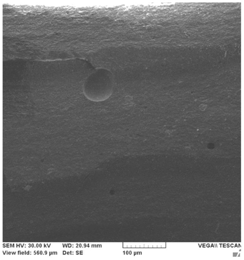
This Scanning Electron Microscopy (SEM) image displays the microstructural topography of a dental restorative composite (Filtek Z250-4). The imaging reveals a heterogeneous surface texture characteristic of a hybrid composite material. A prominent horizontal microcrack, approximately 150 µm in length, is visible in the upper quadrant, where its linear propagation is physically obstructed by a large, spherical micrometer-sized filler particle (approximately 40 µm in diameter). This interaction demonstrates the 'crack-blocking' mechanism which enhances the mechanical properties and fracture toughness of the material. Additional surface irregularities include small dark circular pits, identified as microfiller pull-outs, where filler particles have been dislodged from the resin matrix. The granular background represents the surrounding matrix embedded with varied sizes of filler particles. The image provides clinical relevance regarding the durability and stress-resistance of dental biomaterials under compressive load. A 100 μm scale bar is provided for dimensional reference.
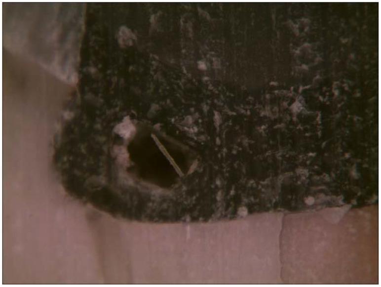
This clinical photograph displays a high-magnification view of a specimen of Short Fiber-Reinforced Composite (SFRC), a material used in restorative dentistry. The image illustrates a significant structural defect: an internal air bubble (void) within the cured composite resin matrix. The material has a dark, mottled, and grainy texture with lighter particulate inclusions. A prominent, dark, circular void is visible, representing an air entrapment that occurred during the placement or polymerization process. Notably, a single reinforcing fiber is seen spanning the diameter of this void, highlighting the material's composition. This visual demonstrates common challenges in restorative procedures using fiber-reinforced bulk-fill materials, where air entrapment can lead to mechanical weaknesses or failure of the restoration. The educational focus is on material science, specifically regarding the handling properties and potential for porosity in direct dental restorations.
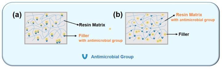
Educational comparison diagram of two antimicrobial dental resin composite strategies: (a) Filler-type and (b) Resin-type. The diagrams illustrate the microscopic structure consisting of a light-blue linear resin matrix and yellow circular filler particles. In the filler-type strategy (a), blue 'U'-shaped icons representing antimicrobial groups are attached specifically to the surface of the yellow filler particles, indicating modified filler surfaces or antimicrobial agent incorporation within the filler. In the resin-type strategy (b), the blue 'U'-shaped antimicrobial groups are covalently bonded directly to the light-blue lines of the resin matrix, as well as associated with the fillers. The diagram illustrates how biocidal moieties like quaternary ammonium methacrylates (QAMs) or MDPB can be integrated into dental materials to provide antibacterial properties. This visual helps dental students and biomaterial researchers understand the difference between surface-released antimicrobial agents (filler-type) and immobilized, long-term antimicrobial properties achieved through copolymerization (resin-type) in restorative dentistry.
Esthetic Restorative Materials & Composite Resins
ESTHETIC RESTORATIVE MATERIALS - Overview
| Material | Examples |
|---|---|
| Composite Resins | Bis-GMA based, UDMA based |
| Glass Ionomer Cements (GIC) | Conventional GIC, RMGIC |
| Resin-Modified GIC | Fuji II LC, Vitremer |
| Compomers | Polyacid-modified composites |
| Porcelain / Ceramics | Feldspathic, IPS Empress |
| Direct Tooth-Colored Inlays | CAD/CAM composites |
COMPOSITE RESTORATIVE MATERIALS (Detailed)
Definition
Composition (3 Main Components)
1. Organic Resin Matrix (Continuous Phase)
- Bis-GMA (Bisphenol A glycidyl methacrylate) - Bowen's resin (1962); high viscosity, low polymerization shrinkage
- TEGDMA (Triethylene glycol dimethacrylate) - diluent monomer; lowers viscosity but increases shrinkage
- UDMA (Urethane dimethacrylate) - flexible, less brittle; used as alternative to Bis-GMA
- Bis-EMA - lower water sorption than Bis-GMA
- Also contains: initiators (camphorquinone for light cure), inhibitors (hydroquinone), accelerators, pigments
2. Inorganic Filler Particles (Dispersed Phase)
- Quartz - hard, chemically inert; good polishability
- Barium/strontium glass - provides radiopacity
- Colloidal silica - microfill particles (0.01-0.04 µm)
- Borosilicate glass
- Filler content: 50-90% by weight / 35-70% by volume
3. Coupling Agent (Silane)
- Organosilane (gamma-methacryloxypropyltrimethoxysilane)
- Bonds the hydrophobic resin matrix to the hydrophilic inorganic fillers
- Prevents filler pull-out, improves mechanical properties, reduces water sorption
4. Activator-Initiator System
- Light-cured (photopolymerization): Camphorquinone (CQ) + amine co-initiator; activated at 468 nm (blue light)
- Self-cured (chemical): Benzoyl peroxide + tertiary amine
- Dual-cured: Combination of both
Classification
A. Based on Filler Particle Size (Most Important Classification)
| Type | Particle Size | Filler % (wt) | Key Properties | Uses |
|---|---|---|---|---|
| Macrofill (Traditional) | 10-100 µm | 70-80% | Strong but rough surface; poor polishability | Posterior; obsolete now |
| Microfill | 0.01-0.1 µm (colloidal silica) | 35-50% | Excellent esthetics and polishability; low strength | Anterior Class III, V |
| Hybrid | 0.1-10 µm (mix of macro + micro) | 75-85% | Balanced strength and esthetics; versatile | Universal - anterior + posterior |
| Microhybrid | 0.01-1 µm | 70-80% | Better polish than hybrid; good strength | Universal |
| Nanofill | 5-100 nm | 78.5% | Excellent polish, wear resistance, low shrinkage | Universal (e.g. Filtek Supreme) |
| Nanohybrid | Nanoclusters + nanomers | ~80% | Best combination of strength + esthetics | Universal |
B. Based on Consistency / Viscosity
- Packable (Condensable) composites - high filler content; used for posterior cavities; higher viscosity
- Flowable composites - low filler, low viscosity; used as liners/base, cervical lesions, small cavities
- Conventional (regular body) - standard handling
C. Based on Curing Method
- Light-cured - most common; requires curing lamp (halogen/LED); depth of cure 1-2 mm per increment
- Self-cured (chemically cured) - two-paste system; useful where light access is limited
- Dual-cured - core build-up materials, luting of indirect restorations
D. Based on Location
- Anterior composites - higher esthetic requirement; microfill or nanofill
- Posterior composites - higher strength requirement; hybrid or packable
- Universal composites - used in both regions
E. Based on Generation / Evolution
- First generation - large particle macrofills
- Second generation - small particle composites
- Third generation - microfills
- Fourth generation - hybrids
- Fifth generation - microhybrids
- Sixth generation - nanofills and nanohybrids
Properties
Physical Properties
- Color and shade matching - available in multiple shades (VITA shade guide); chameleon effect in nanohybrids
- Radiopacity - barium/strontium glass fillers provide radiopacity comparable to enamel
- Thermal properties - higher coefficient of thermal expansion (CTE) than tooth structure; causes microleakage
- Water sorption - TEGDMA increases water sorption; Bis-EMA decreases it
Mechanical Properties
- Compressive strength: 250-300 MPa
- Tensile strength: 30-90 MPa
- Modulus of elasticity: 5-30 GPa
- Wear resistance: Nanohybrids > microhybrids > microfills
- Fracture toughness: Higher with higher filler loading
Polymerization Properties
- Polymerization shrinkage: 1-5% (major drawback) - causes gap formation, microleakage, postoperative sensitivity
- Depth of cure: 1.5-2 mm per increment; must use incremental technique
- Degree of conversion: 55-75%; incomplete polymerization is a concern
Advantages
- Excellent esthetics - tooth colored
- Conservative cavity preparation (adhesive bonding)
- Strengthens remaining tooth structure
- Fluoride release (some types)
- Repairability
- No mercury (unlike amalgam)
- Insulates tooth from thermal changes
- Can bond to both enamel and dentin via adhesive system
Disadvantages
- Polymerization shrinkage - leads to microleakage, sensitivity, secondary caries
- Technique sensitive - moisture contamination compromises bonding
- Wear - less durable in high-stress posterior areas
- Higher cost than amalgam
- Chair time - incremental placement is time consuming
- Staining - surface staining from coffee, tea, tobacco
- Bisphenol-A concern - estrogenic activity of Bis-GMA (limited evidence)
Indications
- Class I to Class V cavity restorations
- Anterior esthetic cases (diastema closure, shape correction)
- Veneers (direct)
- Core build-up
- Sealants (flowable)
- Bonding of orthodontic brackets
- Repair of fractured teeth
Contraindications
- Patients with poor oral hygiene (high caries risk)
- Heavy bruxism / parafunctional habits (posterior)
- Large posterior cavities in high-stress areas (relative)
- Subgingival margins where isolation is impossible
Polymerization Shrinkage - Management
- Incremental placement - layered technique, 2 mm increments
- Bulk-fill composites - modified chemistry allows 4-5 mm depth of cure
- Stress-relieving monomers - silorane-based composites (ring-opening mechanism; < 1% shrinkage)
- Soft-start polymerization - ramped or pulsed curing reduces stress
- Low C-factor cavities - maximize unbonded surfaces
Recent Advances
- Bulk-fill composites (e.g. Filtek Bulk Fill, Tetric EvoCeram Bulk Fill) - 4-5 mm depth cure; reduced visits
- Nanohybrid and Nanocomposites - e.g. Filtek Supreme Ultra
- Self-adhering composites - Vertise Flow; bond without separate adhesive
- Bioactive composites - Activa Bioactive; ion release, remineralization
- Single-shade composites - Omnichroma (Tokuyama); structural color matches any tooth
- Fiber-reinforced composites - polyethylene or glass fibers enhance fracture resistance
- CAD/CAM composites - e.g. Cerasmart, Lava Ultimate for indirect restorations
SEM Microstructure of Composite Resin
Summary Table for Quick Revision
| Feature | Detail |
|---|---|
| Matrix | Bis-GMA, UDMA, TEGDMA |
| Filler | Quartz, barium glass, colloidal silica |
| Coupling agent | Organosilane |
| Curing | Light (camphorquinone, 468 nm) |
| Best polish | Microfill / Nanofill |
| Best strength | Packable hybrid / Nanohybrid |
| Main drawback | Polymerization shrinkage |
| Shrinkage % | 1-5% |
| Universal use | Microhybrid / Nanohybrid |