
Teach me about appendicitis from pre op to post op
appendicitis anatomy appendix inflammation
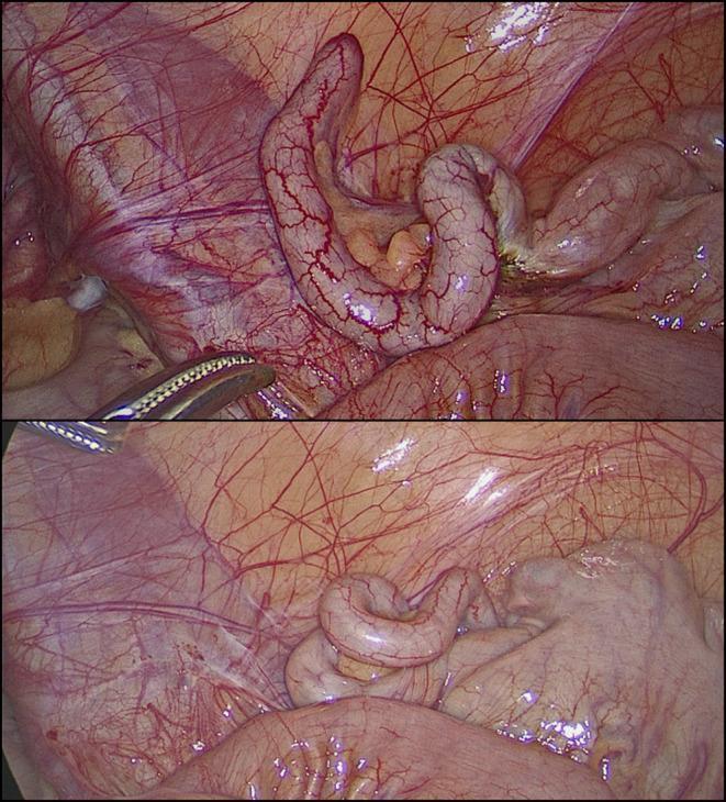
This clinical photograph consists of two laparoscopic views showing a macroscopically normal vermiform appendix during a diagnostic laparoscopy. The appendix appears as an elongated, tubular, and slightly coiled structure with a smooth, pale pink serosal surface. Fine, branching subserosal blood vessels are clearly visible across its length, indicating normal vascularity without the congestion or purulent exudate typical of acute appendicitis. In the upper frame, a laparoscopic grasper is visible, gently manipulating the appendix to facilitate inspection. The surrounding anatomy includes the mesoappendix containing adipose tissue and the glistening peritoneal lining of the right iliac fossa. There are no visual signs of inflammation, perforation, or abscess formation. This image serves as a reference for a 'grossly negative' appendix in the context of clinical suspicion of appendicitis, where symptoms may actually stem from non-inflammatory conditions such as follicular lymphoid hyperplasia or obstructive fecaliths.
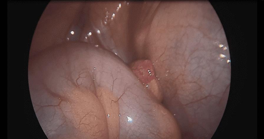
This clinical photograph shows a laparoscopic view of the intra-abdominal cavity, specifically capturing a case of left-sided appendicitis. The distal tip of the appendix is clearly visible, exhibiting distinct signs of inflammation, including erythematous (reddish) discoloration, edema, and a swollen, irregular texture. This inflamed tip is positioned anatomically within the left iliac fossa, a rare variation associated with intestinal malrotation or situs inversus. It is situated between the serosal surfaces of the cecum and the sigmoid colon. The surrounding bowel structures appear relatively healthy with smooth, pinkish-tan surfaces and visible fine vascular markings (vasa recta). The image serves as an educational example of acute appendicitis and the surgical anatomy involved in identifying an ectopic or malpositioned appendix during laparoscopic exploration. Educational focus is on surgical pathology, anatomical variations, and laparoscopic diagnostic findings in generalized peritonitis.
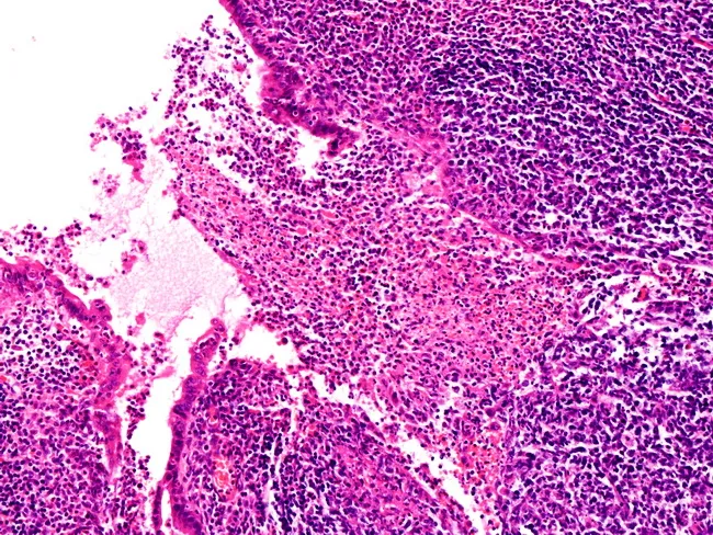
This histopathology image depicts appendiceal tissue at high magnification showing features of early acute appendicitis. Obtained from surgical appendix, the specimen is prepared as a hematoxylin and eosin (H&E) stained section and examined under light microscopy at high magnification (approximately 400x total). The specimen localizes to the vermiform appendix within the right lower quadrant, reflecting gastrointestinal tract anatomy. The mucosa and submucosa display focal neutrophilic infiltration with early cryptitis, subtle architectural distortion, and prominent interstitial edema. The inflammatory infiltrate is predominantly neutrophilic, with dense cellular clustering along the luminal margin and extending into the lamina propria, while goblet cells and overall glandular architecture remain relatively preserved in early disease. Vascular congestion and mild edema contribute to tissue pallor and fragility. No perforation or transmural necrosis is evident at this stage. These histological changes are typical of early inflammatory appendiceal pathology and support a clinical diagnosis of appendicitis when correlated with patient symptoms. Diagnostic significance: confirms early inflammation of the appendix and guides surgical management (appendectomy). Differential considerations include lymphoid hyperplasia-associated inflammation, infectious colitis, or secondary inflammatory responses. Clinically relevant use cases include educational reference, differential diagnosis training, pathology reporting, and correlation with radiologic imaging findings for teaching and research purposes.
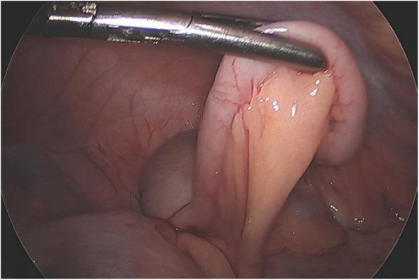
This intraoperative laparoscopic photograph shows a grossly normal vermiform appendix in a pediatric patient. The imaging modality is diagnostic laparoscopy, capturing the right lower quadrant of the abdomen. The appendix is held by a surgical grasper, revealing a smooth, glistening serosal surface with a healthy pinkish-tan hue. There are no visual markers of acute appendicitis, such as luminal distension, erythema, edema, or fibrinopurulent exudate. The mesoappendix and surrounding mesenteric fat appear unremarkable, lacking the hyperemia or 'fat stranding' characteristic of acute inflammation. This image serves as a clinical comparison for medical students to distinguish a healthy appendix from one with inflammatory changes, such as those seen in typical acute appendicitis or cases mimicking it, like Multi-system Inflammatory Syndrome in Children (MIS-C). Educational focus is on the laparoscopic appearance of visceral abdominal anatomy and the visual exclusion of transmural inflammation during a surgical workup for acute abdominal pain.
appendectomy laparoscopic surgical technique trocar placement
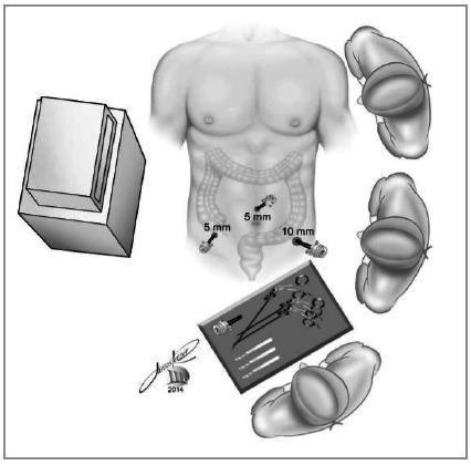
This surgical diagram illustrates the ergonomic setup and port placement for a laparoscopic appendectomy. An overhead view depicts a human torso with the large intestine superimposed for anatomical reference. Three trocars are positioned in the lower abdomen: a 10 mm trocar in the left iliac fossa, a 5 mm trocar in the suprapubic/midline position, and a 5 mm trocar in the right iliac fossa. The surrounding operative environment shows the relative positioning of the surgical team and equipment: a laparoscopic monitor is placed to the patient's right, the surgeon and an assistant stand on the patient's left, and an additional team member or scrub nurse is positioned at the foot of the bed near the instrument tray. The diagram emphasizes the triangulation of instruments and the spatial relationship between the surgical ports, the target pathology (appendix/cecum), and the visualization equipment, serving as an educational guide for standardized laparoscopic positioning.
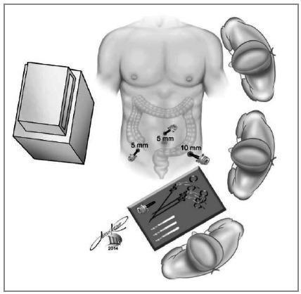
This medical illustration depicts a standard laparoscopic appendectomy surgical setup in a bird's-eye view. The diagram shows a male patient in a supine position with an overlay of the large intestine and appendix. Three trocar ports are identified: a 10 mm trocar located in the left iliac fossa and two 5 mm trocars placed in the right iliac fossa and the suprapubic/midline region. The spatial arrangement of the surgical team and equipment is clearly labeled: the primary surgeon stands on the patient's left side, the assistant is positioned on the patient's right side, and a scrub nurse/technician is at the foot of the table near the instrument (scrub) table. The laparoscopic monitor is positioned on the patient's right side, facing the surgeon. The scrub table contains various laparoscopic instruments including graspers, dissectors, and a spare trocar. This diagram serves as a pedagogical guide for ergonomics and portal placement during minimally invasive abdominal surgery, specifically highlighting a technique for specimen extraction via the 10 mm left iliac fossa port.
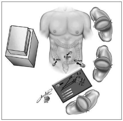
This medical illustration depicts the surgical setup and port placement for a laparoscopic appendectomy. The central focus is a torso diagram showing three trocar sites in the lower abdomen: a 5mm trocar in the right iliac fossa, a 5mm umbilical/suprapubic port, and a larger 10mm trocar in the left iliac fossa. The underlying anatomy highlights the large intestine, particularly the cecum and appendix region, to demonstrate the surgical approach. The surrounding elements provide a birds-eye view of the operating room configuration, featuring the laparoscopic monitor to the patient's right, the surgeon and assistant positioned on the patient's left, and a scrub nurse at the foot of the table. A sterile instrument tray is shown containing laparoscopic graspers, a monopolar hook, and additional trocars. This diagram serves as a clinical guideline for ergonomic positioning and port triangulation during minimally invasive abdominal surgery.
Appendicitis: Pre-Op to Post-Op
1. Anatomy and Pathophysiology
- Luminal obstruction - most often by a fecalith/appendicolith, but also lymphoid hyperplasia (common in children/young adults), parasites, neoplasm, or foreign body
- Intraluminal pressure rises - continued mucosal secretion raises pressure in the closed loop
- By the Law of Laplace, increased wall tension impairs venous and lymphatic return
- Vascular compromise - ischemia, bacterial translocation through the mucosa, inflammation
- Transmural necrosis - leads to perforation, periappendiceal phlegmon/abscess, or diffuse peritonitis
2. Clinical Presentation
- Periumbilical or diffuse abdominal pain (visceral, T10-T11 afferents) that migrates to the right lower quadrant (RLQ) over 12-24 hours as parietal peritoneum becomes involved
- Anorexia (often the first symptom - its absence should make you question the diagnosis)
- Nausea and vomiting (after onset of pain, not before - unlike gastroenteritis)
- Low-grade fever (38-38.5°C)
- Urge to defecate
- Retrocecal appendix: flank or back pain, psoas irritation prominent
- Pelvic appendix: suprapubic pain, pelvic/rectal tenderness, may mimic PID or ovarian pathology
- Children under 5: high perforation rate (51-100%) because diagnosis is delayed; may present with diffuse peritonitis from the start
- Pregnant patients: classic RLQ findings in the first half; appendix displaced but remains in RLQ in >75% of cases even in the third trimester. Physiologic leukocytosis and elevated ESR in pregnancy confuse the picture.
- Elderly: vague symptoms, delayed presentation, high complication rate
3. Physical Examination
- Low-grade pyrexia
- Localized RLQ tenderness
- Muscle guarding
- Rebound tenderness
| Sign | Technique | Mechanism |
|---|---|---|
| McBurney's sign | Deep palpation at McBurney's point (1/3 distance from ASIS to umbilicus) | Direct peritoneal irritation |
| Rovsing's sign | Palpation of LLQ causes pain in RLQ | Transmitted peritoneal stretch |
| Psoas sign | Pain with right hip extension (patient prone or lateral) | Inflamed appendix on psoas muscle |
| Obturator sign | Pain with right hip flexion + internal rotation | Appendix in contact with obturator internus |
| Dunphy's sign | RLQ pain on coughing | Peritoneal irritation |
| Pointing sign | Patient points to where pain started, then migrated | Visceral-to-somatic shift |
Note from Sabiston: These signs indicate localized peritonitis rather than a specific diagnosis - they are indicators, not diagnostic.
4. Investigations
Laboratory Tests
- WBC: Leukocytosis with neutrophilia in ~90% of cases; a normal WBC does not exclude appendicitis (present in 10% of confirmed cases)
- CRP: Most sensitive biomarker but not specific enough alone
- Urinalysis: May show trace pyuria/hematuria due to proximity of inflamed appendix to bladder/ureter - do not be misled into diagnosing a UTI
- Pregnancy test (β-hCG): Mandatory in all women of reproductive age
- Procalcitonin, IL-6: Investigated but not yet standard practice
Imaging
| Modality | Notes |
|---|---|
| Plain X-ray | Low sensitivity/specificity; may show fecalith (<5%), loss of psoas shadow, or free air if perforated |
| Ultrasound (US) | First-line in children and pregnant women; operator-dependent; non-visualization is common; positive if appendix >6mm diameter, non-compressible, with periappendiceal fluid |
| CT (gold standard) | Sensitivity ~95%, specificity ~95%; reduced negative appendectomy rate from 15% to near 5%; shows periappendiceal fat stranding, appendicolith, free fluid |
| MRI | Preferred in pregnancy if US inconclusive; avoids radiation; sensitivity and specificity comparable to CT |
5. Scoring Systems
| Feature | Points |
|---|---|
| Migration of pain to RLQ | 1 |
| Anorexia | 1 |
| Nausea/vomiting | 1 |
| Tenderness in RLQ | 2 |
| Rebound tenderness | 1 |
| Elevated temperature (>37.3°C) | 1 |
| Leukocytosis (WBC >10,000) | 2 |
| Shift to left (neutrophilia) | 1 |
| Total | 10 |
6. Differential Diagnosis
- Mesenteric lymphadenitis (most common mimic in children)
- Ovarian cyst/torsion, ectopic pregnancy, PID (in women)
- Meckel's diverticulitis
- Crohn's disease (terminal ileitis)
- Psoas abscess
- Right ureteric colic
- Cecal carcinoma (especially in the elderly)
- Omental torsion
7. Pre-Operative Management
- Make patient NPO
- IV access and fluid resuscitation - correct dehydration from fever and vomiting
- IV antibiotics: Administer pre-operatively. For simple appendicitis, a single-agent second-generation cephalosporin (e.g., cefoxitin) covers gram-negative rods and anaerobes. Complicated appendicitis requires broader coverage (e.g., piperacillin-tazobactam or cefepime + metronidazole)
- Analgesia: IV opioids do NOT mask the diagnosis and should be given - withholding pain relief is no longer acceptable practice
- Nasogastric tube: Only if there is significant vomiting or obstruction
- Consent: For laparoscopic appendectomy with possibility of conversion to open
8. Operative Management
Laparoscopic Appendectomy (Standard of Care)
- 10-12 mm port at umbilicus (camera)
- 5 mm port in left iliac fossa or suprapubic
- 5 mm port varies by surgeon preference
- Patient supine, Trendelenburg with left lateral tilt (moves bowel away from RLQ)
- Pneumoperitoneum with CO2 to 12-15 mmHg
- Identify the cecum and trace the taenia coli to the appendix base
- Divide the mesoappendix (with harmonic scalpel, monopolar cautery, or clips/stapler)
- Apply two endoloops or a stapler at the appendix base (at least 5mm from cecum)
- Transect the appendix between ligatures
- Specimen is placed in a retrieval bag and removed through the umbilical port
- Irrigate if contamination is present
- Port sites closed (fascial closure at 10mm+ ports)
Open Appendectomy (Grid-Iron/Lanz incision)
- Incision centered over McBurney's point
- Muscle-splitting technique through external oblique, internal oblique, transversalis
- Identify cecum, deliver appendix, ligate mesoappendix, divide appendix
- Stump inversion (purse-string suture) - optional, not mandatory
- Wound lavage if contaminated
Complicated Appendicitis (Perforated/Abscess/Phlegmon)
- Perforation with free peritonitis: Urgent appendectomy (laparoscopic or open) + peritoneal lavage + broad-spectrum antibiotics
- Appendiceal abscess: Image-guided percutaneous drainage + IV antibiotics x4 days, followed by interval appendectomy at 6-8 weeks (if appendicolith present, interval appendectomy is strongly recommended due to high recurrence risk)
- Phlegmon (no drainable abscess): IV antibiotics alone for 4-7 days (IDSA guidelines for intra-abdominal infection); if not improving, re-image to look for evolving abscess
Non-operative Management (Antibiotics Alone)
- ~72.7% of antibiotic-treated patients avoid appendectomy acutely
- However, 39.1% require appendectomy within 5 years
- Recurrence rates range from 8% to 31.8% after non-operative treatment
- Not recommended if fecalith present (much higher failure rate)
- Many surgeons view it as a bridge to elective interval appendectomy
9. Histopathology
- Acute transmural neutrophilic infiltration
- Mucosal ulceration
- Vascular congestion and edema
- In perforation: full-thickness necrosis and periappendiceal exudate
10. Post-Operative Management
Uncomplicated Appendicitis
- Diet: Liquids shortly after recovery from anesthesia; advance to solid diet next day
- Antibiotics: Single pre-op dose only; no post-op antibiotics needed for simple appendicitis
- Pain control: Paracetamol + NSAIDs as first-line; minimize opioids (enhanced recovery pathways now target zero or minimal outpatient opioids)
- Activity: Same-day discharge is possible for many patients with early disease
- Return to full activity: ~2 weeks after laparoscopic appendectomy; ~3 weeks after open
Complicated/Perforated Appendicitis
- Continue IV antibiotics (duration depends on fever, WBC, ileus status, institutional protocol)
- No clear consensus on optimal antibiotic duration; typically guided by clinical response
- Nasogastric tube if ileus develops
- Resume oral intake once ileus resolves (return of flatus/bowel sounds)
- If drain placed: remove when drainage <10-20 mL/day or meets institutional criteria
Post-Op Complications
| Complication | Simple Appendicitis | Complicated Appendicitis |
|---|---|---|
| Wound infection | <1% | 5-20% (much higher with open) |
| Intraabdominal abscess | <1% | 15-30% |
| Bowel obstruction (adhesions) | Rare | Increased risk |
| Fecal fistula | Very rare | Rare |
| Stump appendicitis | Rare | Rare |
| Mortality | <0.1% | 1-4% |
11. Special Populations
- Children (<5 years): Perforation rate approaches 100% due to diagnostic delay and inability to describe symptoms. US and MRI preferred over CT (radiation concern). Same-day discharge possible after laparoscopic appendectomy for early disease.
- Elderly: High complication rate due to comorbidities and atypical presentation. CT is first-line. Higher mortality with perforation.
- Pregnant patients: Appendicitis is the most common surgical emergency in pregnancy. Laparoscopic appendectomy is safe throughout pregnancy. Fetal mortality rises markedly with perforation. A negative appendectomy rate of ~1/3 is accepted because the risk of fetal loss from perforation outweighs the risk of unnecessary surgery.
Key Summary Table
| Phase | Core Actions |
|---|---|
| Presentation | RLQ pain + anorexia + nausea + low-grade fever |
| Exam | McBurney, Rovsing, Psoas, Obturator signs |
| Diagnosis | WBC, CRP, pregnancy test; CT (adults), US/MRI (kids/pregnant) |
| Scoring | AIR or AAS score (preferred); Alvarado for rule-out |
| Pre-op | NPO, IV fluids, analgesia, IV antibiotics, consent |
| Surgery | Laparoscopic appendectomy within 12-24h |
| Complicated | Percutaneous drain if abscess; interval appendectomy at 6-8 wks |
| Post-op simple | Early diet, discharge day 0-1, no post-op antibiotics |
| Post-op complicated | Prolonged antibiotics, watch for abscess/fistula/obstruction |