
Pericardial effusion
"pericardial effusion"[MeSH Terms]
pericardial effusion echocardiogram cardiac tamponade
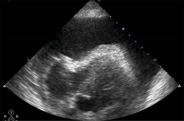
This diagnostic image is a transthoracic echocardiogram (ultrasound) in a subcostal or apical view, demonstrating a large pericardial effusion leading to cardiac tamponade. The most prominent feature is a significant, echo-free (anechoic to hypoechoic) space surrounding the cardiac structures, which represents fluid accumulation within the pericardial sac. Centrally, the heart appears relatively small or compressed within this large volume of fluid. The ultrasound shows the characteristic grayscale appearance of the myocardium and endocardium, though the cardiac chambers are obscured by the surrounding effusion. Clinically, this visual finding is pathognomonic for cardiac tamponade when associated with hemodynamic instability, as the intrapericardial pressure exceeds intracardiac filling pressure, compromising cardiac output. This image serves as a critical educational tool for emergency physicians and cardiologists in identifying life-threatening obstructive shock through point-of-care ultrasound (POCUS).
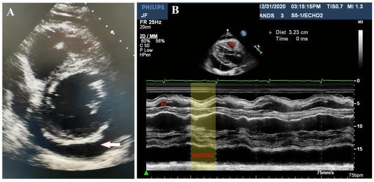
Two-panel echocardiographic study demonstrating pericardial effusion and signs of cardiac tamponade. Panel A shows a 2D transthoracic echocardiogram in a parasternal short-axis view at the basal level of the left ventricle. A large, anechoic (dark) space representing a circumferential pericardial effusion is visible surrounding the cardiac structures; a white arrow specifically highlights the posterior fluid collection. Panel B displays an M-mode echocardiogram with a simultaneous EKG trace. A yellow-shaded region highlights early diastolic collapse of the right ventricular (RV) free wall, a critical diagnostic indicator of increased intrapericardial pressure leading to hemodynamic compromise. The imagery illustrates key pathophysiological findings of cardiac tamponade, specifically the inward motion of the right ventricular wall during diastole when ventricular pressure is lowest, caused by the external pressure of the large effusion.
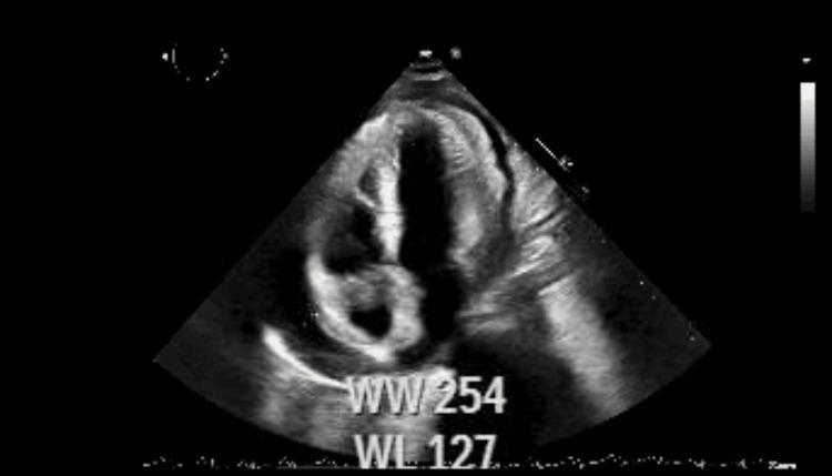
A diagnostic echocardiogram in the apical four-chamber view showing the early hemodynamic signs of cardiac tamponade. The image demonstrates a small-to-moderate circumferential pericardial effusion, visible as an anechoic (dark) space between the hyperechoic pericardium and the epicardium. Key clinical findings include visible compression and diastolic collapse of the right heart chambers, particularly the right atrium and right ventricle, due to increased intrapericardial pressure. There is evidence of abnormal septal motion, often associated with pericardial constriction or tamponade physiology. The overall cardiac silhouette appears slightly distorted by the external fluid pressure. This diagnostic image is intended for intermediate to advanced medical learners to illustrate the pathophysiology of obstructive shock secondary to pericardial effusion and the importance of echocardiography in identifying early signs of cardiac tamponade.
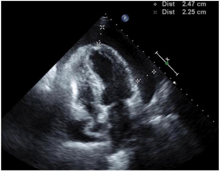
Diagnostic Image: This transthoracic echocardiogram (TTE) in a four-chamber view illustrates a large, circumferential pericardial effusion, visible as a prominent echo-free (anechoic) space surrounding the myocardium. The maximum depth of the effusion is quantified by calipers measuring 2.47 cm and 2.25 cm. Key hemodynamic findings indicative of cardiac tamponade are present, specifically the inward invagination and diastolic collapse of the right atrium and right ventricle. These visual signs suggest that intrapericardial pressure exceeds the filling pressures of the right-sided heart chambers. The image provides critical diagnostic evidence for urgent clinical intervention, such as pericardiocentesis. The educational focus is on identifying sonographic markers of tamponade physiology in the context of oncological complications, such as metastatic adenocarcinoma of the lung.
Pericardial effusion waat is it and how pateint will be risk at heparin