
What is liner and base in dental cements
liner and base dental cements definition purpose types
https://pocketdentistry.com/7-liner-and-bases
dental liner base cavity preparation diagram
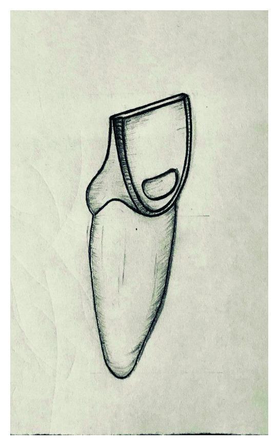
This medical illustration is an anatomical diagram depicting a dental specimen with a Class V cavity preparation, intended for educational use in operative dentistry. The sketch shows a single-rooted tooth, likely a maxillary incisor, from a proximal view. The drawing illustrates a standardized preparation for a composite resin restoration. The cavity is located on the facial surface of the crown, positioned approximately one-third to two-thirds of the crown height. Key anatomical landmarks shown include the clinical crown, the cementoenamel junction (CEJ), and the root. The preparation is characterized by a box-like morphology with rounded internal line angles and distinct, clean margins. A specific clinical detail illustrated is the clearance between the cavity's gingival margin and the CEJ, as well as its relationship to the proximal finish lines. This diagram serves as a schematic for Porcelain Laminate Veneer (PLV) research and dental student training in cavity design and restorative protocols.
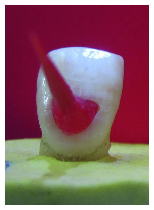
Clinical photograph of a laboratory specimen demonstrating dental cavity preparation and template fabrication. The image shows an extracted human maxillary central incisor crown mounted on a yellow dental stone base. A Class V cavity preparation is visible on the facial surface of the tooth crown, characterized by smooth internal walls and a defined cavosurface margin. A red autopolymerized acrylic resin (GC Pattern Resin) has been applied within the prepared cavity to serve as a standardized template. A slender, cylindrical handling pin or instrument is embedded into the red resin, extending coronally to facilitate the removal and placement of the template. This setup is utilized in restorative dentistry research to ensure uniformity in cavity dimensions across multiple specimens for testing composite filling or porcelain laminate veneer (PLV) protocols. The contrast between the off-white tooth structure and the red resin highlights the precise fit of the guide within the dental preparation.
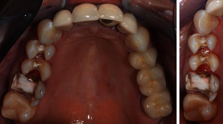
This clinical intraoral photograph displays a series of cavity preparations on the right maxillary posterior teeth (specifically teeth 14, 15, and 16). The preparations follow biomechanical principles for CAD/CAM ceramic restorations, featuring rounded internal line angles and smooth, distinct margins. Within the cavities, a light-colored, opaque composite base material (core build-up) is visible, used to level the cavity floors and protect endodontic access sites. Significant tooth-structure discoloration and staining are present on the prepared molar (tooth 16), likely indicative of previous restorative leakage or long-term endodontic treatment effects. The adjacent gingival tissues appear pink and healthy, with firm adaptation and no signs of acute inflammation or recession. The surrounding dental arch includes existing aesthetic restorations on the anterior and left posterior segments, demonstrating a comprehensive rehabilitative clinical scenario. This image is used to illustrate prosthetic preparation techniques, base placement, and preoperative assessment in restorative dentistry.
Liners and Bases in Dental Cements
Dental Liner
Definition
Functions
- Dentinal sealing - seals dentinal tubules to reduce microleakage, preventing bacterial invasion and irritants from entering
- Pulpal protection - shields the pulp from chemical irritation from restorative materials
- Thermal insulation - provides a thin barrier against temperature changes
- Biological/therapeutic effect - some liners (e.g., calcium hydroxide) stimulate formation of irregular secondary (tertiary) dentine and can be used for direct/indirect pulp capping
Materials Used as Liners
- Calcium hydroxide (most common therapeutic liner)
- Glass ionomer cement
- Flowable resin composite
- Cavity varnish (copal varnish - largely replaced by dentine bonding agents today)
- Zinc phosphate (in thin application)
Dental Base
Definition
Functions
- Thermal insulation - provides significant protection against temperature fluctuations, especially under large metallic restorations (e.g., amalgam)
- Mechanical support - absorbs and distributes occlusal forces; must be strong enough to support the restorative material during placement and function
- Chemical protection - prevents infiltration of hazardous chemicals from dental materials to the pulp
- Electrical insulation - under metallic restorations to reduce galvanism
- Fills undercuts - used to block out undercut areas for indirect restorations (e.g., gold or composite inlays)
- Bacteriostatic effect - some bases inhibit residual bacteria in deep carious cavities
Materials Used as Bases
| Material | Key Properties |
|---|---|
| Zinc oxide eugenol (ZOE) | Sedative/obtundent effect on pulp; 4 types (Type I: temporary cement; Type II: permanent cement; Type III: temp restorations; Type IV: cavity liner) |
| Zinc phosphate cement | High compressive strength; good thermal insulation; slightly acidic initially |
| Glass ionomer cement | Adhesive to tooth structure; fluoride release; good biocompatibility |
| Zinc polycarboxylate | Adhesive to tooth structure; mild on pulp; good compressive strength |
| Flowable resin | Used under composite restorations |
Key Differences: Liner vs. Base
| Feature | Liner | Base |
|---|---|---|
| Thickness | Thin (~0.5 mm) | Thick (>0.5 mm, up to 2 mm) |
| Primary role | Pulpal protection, dentinal sealing, therapeutic | Thermal insulation, mechanical support |
| Strength required | Low | High (must withstand condensation forces) |
| Placement | Walls and floor of cavity | Primarily the floor (pulpal wall) |
| Examples | Calcium hydroxide, cavity varnish | Zinc phosphate, ZOE, GIC, polycarboxylate |
Ideal Properties of Both
- No adverse effects on the pulp
- High compressive and tensile strengths (especially bases)
- Radiopacity (for radiographic visibility)
- Easy mixing and rapid setting
- Adhesive to enamel and dentine (but not to instruments)
- Bacteriostatic in the presence of residual caries
- Minimal film thickness (for luting applications)