
NEONATAL EMERGENCIES MANAGEMENT MASTER GUIDE FROM BASIC TO MASTER LEVEL WITH REASONING WITH MNEMONICS
Neonatal emergencies: map of topics
- Resuscitation at birth (NRP, the ABC approach, drugs, when to stop)
- Respiratory emergencies (RDS, TTN, meconium aspiration, pneumothorax, CDH, PPHN, apnea)
- Neonatal sepsis and meningitis (early vs late onset, empiric antibiotics)
- Cardiac emergencies (duct-dependent lesions, cyanotic vs shock, PGE1, SVT)
- Metabolic emergencies (hypoglycemia, hypocalcemia, inborn errors of metabolism, hyperammonemia)
- Neonatal seizures (causes, EEG, phenobarbitone and second-line drugs, HIE and cooling)
- Jaundice and hemolytic disease (phototherapy and exchange transfusion thresholds, kernicterus)
- Surgical emergencies (NEC, esophageal atresia/TEF, intestinal atresia, malrotation/volvulus, gastroschisis/omphalocele, anorectal malformation)
- Hematologic emergencies (vitamin K deficiency bleeding, polycythemia, severe anemia, DIC)
- Thermoregulation, fluids, electrolytes and shock (hypothermia, hypernatremia, shock types)
Overview
- Stabilize first. Airway, breathing, circulation, temperature and glucose.
- Treat the common, treatable causes empirically. Sepsis and hypoglycemia come before you have a diagnosis.
- Then find the cause.
- TACTIC: the causes of respiratory distress.
- TORCH: congenital infections.
- I-ONE-GLUC (my own working aid, not a standard textbook mnemonic): Infection, Oxygen (hypoxia), Neurologic, Electrolytes, Glucose, Lungs, Unusual metabolic, Cardiac.
- NRP "HAD" sequence: the order of steps in resuscitation.
- SOAP-ME: equipment check before a resuscitation.
- Neonatal seizure causes: a "HIE, ICH, infection, metabolic" stack.
- Is the baby blue, pale, floppy or bilious? Each points to a different system.
- Does it respond to oxygen? The hyperoxia test separates lung disease from cyanotic heart disease.
- Is the problem duct-dependent? If it is, give prostaglandin E1 (PGE1) early.
- Is it surgical? Bilious vomiting is a surgical emergency until proven otherwise.
Which topic should I cover in full depth first (basic to master level, with reasoning and mnemonics)?
Neonatal emergencies: map of topics
- Resuscitation at birth (NRP, the ABC approach, drugs, when to stop)
- Respiratory emergencies (RDS, TTN, meconium aspiration, pneumothorax, CDH, PPHN, apnea)
- Neonatal sepsis and meningitis (early vs late onset, empiric antibiotics)
- Cardiac emergencies (duct-dependent lesions, cyanotic vs shock, PGE1, SVT)
- Metabolic emergencies (hypoglycemia, hypocalcemia, inborn errors of metabolism, hyperammonemia)
- Neonatal seizures (causes, EEG, phenobarbitone and second-line drugs, HIE and cooling)
- Jaundice and hemolytic disease (phototherapy and exchange transfusion thresholds, kernicterus)
- Surgical emergencies (NEC, esophageal atresia/TEF, intestinal atresia, malrotation/volvulus, gastroschisis/omphalocele, anorectal malformation)
- Hematologic emergencies (vitamin K deficiency bleeding, polycythemia, severe anemia, DIC)
- Thermoregulation, fluids, electrolytes and shock (hypothermia, hypernatremia, shock types)
Overview
- Stabilize first. Airway, breathing, circulation, temperature and glucose.
- Treat the common, treatable causes empirically. Sepsis and hypoglycemia come before you have a diagnosis.
- Then find the cause.
- TACTIC: the causes of respiratory distress.
- TORCH: congenital infections.
- I-ONE-GLUC (my own working aid, not a standard textbook mnemonic): Infection, Oxygen (hypoxia), Neurologic, Electrolytes, Glucose, Lungs, Unusual metabolic, Cardiac.
- NRP "HAD" sequence: the order of steps in resuscitation.
- SOAP-ME: equipment check before a resuscitation.
- Neonatal seizure causes: a "HIE, ICH, infection, metabolic" stack.
- Is the baby blue, pale, floppy or bilious? Each points to a different system.
- Does it respond to oxygen? The hyperoxia test separates lung disease from cyanotic heart disease.
- Is the problem duct-dependent? If it is, give prostaglandin E1 (PGE1) early.
- Is it surgical? Bilious vomiting is a surgical emergency until proven otherwise.
All
"Infant, Newborn"[MeSH Terms] AND "Resuscitation"[MeSH Terms]
NRP 9th edition 2025 neonatal resuscitation algorithm changes AHA AAP guidelines
https://journalfeed.org/article-a-day/2025/2025-new-aha-nrp-…
Neonatal emergencies master guide
PART 0. The core logic of the sick neonate
- Airway, Breathing, Circulation
- Dextrose (check glucose in every sick neonate)
- Expose and keep Euthermic (36.5 to 37.5°C)
- Fever or Fluid status
- Gas (blood gas, lactate)
- Sepsis cover (cultures, then antibiotics without delay)
- What colour is the baby: blue, pale or grey, jaundiced, or mottled?
- What is the vomit: bilious means surgical until proven otherwise.
- Does it respond to oxygen? Lung disease responds. Cyanotic heart disease does not.
- Is it duct-dependent? Think of this in any baby who is shocked or cyanosed in the first 2 weeks.
- What is the timing? Hours point to birth events, days to infection, metabolic disease or heart disease, and weeks to cholestasis or pyloric stenosis.
PART 1. Resuscitation at birth (NRP, 2025 update)
- Delayed cord clamping. The 2025 update recommends deferring clamping for at least 60 seconds in term and preterm babies who need no immediate resuscitation. Intact cord milking may be reasonable for babies of 28 to 36+6 weeks when delayed clamping is not possible. Do not do it below 28 weeks because of the risk of severe intraventricular hemorrhage (IVH).
- Initial steps. Warm, dry, stimulate, position the airway. Suction only if the airway is obstructed.
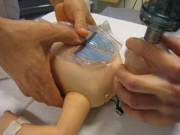
- HR <100, apnea or gasping. Start positive pressure ventilation (PPV) at 30 to 60 breaths/min. The primary feedback is a rising heart rate.
- Oxygen. Start at 21% for babies over 35 weeks. Use 21 to 30% for 32 to 34+6 weeks. Very preterm babies below 32 weeks may need 30 to 100%. Titrate to the pulse oximeter target.
- Ventilation not working: "MR. SOPA" (a standard NRP mnemonic)
- Mask adjustment
- Reposition the airway
- Suction
- Open the mouth
- Pressure increase
- Alternative airway. The laryngeal mask is now an earlier option for babies ≥34 weeks.
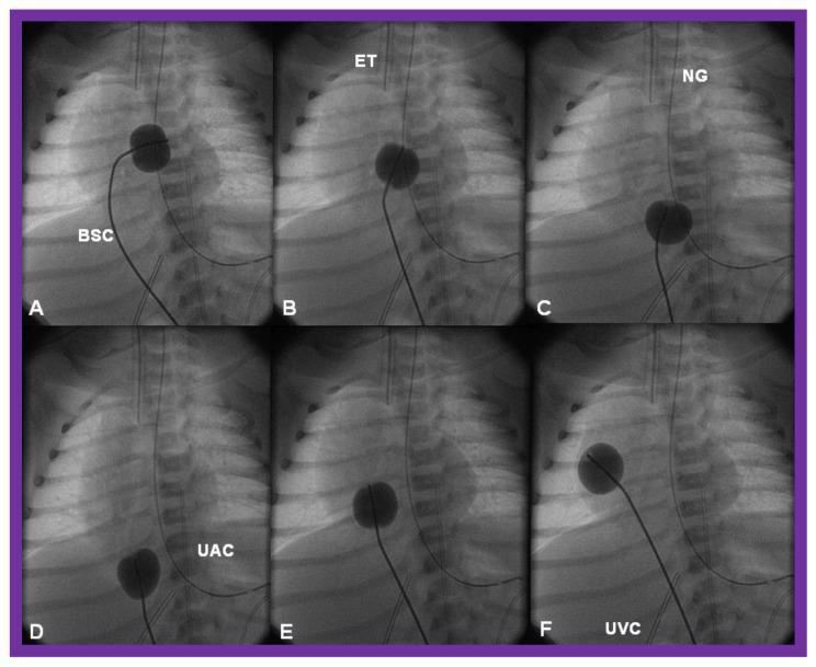
- HR <60 despite effective ventilation with an advanced airway. Start chest compressions at 3:1 with the two-thumb encircling technique (90 compressions plus 30 breaths per minute). Raise FiO2 to 100% during compressions.
- HR still <60. Give epinephrine, preferably IV or via an umbilical venous catheter. IO access is acceptable if the umbilical vein cannot be used. The endotracheal route is only a bridge while access is secured.
- Volume. Give normal saline or blood if hypovolemia is suspected and HR remains <60.
- Stopping. Consider redirecting care if there is no detectable heart rate after 20 minutes of high-quality resuscitation.
- Epinephrine 1:10,000: IV/UVC 0.01 to 0.03 mg/kg (0.1 to 0.3 mL/kg), repeated every 3 to 5 min. Endotracheal 0.05 to 0.1 mg/kg (0.5 to 1 mL/kg).
- Normal saline 10 mL/kg over 5 to 10 min.
- APGAR: Appearance (colour), Pulse, Grimace (reflex), Activity (tone), Respiration. The score is not used to decide whether to start resuscitation.
- SOAP-ME (equipment check before delivery): Suction, Oxygen, Airway equipment, Pharmacy (drugs), Monitor, Equipment.
- "Ventilation is king": nearly all newborn bradycardia is hypoxic. Fix the airway before reaching for compressions or drugs.
PART 2. Respiratory emergencies
- Sepsis or pneumonia
- Structural: choanal atresia, congenital diaphragmatic hernia (CDH), tracheoesophageal fistula (TEF), airway anomalies
- Shunt (heart disease)
- Sugar and metabolic (hypoglycemia, acidosis from IEM)
- Lung: respiratory distress syndrome (RDS), TTN, MAS, pneumothorax, persistent pulmonary hypertension of the newborn (PPHN)
| Condition | Clue | Key management |
|---|---|---|
| RDS | Preterm, ground-glass lungs with air bronchograms | CPAP early, surfactant, caffeine, minimal ventilation |
| TTN | Term or C-section, fluid in fissures, resolves in 24 to 72 h | Oxygen, observe, rule out sepsis |
| MAS | Post-term, meconium-stained liquor, hyperinflation | Ventilate if apneic, no routine suction. Watch for PPHN and air leak |
| Pneumothorax | Sudden deterioration, one side hyperlucent, transillumination | Needle decompression (2nd intercostal space mid-clavicular line, or 4th to 5th anterior axillary line), then chest tube |
| CDH | Scaphoid abdomen, bowel sounds in chest | Avoid bag-mask ventilation. Intubate, place an orogastric tube, treat PPHN |
| PPHN | Pre/post-ductal SpO2 gap >10%, labile hypoxia | Avoid hypoxia and acidosis, inhaled nitric oxide, gentle ventilation |
| Choanal atresia | Blue at rest, pink when crying | Oral airway |
| Apnea | Preterm: prematurity. Term: always look for a cause | Caffeine for prematurity. Work up sepsis, seizures, hypoglycemia, ICH |
PART 3. Neonatal sepsis and meningitis
- Early onset (<7 days): fulminant, linked to maternal risk factors (fever, group B streptococcus (GBS), prolonged rupture of membranes). It is more often septic shock and neutropenia.
- Late onset (>7 days): gradual, with meningitis more common - Tintinalli's.
- Organisms: "GEL" (my aid): GBS, E. coli and enterics, Listeria. Add HSV in a sick neonate with seizures, vesicles or a hepatitis picture.
- Signs: "T-C-R-F-J-R" (my aid): Temperature instability, CNS signs, Respiratory distress, Feeding problems, Jaundice, Rash (Table 116-3).
- Work-up: CBC, blood culture, catheter urine culture, lumbar puncture (LP) if stable, CRP or procalcitonin, and a chest X-ray if there are respiratory signs. Nuchal rigidity is absent in most neonates with meningitis.
- Empiric treatment: ampicillin (about 50 mg/kg) plus gentamicin (2.5 mg/kg) - Tintinalli's. Add cefotaxime for meningitis. Avoid ceftriaxone in neonates because it displaces bilirubin and precipitates with calcium (my knowledge, the retrieved text was truncated).
- Add acyclovir (20 mg/kg/dose per Harriet Lane) if HSV is plausible - Red Book Table 4.12 and Harriet Lane.
- Reasoning: never delay antibiotics for an LP in an unstable baby. Give antibiotics within the hour.
PART 4. Cardiac emergencies
- Cyanotic lesions present in the first days.
- Left-sided obstructive (duct-dependent) lesions present at about days 5 to 14 with feeding difficulty and shock, after a day or two of poor feeding or lethargy - Tintinalli's.
- Suspect any shocked neonate with hepatomegaly, weak or unequal femoral pulses, or no response to fluids of having a duct-dependent lesion.
- Start prostaglandin E1 (PGE1) early. The usual start is 0.05 to 0.1 mcg/kg/min, then titrate down to the lowest effective dose (verify). Be ready for apnea and intubate if needed.
- Exception: obstructed TAPVR, where PGE1 can worsen the obstruction - Creasy & Resnik.
- Use small fluid boluses of 10 mL/kg with reassessment. Avoid excess oxygen in left-sided lesions.
- Supraventricular tachycardia (SVT, HR >220): ice-water to the face, then adenosine 0.1 mg/kg rapid IV push. Use synchronized cardioversion at 0.5 to 1 J/kg if unstable (verify).
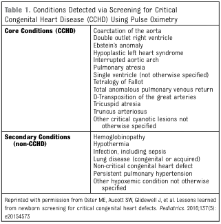
- Screening: pulse-oximetry screening at 24 hours or more.
PART 5. Metabolic emergencies
- Treat when the glucose is <40 mg/dL with symptoms (jitteriness, apnea, seizures, lethargy, poor feeding). Give 2 to 4 mL/kg of 10% dextrose, then start an infusion - Rosen's, Medications. Tintinalli's Table 146-1 lists a larger neonatal D10 bolus of about 5 mL/kg, so local protocols differ.
- Persistent hypoglycemia means hyperinsulinism, adrenal insufficiency or an IEM. Take a critical sample before treating if it does not delay treatment.
- Clue: a baby who was well for 1 to 3 days after birth and then deteriorates after feeds start, looking like sepsis (poor feeding, vomiting, lethargy, tachypnea, seizures).
- Tests: glucose, gas, anion gap, lactate, ammonia, ketones, urine reducing substances, plasma amino acids, urine organic acids.
- Management:
- Stop protein feeds and give high-rate glucose (D10 infusion).
- Give fluid boluses carefully, and reassess after each one because heart disease can look similar - Tintinalli's.
- Treat hyperammonemia with ammonia scavengers and early dialysis in a metabolic center.
- Reasoning pattern: hyperammonemia with respiratory alkalosis suggests a urea cycle defect. Metabolic acidosis with a high anion gap suggests an organic acidemia.
PART 6. Neonatal seizures
- Hypoxic-ischemic encephalopathy (day 0 to 2, most common)
- Intracranial hemorrhage or infarct
- Sepsis or meningitis (including HSV)
- Sugar (hypoglycemia)
- Minerals: calcium, magnesium, sodium
- Errors of metabolism (including pyridoxine dependency)
- Narcotic or drug withdrawal
- Developmental or genetic (channelopathies, benign familial seizures)
- ABC, check glucose and give dextrose if low.
- Check calcium, magnesium, sodium and a gas. Do a sepsis work-up.
- Phenobarbital 20 mg/kg IV is the usual first line (verify).
- Second line: levetiracetam, phenytoin or midazolam.
- Pyridoxine trial (100 mg IV, verify) if the seizures are refractory.
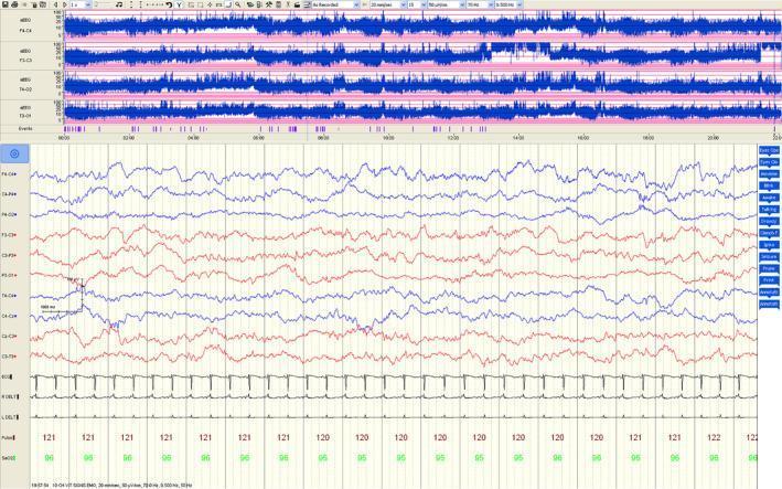
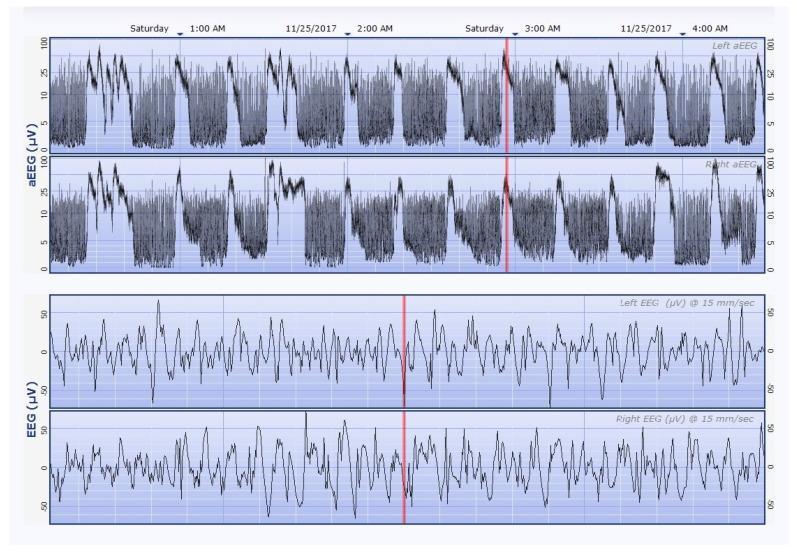
- Seizures can be subtle (eye deviation, lip smacking, cycling movements, apnea). EEG is the gold standard, and EEG-clinical dissociation after phenobarbital is common.
- Cool for HIE if criteria are met.
PART 7. Neonatal jaundice
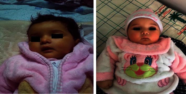
- <24 hours: always pathologic. Think hemolysis (Rh, ABO, G6PD), congenital infection or sepsis.
- Day 2 to 3: physiologic peak, but also ABO disease and sepsis.
- Week 1: breastfeeding jaundice and breast milk jaundice, sepsis, hypothyroidism.
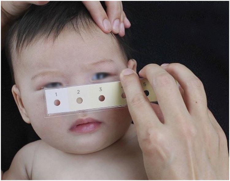
- >2 weeks, or direct (conjugated) bilirubin >1 mg/dL: cholestasis. Look for biliary atresia (pale stools, dark urine) and refer urgently. A Kasai procedure before about 60 days gives the best outcome.
- Treat using the 2022 AAP phototherapy and exchange thresholds, which depend on gestational age, hours of life and neurotoxicity risk factors (gestation <38 weeks, low albumin, isoimmune hemolysis or G6PD deficiency, sepsis, clinical instability). Use a tool such as BiliTool.
- Escalate care when the bilirubin is within 2 mg/dL of the exchange threshold. Escalation means intensive phototherapy, IV fluids, a repeat bilirubin every 2 hours and NICU transfer.
- IVIG (0.5 to 1 g/kg) for isoimmune hemolysis. Exchange transfusion for failure of intensive phototherapy or signs of bilirubin encephalopathy.
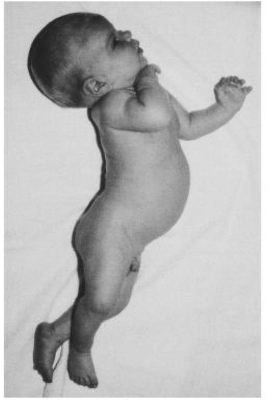
- Warning signs of encephalopathy: lethargy, hypotonia changing to hypertonia, arching (opisthotonos), high-pitched cry, and fever. Treat as an emergency.
PART 8. Surgical emergencies
| Condition | Clues | Initial management |
|---|---|---|
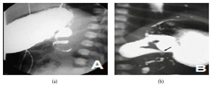
| Malrotation with volvulus | Bilious vomiting, abdominal tenderness, shock | Resuscitate, urgent upper GI contrast study, then Ladd procedure. Do not wait if the baby is unstable |
| NEC | Preterm, feed intolerance, bloody stool, distension. Pneumatosis, portal venous gas, pneumoperitoneum | NPO, nasogastric decompression, broad-spectrum antibiotics, fluids, serial X-rays, surgical review |
| Esophageal atresia with TEF | Drooling, choking with feeds, orogastric tube coils, gasless abdomen in pure atresia | Head-up, continuous upper pouch suction (Replogle), avoid PPV if possible, look for VACTERL |
| Duodenal atresia | "Double bubble", Down syndrome | Orogastric tube decompression, fluids, surgery |
| Gastroschisis | Defect to the right of the cord, no sac | Cover bowel with warm saline-soaked gauze or a plastic bowel bag, lay the baby on the side, give extra fluids, orogastric tube |
| Omphalocele | Sac present, associated anomalies and syndromes | Protect the sac, keep warm, look for cardiac and chromosomal anomalies |
| Hirschsprung disease | No meconium by 48 hours | Rectal washouts, antibiotics if enterocolitis |
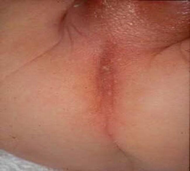
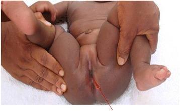
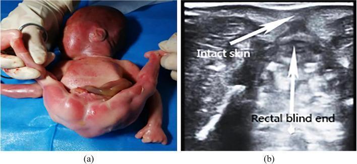
| Anorectal malformation | No anus on inspection | Look for VACTERL, check urine for meconium |
PART 9. Hematologic emergencies
- Vitamin K deficiency bleeding: give vitamin K 1 mg IM at birth (0.5 mg if <1.5 kg, verify). For active bleeding, give IV vitamin K plus FFP or PCC. Intracranial hemorrhage can be the first sign - Tintinalli's.
- Polycythemia: venous hematocrit >65%. Partial exchange transfusion if symptomatic.
- Severe anemia: think fetomaternal hemorrhage, twin-twin transfusion, hemolysis. Transfuse.
- Thrombocytopenia: immune (NAIT or maternal ITP) or sepsis, DIC. Treat the cause. Give matched platelets for severe bleeding.
PART 10. Shock, fluids, temperature and electrolytes
- Shock types: hypovolemic (dehydration, hemorrhage), septic, cardiogenic (duct-dependent lesions, SVT, myocarditis), obstructive (tension pneumothorax) and adrenal crisis.
- Fluids: crystalloid boluses of 10 to 20 mL/kg in neonates, with reassessment after each - Tintinalli's. Use smaller boluses (10 mL/kg) if cardiac disease is possible.
- Vasoactives: dopamine or epinephrine early if fluid-refractory. Give hydrocortisone for catecholamine-resistant shock.
- Hypernatremic dehydration: exclusively breastfed babies with poor intake and weight loss. Correct slowly.
- Weight loss: normal loss is up to 10 to 12% in the first 3 to 7 days, and babies gain 20 to 30 g/day in the first 3 months - Tintinalli's.
- Temperature: cold stress raises oxygen and glucose consumption. Prevent it with skin-to-skin contact, hats, plastic wrap for preterm babies and warmed rooms.
PART 11. Master-level quick map
| Presentation | First thought | Fast action |
|---|---|---|
| Blue, no response to O2 | Cyanotic heart disease, PPHN | PGE1, echo |
| Pale or grey with shock, day 5 to 14 | Duct-dependent left heart lesion, sepsis | PGE1 and antibiotics both |
| Bilious vomiting | Malrotation with volvulus | Surgical review, contrast study |
| Seizure | HIE, glucose, calcium, sepsis | Dextrose, phenobarbital, work-up |
| Jaundice <24 h | Hemolysis | Phototherapy, bilirubin and Coombs |
| Poor feeding after a well interval | IEM, sepsis | Glucose, ammonia, antibiotics |
| Scaphoid abdomen with respiratory distress | CDH | Intubate, no bag-mask |
| Drooling, choking with feed | Esophageal atresia | Replogle tube, no oral feeds |
| Ambiguous genitalia with vomiting | CAH | Saline, hydrocortisone |
- Treat as one: sepsis and duct-dependent heart disease can look identical. Cover both until separated.
- Always check glucose in any sick neonate.
- Bilious vomiting and weak femoral pulses are red flags.
- Preterm babies behave differently: NEC, IVH, apnea of prematurity and RDS apply.
- Neonates tolerate fluids and drugs poorly. Dose by weight, reassess often and avoid nephrotoxins and ceftriaxone.
With textbook figures and images
neonatal resuscitation algorithm
respiratory distress syndrome newborn chest radiograph ground glass air bronchograms
congenital diaphragmatic hernia chest radiograph neonate bowel in thorax
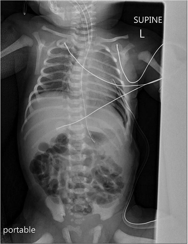
A postoperative portable anteroposterior (AP) radiograph of a neonate in the supine position, capturing the chest, abdomen, and pelvis. The image shows the lungs are well-expanded with the mediastinum and cardiac shadow in a midline position. A pigtail chest tube is visible in the left hemithorax, placed to manage a small subpulmonic pneumothorax. Additional medical support devices include an endotracheal tube, a nasogastric tube extending into the stomach, and multiple external monitoring leads and cables across the thorax. The abdomen demonstrates a typical neonatal bowel gas pattern with air-filled loops visible in the lower quadrants. Skeletal structures, including the ribs, spine, and pelvis, appear unremarkable for the age group. The image illustrates postoperative management and device placement following surgical correction of a congenital diaphragmatic hernia, serving as a pedagogical tool for neonatal radiology and critical care monitoring.
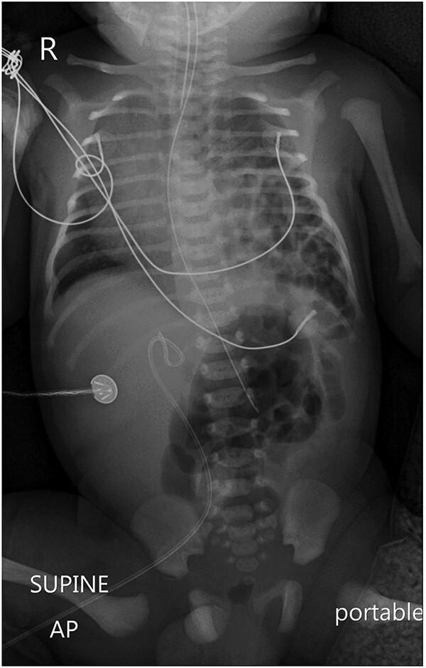
A portable anteroposterior (AP) supine chest and abdominal radiograph of a neonate demonstrating a classic presentation of a left-sided congenital diaphragmatic hernia (CDH). The left hemithorax is occupied by multiple radiolucent, air-filled loops of bowel, which have herniated from the abdomen into the thoracic cavity. This mass effect has resulted in the total collapse of the left lung and a significant mediastinal shift toward the right, as evidenced by the displacement of the cardiac silhouette and trachea. The right lung appears partially collapsed due to the contralateral pressure. Iatrogenic structures are visible, including an endotracheal tube and a nasogastric tube (the latter seen following an abnormal course toward the herniated stomach/bowel). An ECG lead is noted on the right abdominal wall. This diagnostic image is a critical educational example of neonatal respiratory distress caused by anatomical defects, illustrating the hallmark radiological features of CDH, including a 'scaphoid' appearance of the lower abdominal gas pattern due to the displacement of abdominal contents into the thorax.
necrotizing enterocolitis pneumatosis intestinalis abdominal radiograph
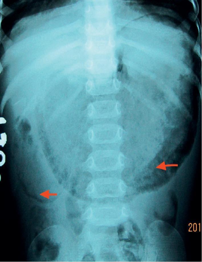
This diagnostic image is a plain anteroposterior abdominal radiograph of a pediatric patient, demonstrating significant pathology of the upper gastrointestinal tract. The primary finding is a grossly distended, gas-filled stomach and proximal duodenum, occupying much of the upper and middle abdomen. Key diagnostic features include gastroduodenal pneumatosis (pneumatosis intestinalis), characterized by linear and curvilinear radiolucent gas streaks within the walls of the distended stomach and duodenum, highlighted by red arrows. This intramural air gives the gastric and duodenal walls a bubbly or mottled appearance. The distal bowel appears relatively gas-less, suggesting a proximal high-grade obstruction. These findings are clinically significant for identifying conditions such as duodenal web, stenosis, or necrotizing enterocolitis involving the proximal gut. This radiograph serves as a critical educational example of pneumatosis intestinalis and gastric emphysema, highlighting the visual cues for identifying gas within organ walls as distinct from intraluminal gas.
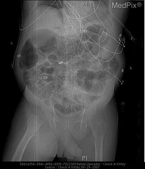
**Imaging Modality:** Anteroposterior (AP) abdominal radiograph (X-ray).
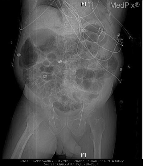
**Imaging Modality:** Anteroposterior (AP) abdominal radiograph (plain film/X-ray).
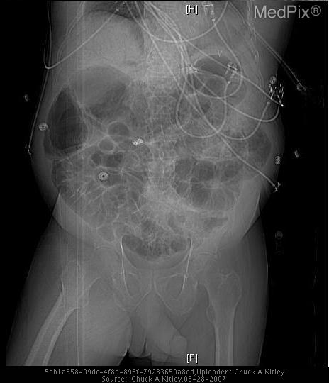
**Imaging Modality:** Abdominal Radiograph (X-ray), AP view/scout film.
esophageal atresia tracheoesophageal fistula radiograph coiled tube
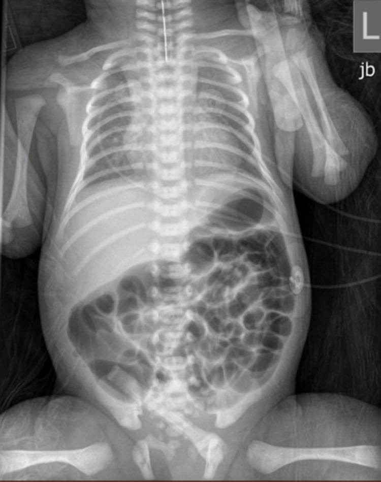
Anteroposterior (AP) neonatal chest and abdominal radiograph demonstrating diagnostic findings for esophageal atresia (EA) with an associated tracheoesophageal fistula (TEF). The primary finding is a radiopaque orogastric feeding tube that terminates abruptly in the proximal esophagus, visualized as a blind pouch in the upper mediastinum, indicating atresia. Despite the esophageal obstruction, there is significant bowel gas distributed throughout the stomach and small intestine in the abdomen. This presence of distal air confirms the existence of a distal tracheoesophageal fistula, allowing air to pass from the trachea into the lower gastrointestinal tract. The skeletal structures show normal neonatal development, and the abdominal gas pattern suggests patency distal to the stomach. This combination of a coiled or superiorly terminating feeding tube and subdiaphragmatic air is a classic radiological sign used in pediatric surgery to classify Type C esophageal atresia. The image is a critical educational example for neonatal intensive care and pediatric radiology training.
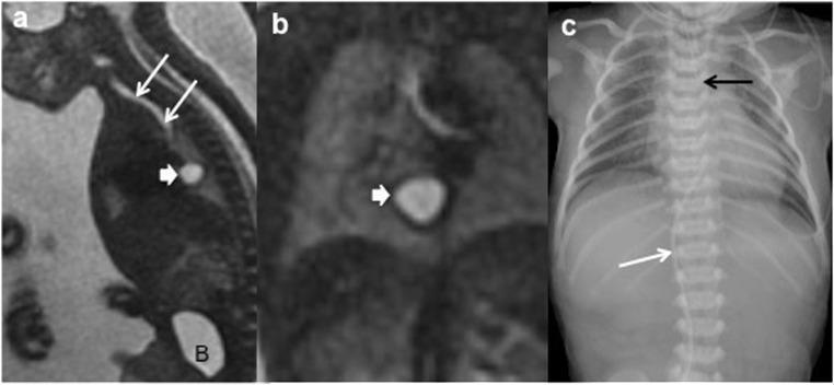
This composite figure illustrates the prenatal and postnatal imaging findings of a 21-week fetus with esophageal atresia. Image (a) is a sagittal fetal MRI using the FIESTA sequence, demonstrating a well-defined, hyperintense mediastinal cyst (thick arrow) posterior to the trachea (thin arrows). The fetal bladder is marked with 'B'. Image (b) provides a coronal HASTE fetal MRI view, showing a detailed perspective of the same hyperintense mediastinal duplication cyst (thick arrow). Image (c) is a postnatal anteroposterior thoraco-abdominal radiograph. It reveals a gastric tube coiled in the upper esophageal pouch (black arrow), characteristic of esophageal atresia. Notably, there is a total absence of gas in the bowel and abdomen, indicating a type I or II esophageal atresia without a distal tracheoesophageal fistula. A radiopaque venous umbilical catheter is also visible (white arrow). The constellation of findings—polyhydramnios, absent gastric bubble on ultrasound, and the identification of a mediastinal cyst—serves as critical diagnostic indicators for enteric duplication cysts and associated esophageal anomalies.
gastroschisis omphalocele newborn
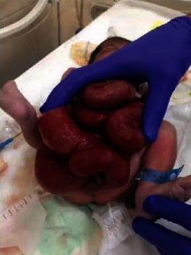
This clinical photograph displays a neonate with gastroschisis, a congenital ventral abdominal wall defect. The image shows a full-term newborn lying on a medical surface shortly after delivery. A significant periumbilical defect is visible, through which multiple loops of small intestine have herniated. The eviscerated bowel appears erythematous and edematous, lacking a protective peritoneal sac (distinguishing it from omphalocele). A healthcare provider, wearing blue sterile surgical gloves, is positioned to stabilize the exposed viscera. Visible neonate details include a blue identification band on the right ankle and standard newborn limbs. The image serves as a clinical example of a major abdominal wall defect requiring immediate neonatal intensive care and surgical intervention. It illustrates the typical presentation of gastroschisis where the bowel is exposed directly to amniotic fluid in utero, leading to its characteristic thickened and inflamed appearance at birth.
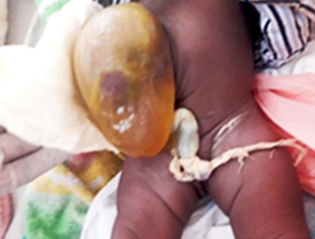
This clinical photograph shows a large omphalocele in a newborn infant, representing a midline abdominal wall defect. The protrusion is encased in a smooth, translucent amnioperitoneal sac. The sac exhibits a yellowish-brown hue with a focal area of dark purple discoloration, suggestive of bruising or underlying visceral congestion. Small, white, bubble-like surface irregularities are noted on the membrane. The umbilical cord is visible at the inferior aspect of the sac, appearing as a pale, whitish, fibrous structure with irregular texture against the reddish-brown skin of the abdominal wall. This image demonstrates the initial presentation of a congenital omphalocele before conservative treatment with eschar-inducing agents (e.g., aqueous eosin). Key educational features include the distinction between omphalocele (covered by a sac) and gastroschisis, and the assessment of sac integrity for pediatric surgical or neonatal management.
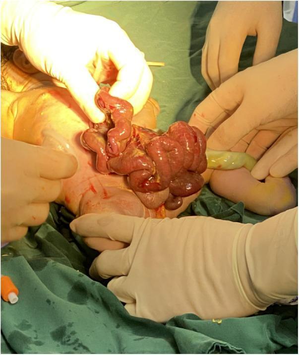
A clinical photograph of a newborn in an operating room setting illustrating gastroschisis, a congenital abdominal wall defect. The image shows a large evisceration of abdominal contents, including loops of the small and large intestines and a portion of the stomach, protruding through a right-sided paraumbilical defect. The herniated bowel loops exhibit significant edema, characteristic of prolonged exposure to amniotic fluid (matted, thickened appearance), and appear erythematous with a moist, glistening surface. The umbilical cord is visible and intact to the left of the defect. Gloved surgical hands are shown assessing the abdominal wall fissure and the visceral mass. This clinical finding is a hallmark of gastroschisis, where the bowel is not covered by a peritoneal sac, distinguishing it from an omphalocele. The case demonstrates the immediate postnatal presentation prior to the implementation of silo bag placement or primary surgical closure.
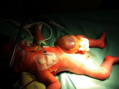
This clinical photograph displays a newborn infant in a neonatal care setting with a large, midline abdominal wall defect characteristic of a giant omphalocele. The defect presents as a substantial, hemispherical sac originating from the umbilical region, estimated at approximately 8 cm in diameter. The sac is covered by a thin, translucent peritoneal-amniotic membrane, through which the underlying visceral contents—primarily a significant portion of the liver and intestinal loops—are visible. Parts of the liver show a dark, purplish hue through the membrane. Concurrent medical management is visible, including a nasogastric tube for decompression, several adhesive dressings on the chest and upper abdomen, and white gauze packing at the base of the omphalocele sac. The image illustrates a major congenital anomaly requiring staged surgical closure and neonatal intensive care. Key educational concepts include midline abdominal wall defects, differentiation from gastroschisis by the presence of a covering sac, and initial neonatal stabilization.
duodenal atresia double bubble sign
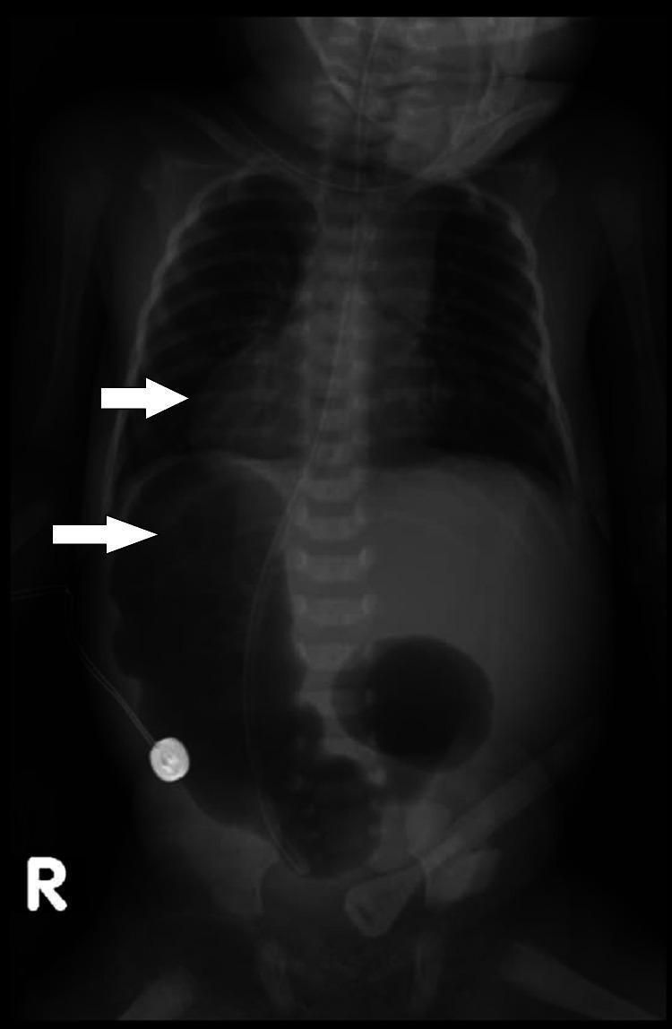
A plain anteroposterior (AP) X-ray of a neonate's chest and abdomen demonstrating situs inversus totalis and duodenal atresia. The cardiac silhouette is predominantly located in the right hemithorax (dextrocardia). In the abdomen, the liver shadow is positioned on the left side, while the stomach gas bubble is located on the right. A classic 'double bubble' sign is visible in the upper right quadrant, representing a dilated, gas-filled stomach and proximal duodenum, characteristic of duodenal atresia. A radiopaque nasogastric tube (NGT) is seen traversing the midline and terminating in the right-sided gastric bubble. A circular metallic temperature probe is visible on the right lateral abdominal wall. The image illustrates the complete reversal of thoracic and abdominal organs in a neonatal patient presenting with signs of intestinal obstruction.
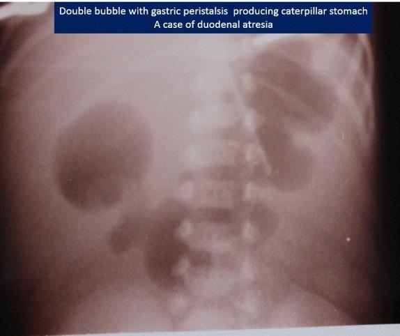
This abdominal X-ray of a neonate demonstrates the classic 'double bubble' sign, highly indicative of duodenal atresia. Two large, air-filled radiolucencies are visible in the upper abdomen: a larger gastric bubble on the patient's left and a smaller duodenal bubble on the right of the midline. A notable feature in this specific image is the 'caterpillar sign,' where the dilated gastric wall shows multiple waves of peristalsis (undulating borders), suggesting an attempt by the stomach to overcome the distal obstruction. There is a complete absence of bowel gas distal to the duodenum throughout the rest of the abdomen, signifying high intestinal obstruction. This diagnostic image is a key educational resource for pediatric radiology and neonatal surgery, illustrating the pathophysiology of congenital intestinal malformations and the radiographic markers used to differentiate them from other causes of neonatal emesis.
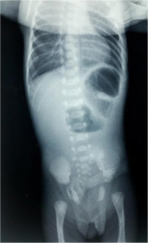
This diagnostic image is an anteroposterior plain radiograph of an infant's abdomen and thorax. The primary finding is the pathognomonic 'double-bubble' sign, characterized by two large, air-filled lucencies in the upper abdomen representing a dilated stomach (left) and a dilated proximal duodenum (right). There is a complete absence of gas throughout the distal intestinal tract, which indicates a high-grade proximal bowel obstruction. These radiographic features are classically associated with congenital duodenal atresia. The skeletal structures, including the rib cage, vertebral column, pelvis, and proximal femurs, appear appropriate for gestational age without obvious deformities or secondary complications. The image serves as a critical educational example of neonatal intestinal obstruction and the classic presentation of duodenal atresia in pediatric radiology.
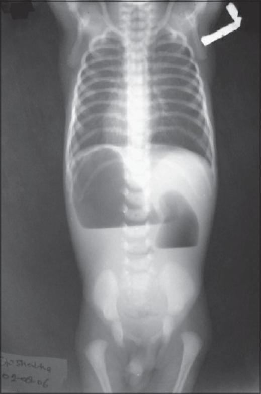
This diagnostic image is an anteroposterior babygram (plain film X-ray of the chest and abdomen) of a neonate. The thoracic cavity demonstrates dextrocardia, where the cardiac silhouette is predominantly positioned in the right hemithorax. In the upper abdomen, there is a prominent 'reverse double bubble' sign, characterized by two distinct gaseous shadows with air-fluid levels. The larger bubble (stomach) is located on the right side, and the smaller bubble (proximal duodenum) is located on the left, indicating situs inversus associated with duodenal obstruction. There is a complete absence of gas in the distal intestinal segments, suggesting a high-grade intestinal obstruction such as duodenal atresia or a perforated duodenal diaphragm. The clinical relevance of this visual includes the recognition of heterotaxy syndromes or situs inversus totalis in the context of neonatal bowel obstruction. This material is suitable for medical students and pediatric residents focusing on neonatal radiology and congenital gastrointestinal anomalies.
transposition of the great arteries egg on a string chest radiograph newborn cyanotic heart disease
neonatal jaundice kernicterus phototherapy bilirubin
Neonatal emergencies: illustrated companion to the master guide
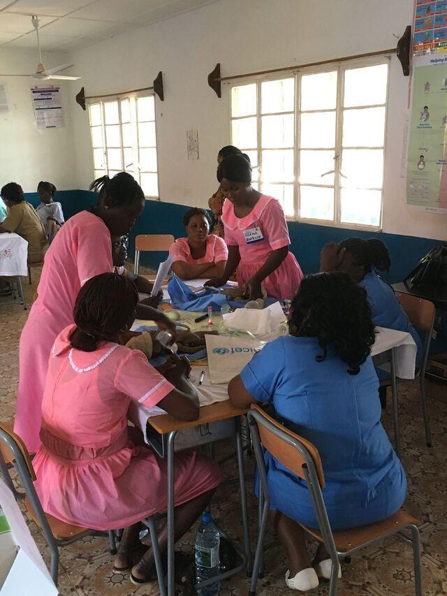
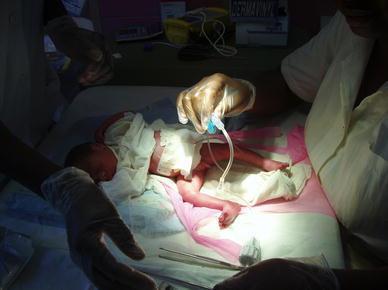
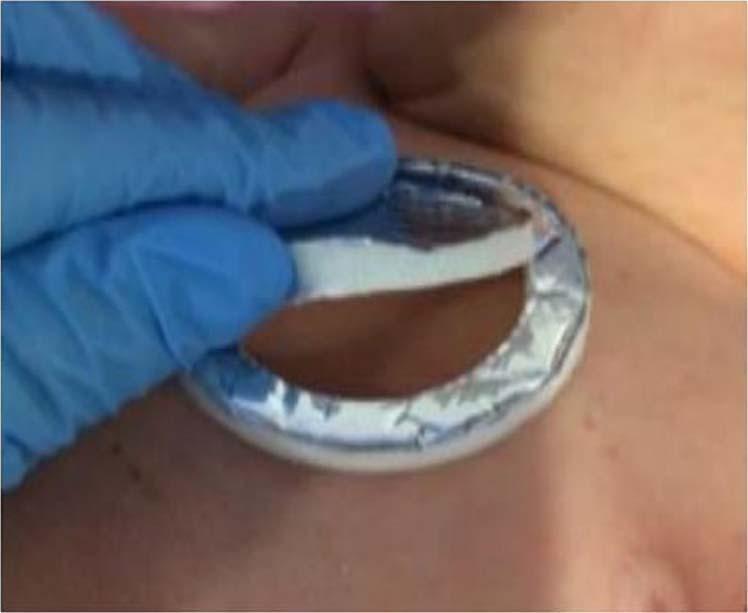
Part 1. Resuscitation and access
Part 2. Respiratory emergencies: reading the neonatal chest X-ray
RDS (preterm, surfactant deficiency)
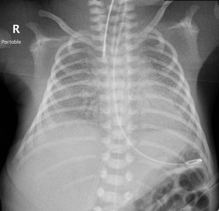
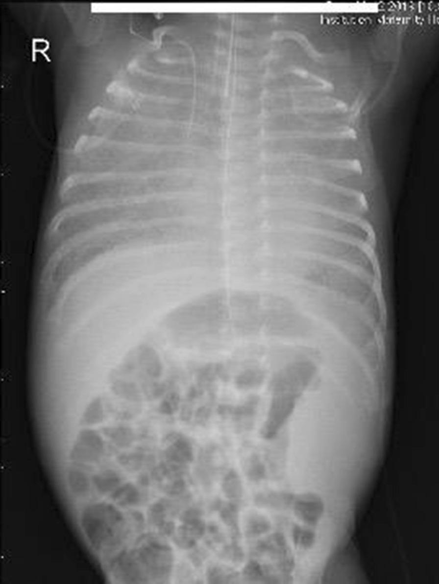
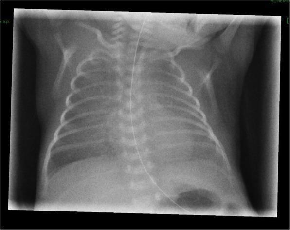
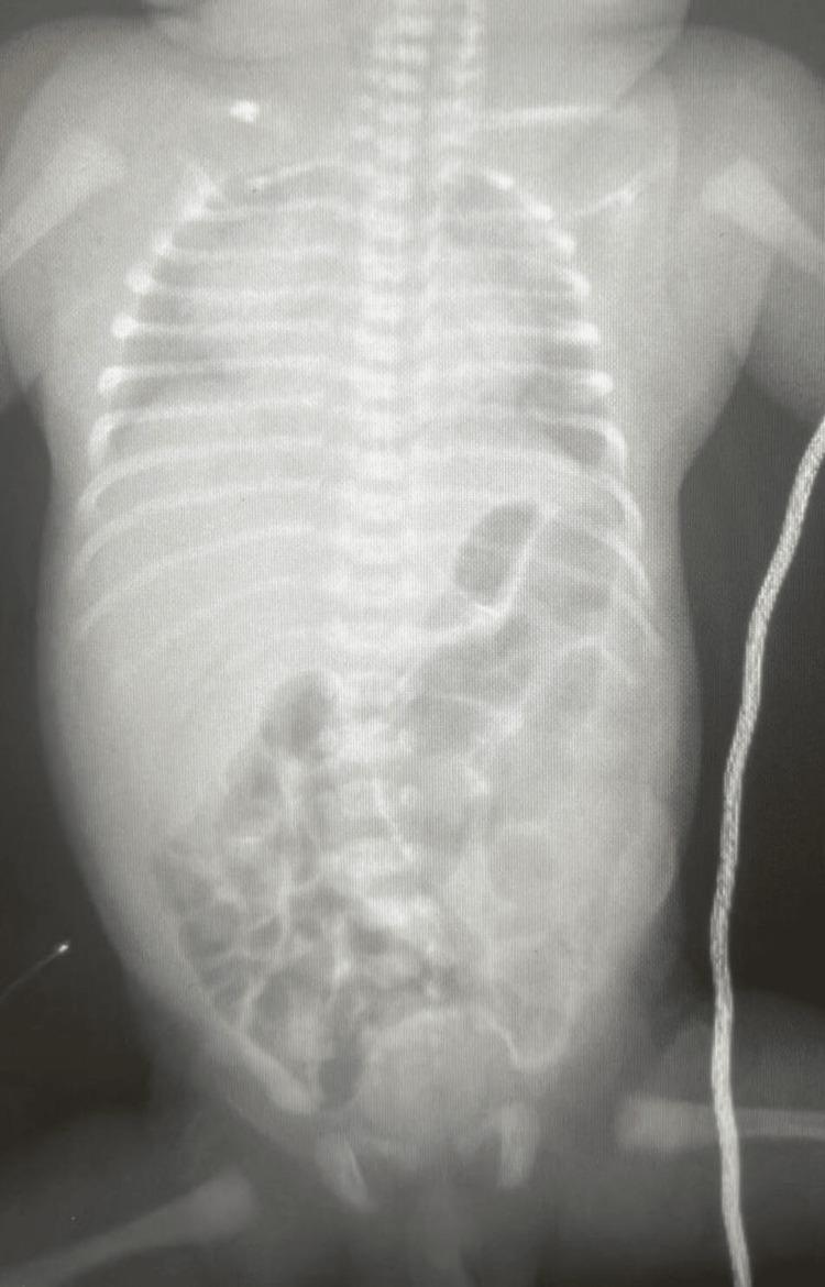
TTN (term or late preterm, often after C-section)
Meningeal aspiration with air leak (MAS + pneumothorax)
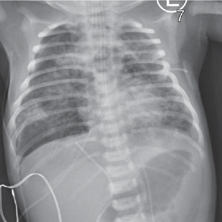
Pneumothorax
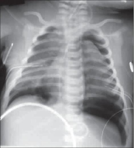
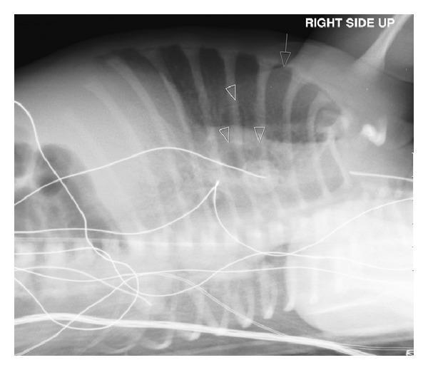
Congenital diaphragmatic hernia (CDH)
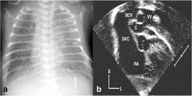
Part 4. Cardiac emergencies
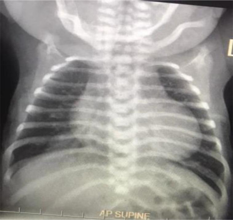
Part 6. Neonatal seizures: monitoring
Part 7. Jaundice
Part 8. Surgical emergencies
Malrotation with midgut volvulus
Intramural gas (NEC family)
Esophageal atresia with TEF
Duodenal atresia (double bubble)
Gastroschisis versus omphalocele
Anorectal malformations
One-page visual pattern recognition
| What you see | Think | First action |
|---|---|---|
| Ground-glass, low volumes, preterm | RDS | CPAP, surfactant |
| Hazy lungs, term or C-section | TTN | Oxygen, observe, exclude sepsis |
| Coarse patchy, hyperinflated, post-term | MAS, watch for air leak | Ventilate gently, check for pneumothorax |
| Bowel in chest, shift, scaphoid abdomen | CDH | Intubate, orogastric tube, no mask ventilation |
| Egg-on-a-string heart, cyanosis | TGA | PGE1, urgent cardiology |
| Ground-glass with cardiomegaly in first days | Obstructed TAPVC | Avoid PGE1, urgent surgery |
| Corkscrew duodenum on contrast | Midgut volvulus | Operating room |
| Double bubble, gasless distal bowel | Duodenal atresia | Decompress, check for Down syndrome and cardiac anomalies |
| Tube coils in upper chest, gas in abdomen | EA with distal TEF | Replogle suction, head-up |
| Defect right of cord, no sac | Gastroschisis | Cover the bowel, side-lying, extra fluids |
| Defect with a sac | Omphalocele | Protect the sac, search for associated anomalies |
| Arching in a jaundiced baby | Bilirubin encephalopathy | Emergency exchange transfusion assessment |
Neonatal emergencies master guide With textbook figures and images