
Anatomy and physiology of hair
Reading File
Finding Sources
Finding Sources
Finding Sources
Searching Images
hair follicle structure anatomy diagram cross section
Reading File
Reading File
Reading File
Reading File
Reading File
Reading File
Reading File
Anatomy and Physiology of Hair
1. Overview
Hair is a keratinized skin appendage produced by the hair follicle, a complex "mini-organ" embedded in the dermis and subcutis. Each follicle cycles independently through growth, regression, and rest phases throughout life.
2. Anatomy
Hair types
- Lanugo - fine, unpigmented hair present in the prenatal period, shed before or shortly after birth.
- Vellus hair - short (<2 cm), fine (<30 μm diameter), unmedullated, poorly pigmented hair. Its bulb sits in the reticular dermis. Covers most of the body before puberty.
- Terminal hair - coarse (>60 μm diameter), long (can exceed 100 cm), medullated, pigmented hair. Its bulb lies in the subcutaneous fat. Present on the scalp, eyebrows, and eyelashes at birth; androgens convert vellus follicles to terminal follicles at puberty in the axillae, genital area, and (in men) beard/chest. (Fitzpatrick's Dermatology, p. 121-122)
Hair follicle structure
The follicle is divided into a permanent upper portion (infundibulum + isthmus) and a cycling lower portion (suprabulbar region + bulb). From outermost to innermost, the compartments are:
- Connective tissue (perifollicular) sheath - an inner glassy/vitreous basement membrane continuous with the epidermal basement membrane, and an outer type III collagen sheath. It thickens and disintegrates during catagen and can itself induce new follicle formation when transplanted.
- Outer root sheath (ORS) - continuous with the epidermis; keratinizes with a granular layer in the infundibulum but undergoes trichilemmal keratinization (no granular layer) in the isthmus. The ORS at the base of the isthmus forms the bulge, the niche for epithelial and melanocyte stem cells and the attachment site of the arrector pili muscle.
- Inner root sheath (IRS) - Henle's layer, Huxley's layer, and the IRS cuticle; made of soft keratin, breaks down at the isthmus as sebum enters the canal, and does not emerge with the hair shaft.
- Hair shaft, composed of three layers:
- Cuticle - outermost, overlapping keratinized squamous cells (like roof tiles), protects the shaft and determines porosity.
- Cortex - ~80% of hair mass, packed with hard keratin intermediate filaments cross-linked by keratin-associated proteins (KAPs); determines texture, elasticity, and color (melanin from hair-bulb melanocytes is deposited here).
- Medulla - central core of loosely packed, soft-keratin cells; present only in thick/terminal hairs. (Fitzpatrick's Dermatology, p. 123; Histology: A Text and Atlas, p. 1351)
Associated structures
- Dermal papilla - mesenchymal core at the base of the bulb that signals to the overlying hair matrix and dermal-epithelial crosstalk drives follicle growth.
- Hair matrix - the germinative epithelium surrounding the papilla; gives rise to seven cell lineages (three of the hair shaft, four of the IRS).
- Sebaceous gland - opens into the follicular infundibulum (pilosebaceous unit).
- Arrector pili muscle - smooth muscle attaching to the bulge; contraction erects the hair ("goose bumps") and can also help express sebum.
- Innervation - myelinated sensory fibers form a network around follicles with free nerve endings (pain), lanceolate endings (movement/acceleration), Merkel cells (pressure), and pilo-Ruffini corpuscles (tension). Perifollicular nerves release neuropeptides (substance P, CGRP) that also modulate the hair cycle.
- Melanocyte stem cells reside in the bulge and repopulate hair-bulb melanocytes each anagen cycle; their depletion (e.g., via Bcl2 loss) underlies hair graying. (Fitzpatrick's Dermatology, p. 128-130)
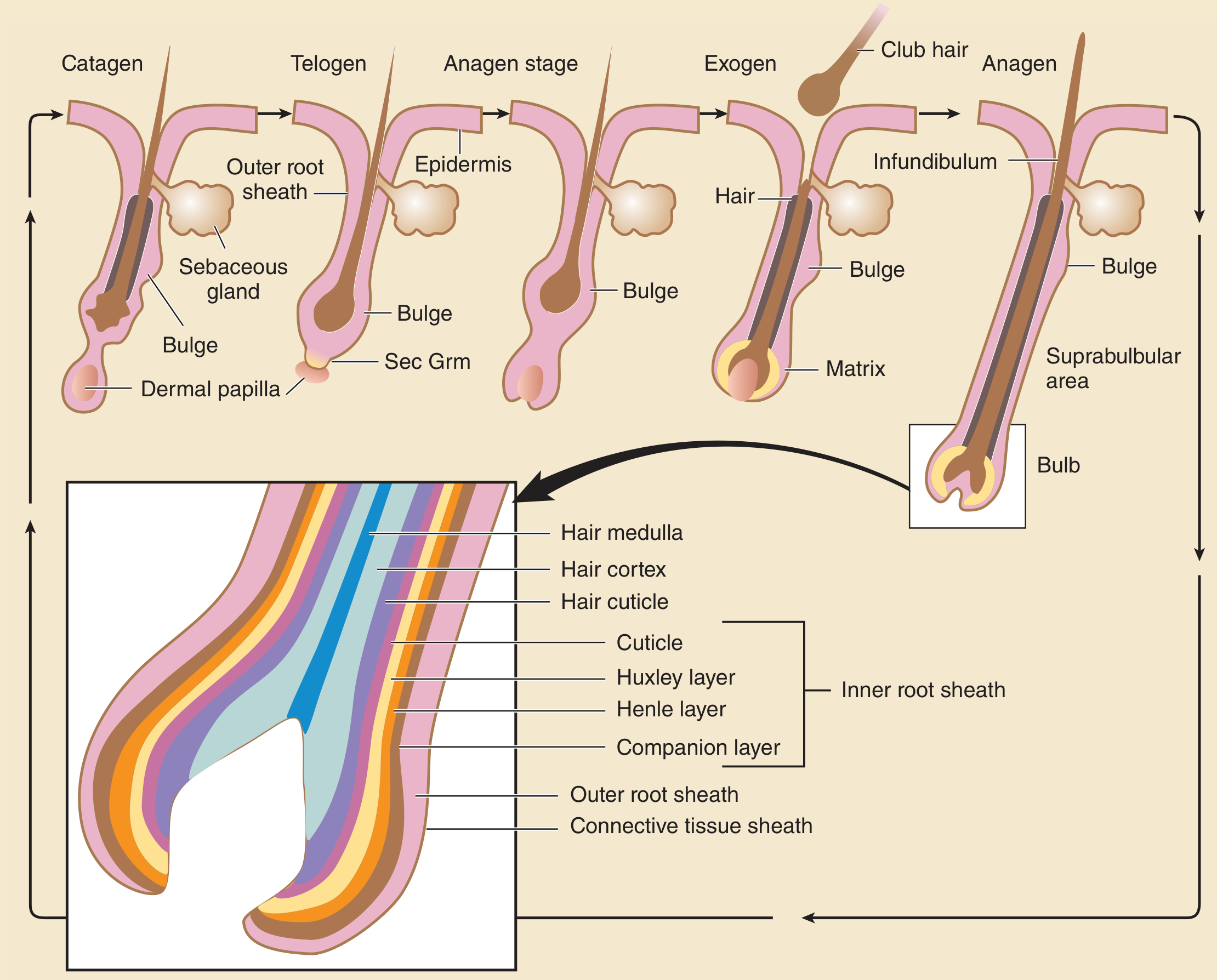
3. Physiology - the Hair Growth Cycle
Each follicle cycles independently and asynchronously through:
- Anagen (growth phase) - active proliferation of matrix keratinocytes; determines the ultimate length of the hair. Duration is site-dependent: scalp 2-8 years; eyelashes/eyebrows and other body sites only 1-6 months. About 85-90% of scalp hairs are in anagen at any time.
- Catagen (regression/involution) - a brief (~2-3 week) apoptosis-driven phase in which the lower follicle regresses; the basement membrane thickens and then disintegrates.
- Telogen (rest) - a quiescent phase (roughly 2-3 months on the scalp) during which the club hair is retained in a shortened follicle before shedding.
- Exogen (shedding) - active release of the telogen club hair, which may be followed by a kenogen lag period (empty follicle) before a new anagen hair emerges. (Fitzpatrick's Dermatology, p. 124; Andrews' Diseases of the Skin; Goldman-Cecil Medicine)
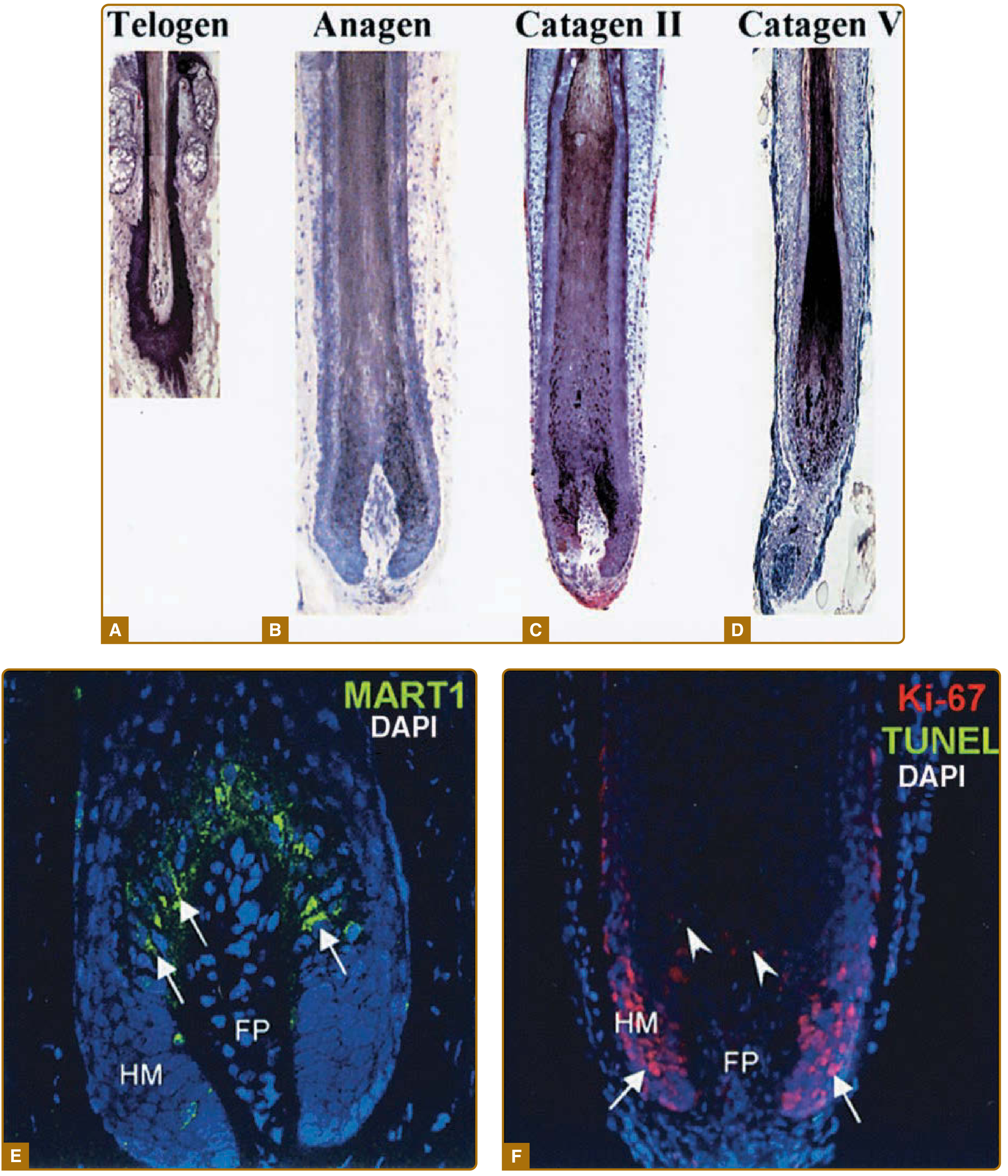
Regulation of the cycle
- Molecular signaling: Wnt/β-catenin, Sonic hedgehog (SHH), BMP, Notch, EDA/EDAR, and FGF pathways coordinate follicle morphogenesis and cycling; SHH and its receptor Patched1 are required for dermal papilla and hair germ formation.
- Stem cells: Bulge epithelial stem cells regenerate the lower follicle at each anagen onset; bulge melanocyte stem cells (expressing c-kit, Sox10, Mitf) regenerate pigment-producing melanocytes.
- Hormones: Androgens convert vellus to terminal follicles at androgen-dependent sites (beard, axilla, pubic region) at puberty, but paradoxically can miniaturize terminal scalp follicles in genetically susceptible individuals (androgenetic alopecia).
- Neural input: Perifollicular nerve-derived neuropeptides and Merkel cell-derived factors influence cycling in a hair-cycle-dependent manner.
Functions of hair
- Thermoregulation - insulation and, via arrector pili contraction, trapping of an air layer.
- Sensory function - follicles are richly innervated mechanoreceptors that detect touch, air movement, and pressure before it reaches the skin surface.
- Photoprotection and physical protection - scalp hair, eyebrows, and eyelashes shield the skin/eyes from UV light, sweat, and foreign particles; nasal and ear hair filter airborne particles.
- Social/sexual signaling - secondary sexual hair patterns (beard, axillary, pubic hair) develop under androgen influence at puberty.
- Reservoir for epidermal regeneration - the follicle's abundant stem cell populations contribute to re-epithelialization after skin injury, which is why hair-bearing skin (face, scalp) heals faster than adnexa-poor skin (back).
Recent evidence check
A quick search for recent meta-analyses/systematic reviews on hair follicle biology did not surface findings that contradict this core anatomy/physiology - the underlying cycle biology (anagen/catagen/telogen/exogen) and follicle structure are stable, well-established science rather than an area of shifting evidence. Most current research (e.g., on androgenetic alopecia treatments, JAK inhibitors for alopecia areata) concerns clinical management rather than basic anatomy/physiology, so no correction is needed here.
If you want, I can go deeper into a specific area next - for example, the molecular biology of the hair cycle, the pathophysiology of hair loss (androgenetic alopecia, telogen effluvium, alopecia areata), or histology slides of the follicle at different cycle stages.
This is a shared conversation. Sign in to Orris to start your own chat.