
Expalin me in detail about perineal ab ssess from Bailey and love completely in detail
perianal abscess types ischiorectal supralevator intersphincteric diagram
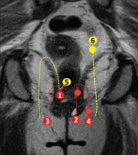
**Imaging Modality:** Coronal T2-weighted Magnetic Resonance Imaging (MRI) of the pelvis with superimposed schematic diagrams. **Anatomical Region:** The perianal and pelvic floor region, specifically showing the anal canal, internal and external anal sphincters, ischioanal/ischiorectal fossae, and the levator ani muscle complex. **Content Description:** This image provides a comparative schematic of perianal fistulous tracts according to the St. James’s University Hospital MRI classification. Numbered annotations (1–6) and color-coded dashed lines delineate different grades of fistula complexity: * **Grade 1 & 2 (Intersphincteric):** Red dashed lines confined between the internal and external sphincters, including simple tracks and those with associated abscesses. * **Grade 3 & 4 (Transsphincteric):** Lines extending through the external sphincter into the ischioanal fossa, with Grade 4 illustrating secondary tracks or abscesses. * **Grade 5 (Supralevator):** Yellow dashed line extending superior to the levator ani muscle. * **Grade 6 (Extrasphincteric):** Yellow dashed line illustrating a track originating from the pelvic cavity/rectum, bypassing the sphincter complex entirely. **Clinical Significance:** This educational schematic serves as a reference for grading fistula-in-ano, identifying relationship to the sphincter mechanism, and detecting secondary complications like abscess formation or supralevator extension.
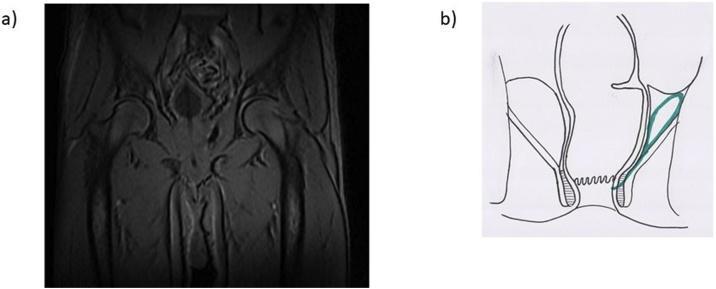
This composite educational graphic details a supralevator abscess through diagnostic imaging and anatomical illustration. Image (a) is a coronal T2-weighted magnetic resonance image (MRI) of the male pelvis, showing a hyperintense fluid collection measuring approximately 6x2x3 cm located superior to the levator ani muscle. This collection is positioned laterally to the rectum and adjacent to the prostatic region and internal obturator muscle, with an associated inter-sphincteric fistula tract extending inferiorly. Image (b) provides a schematic coronal diagram of the anorectal region, highlighting the abscess in green. The illustration depicts the anatomical relationship between the abscess, the levator ani muscle, and the anal sphincters. It clearly shows the fistulous communication originating from the supralevator space and tracking through the inter-sphincteric plane toward the anal canal. These images together demonstrate a complex perianal infection, which is a critical diagnostic finding in colorectal surgery for determining surgical drainage approaches and identifying potential internal fistula openings.
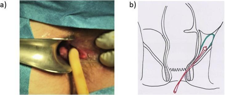
A composite image illustrating the surgical management of a complex perianal abscess. Image (a) is a clinical photograph of a perianal surgical site. A metallic anal retractor is used to visualize the rectal lumen and fistula opening. A yellow, mushroom-tip Pezzer drain is shown inserted through an inter-sphincteric fistula tract to facilitate continuous drainage of deeper infection. Image (b) is a coronal anatomical diagram of the anorectal region. The illustration depicts the rectum, internal and external anal sphincters, and the supralevator space. A green-contoured area highlights a supralevator abscess. A red line represents the Pezzer drain, showing its path originating externally, traversing the inter-sphincteric plane, and terminating within the abscess cavity. The educational focus is on the surgical treatment of supralevator abscesses and the specific placement of self-retaining drains for long-term management of complex fistulous tracts in colorectal surgery.
Perianal (Anorectal) Abscess - Bailey and Love's Short Practice of Surgery, 28th Edition
Anatomy - Sites of Abscess Formation
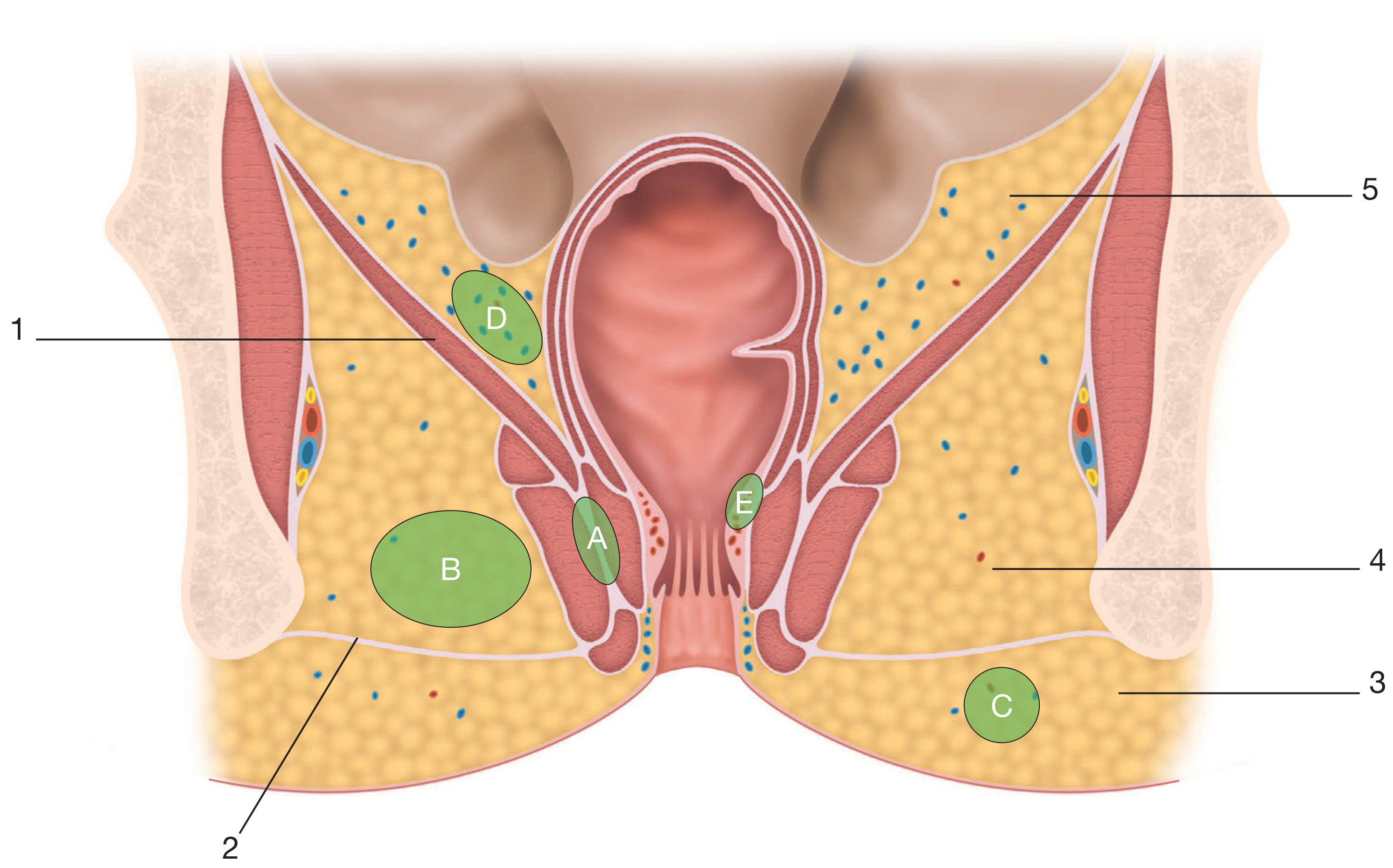
- 1 - Levator ani muscle
- 2 - Superficial perineal fascia
- 3 - Superficial perianal space
- 4 - Ischiorectal space
- 5 - Supralevator space
- A - Intersphincteric abscess
- B - Ischiorectal abscess
- C - Superficial perianal abscess
- D - Supralevator abscess
- E - Submucosal abscess
Aetiology
The Cryptoglandular Theory (Parks)
- Infection begins in the intersphincteric anal glands at the level of the dentate line
- Pus travels along the path of least resistance and may spread in several directions:
- Caudally - to present as a perianal abscess (most common - 60%)
- Laterally across the external sphincter - to form an ischiorectal abscess (~30%)
- Superiorly above the anorectal junction - to form a supralevator intermuscular or pararectal abscess (rare), depending on its relation to the longitudinal muscle
- Circumferentially in any of the three planes: intersphincteric/intermuscular, ischiorectal, or pararectal supralevator - giving rise to horseshoe abscesses
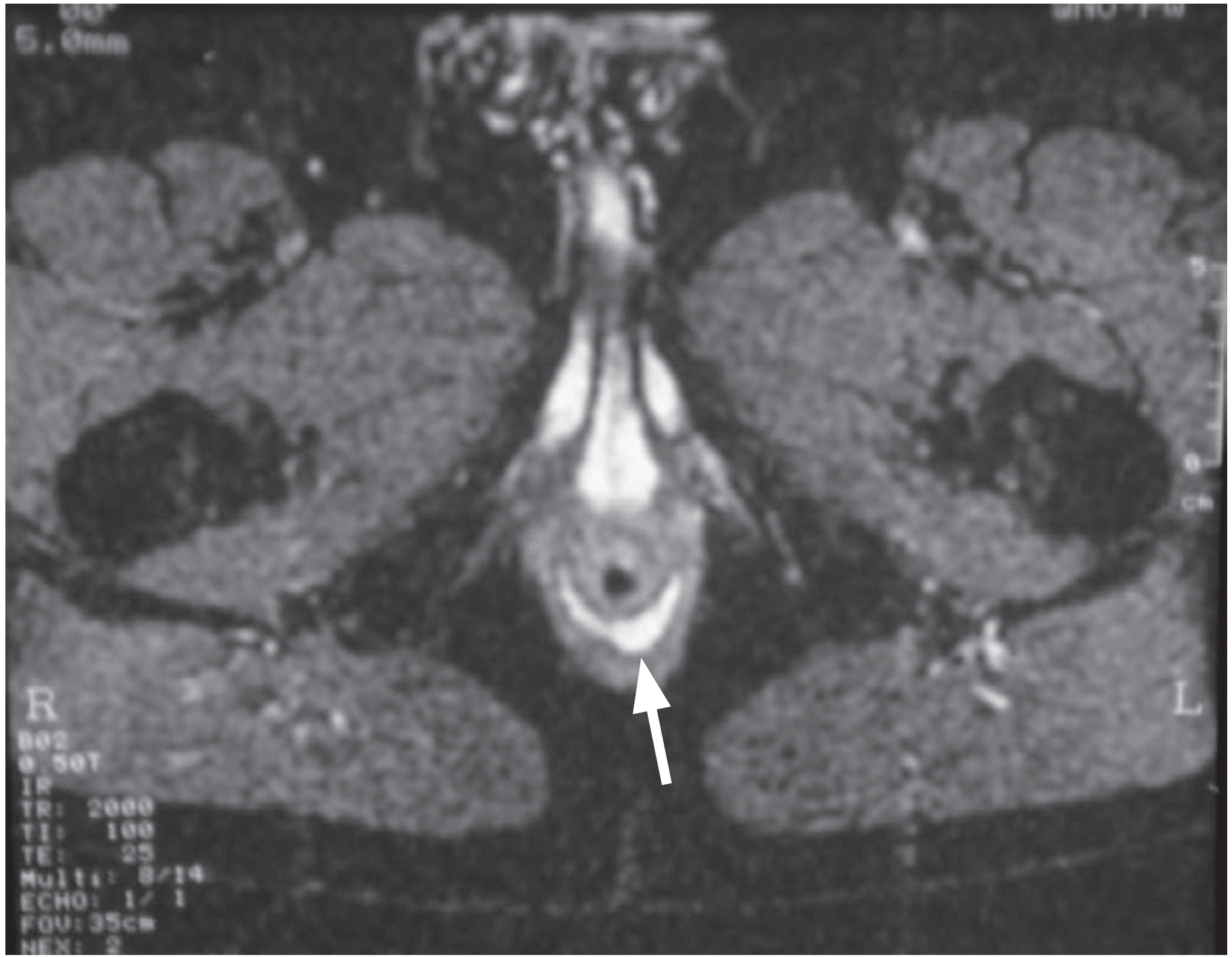
Other Causes of Anorectal Sepsis (NOT from anal gland infection)
| Site | Cause |
|---|---|
| Submucosal abscess | Following haemorrhoidal sclerotherapy (usually resolves spontaneously) |
| Mucocutaneous / marginal abscess | Infected haematoma |
| Ischiorectal abscess | Foreign body, trauma, deep skin-related infection |
| Pelvirectal supralevator sepsis | Originating from pelvic disease |
- Rectal neoplasm and particularly Crohn's disease may be the underlying cause
- Immunosuppressed patients, those with diabetes or AIDS may present with perianal or pelvirectal sepsis that can run an aggressive course
Classification of Anorectal Abscesses
| Type | Location | Incidence |
|---|---|---|
| Perianal (superficial perianal) | Confined by terminal extensions of longitudinal muscle, at the anal margin | 60% |
| Ischiorectal | Within the fatty ischiorectal space, lateral to the anal canal | ~30% |
| Supralevator | Above the levator ani - intermuscular or pararectal | Rare |
| Submucosal | Under the rectal mucosa | Uncommon |
| Intersphincteric | Between the internal and external sphincters | Less common |
| Horseshoe | Circumferential spread in any of the three planes | Uncommon |
Clinical Presentation
Perianal Abscess (most common)
- Short (2-3 day) history of increasingly severe, well-localised pain
- Palpable tender lump at the anal margin
- Examination: indurated, hot, tender perianal swelling
- Confined by the terminal extensions of the longitudinal muscle
Ischiorectal Abscess
- Located in the larger, fatty-filled ischiorectal space where tissue tension is much lower
- Usually presents later with less well-localised symptoms
- More constitutional upset and fever
- Examination: the affected buttock is diffusely swollen with widespread induration and deep tenderness
Supralevator Abscess
- If sepsis is higher (supralevator), the presentation is different:
- Deep rectal pain
- Fever
- Sometimes disturbed micturition (urinary symptoms)
- Nothing evident on external examination
- But: tender supralevator induration palpable on digital rectal examination above the anorectal junction
Differential Diagnosis
- Abscess connected with a pilonidal sinus
- Bartholin's gland abscess (in women)
- Cowper's gland abscess
Management
General Principle
- Careful examination under anaesthesia (EUA)
- Sigmoidoscopy and proctoscopy
- Adequate drainage of pus
Drainage of Perianal and Ischiorectal Abscess (60% + 30%)
- Drainage is through the perineal skin
- Traditionally: a cruciate incision over the most fluctuant point, with excision of skin edges to deroof the abscess
- However, Bailey and Love emphasizes: skin preservation is important - wide excision of otherwise healthy tissue should be avoided
- A gentle search may be made for an underlying fistula if the surgeon is experienced; if obvious, a loose draining seton may be passed
- Injudicious probing in the acute stage is potentially dangerous and may lead to a much more difficult situation
- Unless by highly experienced hands, immediate fistulotomy should NOT be performed
- Despite lack of evidence, packing the abscess cavity is commonplace
Management of Supralevator Sepsis
- Dependent on its origin:
- Sepsis originating in pelvic disease (appendiceal, gynaecological, diverticular, Crohn's disease, malignancy) - requires management of the underlying cause; intrarectal drainage may be appropriate to avoid creation of an extrasphincteric fistula
Summary Box 80.11 - Anorectal Abscess (Bailey and Love)
- Presents as a painful, throbbing swelling in the anal region with associated pyrexia
- Classified according to anatomical site
- Treatment is drainage of pus and appropriate systemic antibiotics
- Consider underlying diagnosis: fistula-in-ano, Crohn's disease, diabetes, immunosuppression
Important Clinical Points (Bailey and Love)
-
Fistula-in-ano relationship: Most anorectal abscesses arise from cryptoglandular infection and ~30-50% will develop a fistula-in-ano after drainage. Do NOT perform immediate fistulotomy unless highly experienced.
-
Horseshoe abscesses: Can track circumferentially in intersphincteric, ischiorectal, or supralevator planes - these require careful EUA and sometimes staged drainage.
-
Immunosuppressed / diabetic patients: May present with aggressive, rapidly spreading sepsis (including necrotising fasciitis) - perianal abscess is listed as a predisposing condition for Fournier's gangrene (necrotising fasciitis of perineum).
-
Crohn's disease: Perianal abscess in Crohn's presents with swelling, redness, pain, then discharge of pus or faecal drainage (fistulous track to perianal skin or vagina). Fistulotomy should generally be avoided in Crohn's disease as wound edges heal poorly.
-
Antibiotics: Systemic antibiotics (e.g., metronidazole + ciprofloxacin) are used alongside drainage, particularly in perianal Crohn's disease and immunosuppressed patients. Long-term metronidazole should be avoided due to peripheral neuropathy risk.