

What does this mean? Can you give pictorial representation of each
physical examination percussion
clinical percussion examination shifting dullness tidal percussion Traube's space Kronig isthmus
"percussion myokymia" physical examination
https://www.ncbi.nlm.nih.gov/books/NBK459220
clinical diagram chest percussion landmarks cardiac dullness liver dullness Traube space
Educational clinical-examination infographic showing an anterior adult torso with left and right lungs, clavicles, intercostal spaces, heart, liver, stomach gas bubble, diaphragm and abdomen. Mark the percussion landmarks named: Kronig's isthmus over each shoulder apex, clavicular percussion, intercostal lung fields, right liver dullness, left cardiac dullness, left Traube's space, and lower abdominal flanks for shifting dullness. Include small inset diagrams for tidal percussion at the right midclavicular line and a patient turning on side showing ascitic fluid shifting. Use clear accurate labels for medical students.
- Resonant: normal air-filled lung
- Hyperresonant: too much air, for example pneumothorax or emphysema
- Dull: solid organ, fluid, or consolidated lung
- Tympanic: hollow, air-filled stomach or bowel, like a drum
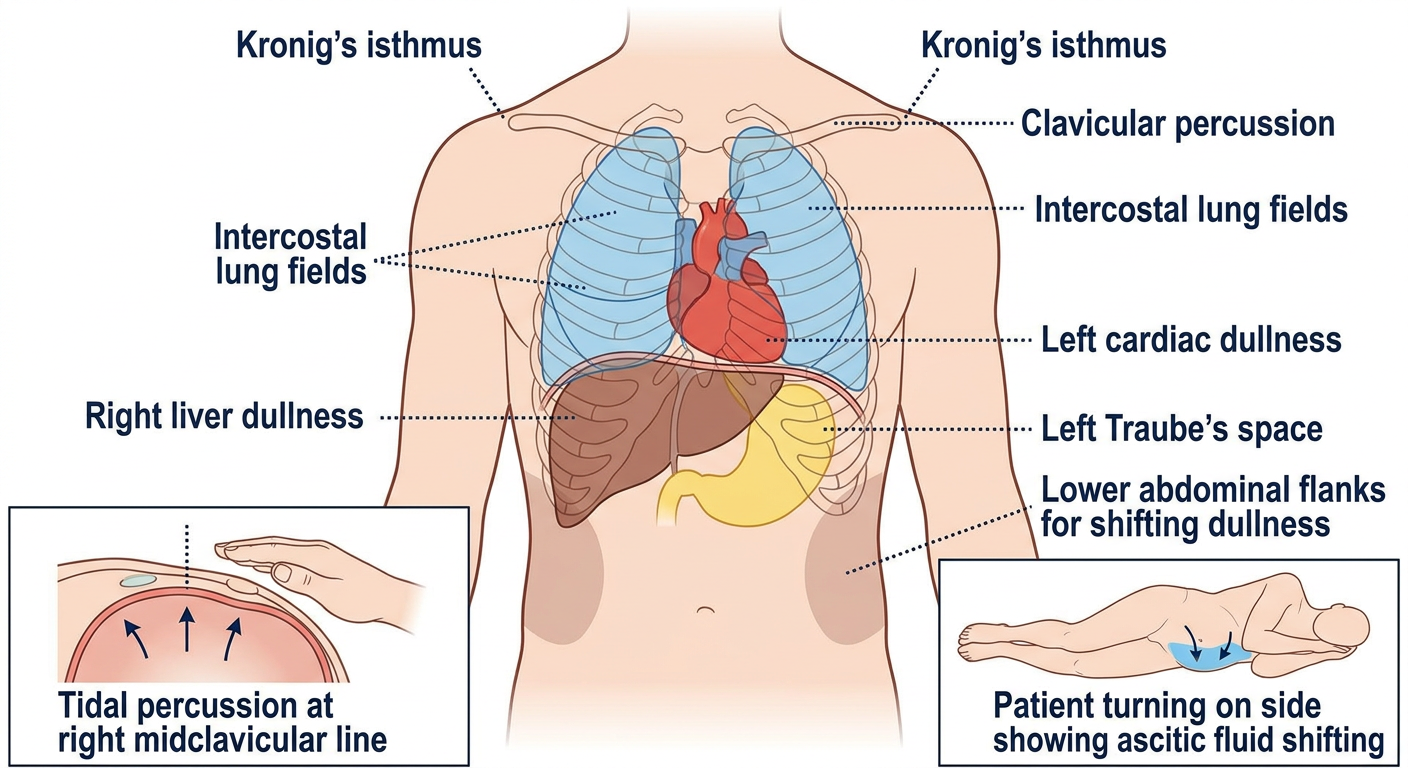
Simple anterior body map
                FRONT VIEW

             Shoulder        Shoulder
          [Kronig]          [Kronig]
                 \          /
          clavicle \      / clavicle
          ────────────────
          Lung: resonant   Lung: resonant
          intercostal      intercostal
          spaces           spaces

             Heart area
          left cardiac
             dullness

                           Right liver:
                           dullness

          Left upper abdomen:
          Traube's space
          usually tympanic/resonant

       Abdomen:
       centre often tympanic
       flanks may become dull in ascites
Terms in your list
| Term | Where / what it means | Normal finding | Why it is checked |
|---|---|---|---|
| Kronig’s isthmus | A narrow band of resonant lung sound over the top of each shoulder, between the neck and shoulder. | Resonant and roughly equal on both sides. | Narrowing may suggest fibrosis or an apical lung lesion; widening may occur with hyperinflation. This is a traditional exam sign. |
| Clavicular percussion | Tapping over and just below each collarbone to compare the lung apices. | Similar resonance on both sides. | Detects apical lung disease, such as upper-lobe consolidation, fibrosis, mass, or pneumothorax. |
| Intercostal resonance | Percussion over the spaces between the ribs across both lung fields. | Resonant bilaterally. | Dullness can occur with pneumonia, collapse, tumor, or pleural fluid. Hyperresonance can occur with excess air, especially pneumothorax. |
| Liver dullness | Right lower chest / right upper abdomen, where the liver lies under the diaphragm. | Dull sound over the liver. | Helps estimate liver span. Loss of expected liver dullness, with resonance instead, can indicate free gas under the diaphragm in the appropriate acute-abdomen setting. |
| Cardiac dullness | Left front of chest where the heart lies behind the chest wall. | Relative dullness compared with adjacent air-filled lung. | Historically used to estimate cardiac enlargement or displacement. It is less reliable than imaging and echocardiography. |
| Traube’s area (Traube’s space) | Semilunar region in the left lower chest / left upper abdomen. Its borders are roughly the left sixth rib above, left anterior or mid-axillary line laterally, and left costal margin below. | Usually tympanic or resonant due to the stomach gas bubble. | Dullness may support splenic enlargement, but can also occur after food, with obesity, or colonic contents. Stanford’s spleen examination guide explains that it is a screening clue, not confirmation. |
| Tidal percussion | Percussion at the right lower chest during full expiration and then deep inspiration. | The lower border of lung resonance moves downward with inspiration, reflecting diaphragm movement. | Assesses diaphragmatic excursion. Reduced movement may occur with diaphragmatic weakness, pleural disease, lung hyperinflation, or a subdiaphragmatic process. |
| Shifting dullness | Abdominal percussion performed supine, then repeated after turning the patient to one side. | Usually no shift in a normal abdomen. | A shifting area of dullness suggests free fluid in the abdomen, called ascites. Fluid goes to the dependent side while gas-filled bowel rises. Clinical Methods describes the usual pattern as central tympany with flank dullness when the patient is supine. |
| Percussion myokymia | Tapping over a muscle or nerve produces visible fine, rippling, involuntary muscle movements. | Absent. | A rare neurological sign of nerve hyperexcitability. “Myokymia” literally refers to wave-like muscle twitching. It is not a routine chest-percussion finding and may be included as an advanced/rare examination sign. |
| Skodaic resonance | An area of increased resonance just above a pleural effusion. | Absent. | With fluid at the lung base, the compressed lung immediately above it may sound unusually resonant. This is an older but classically described sign. Harrison’s describes it as “an arc of hyperresonance above a pleural effusion.” |
Key picture for shifting dullness
SUPINE: ascites fluid spreads to flanks

      gas-filled bowel
          TYMPANY
       ┌───────────┐
 DULL  │           │  DULL
fluid  └───────────┘ fluid


LEFT LATERAL POSITION: fluid moves downward

       upper side: more resonant
             ↑ bowel floats

       lower/dependent side: DULL
             ↓ fluid collects
Key picture for tidal percussion
Right mid-clavicular line

Expiration:  lower lung border here
Inspiration: lower lung border moves DOWN

        lung
        ↓
  ─────────────  expiration border
       ↓↓
  ─────────────  inspiration border

Distance moved = diaphragmatic excursion
Rales
pulmonary crackles auscultation
rales crackles lung auscultation fine coarse definition clinical
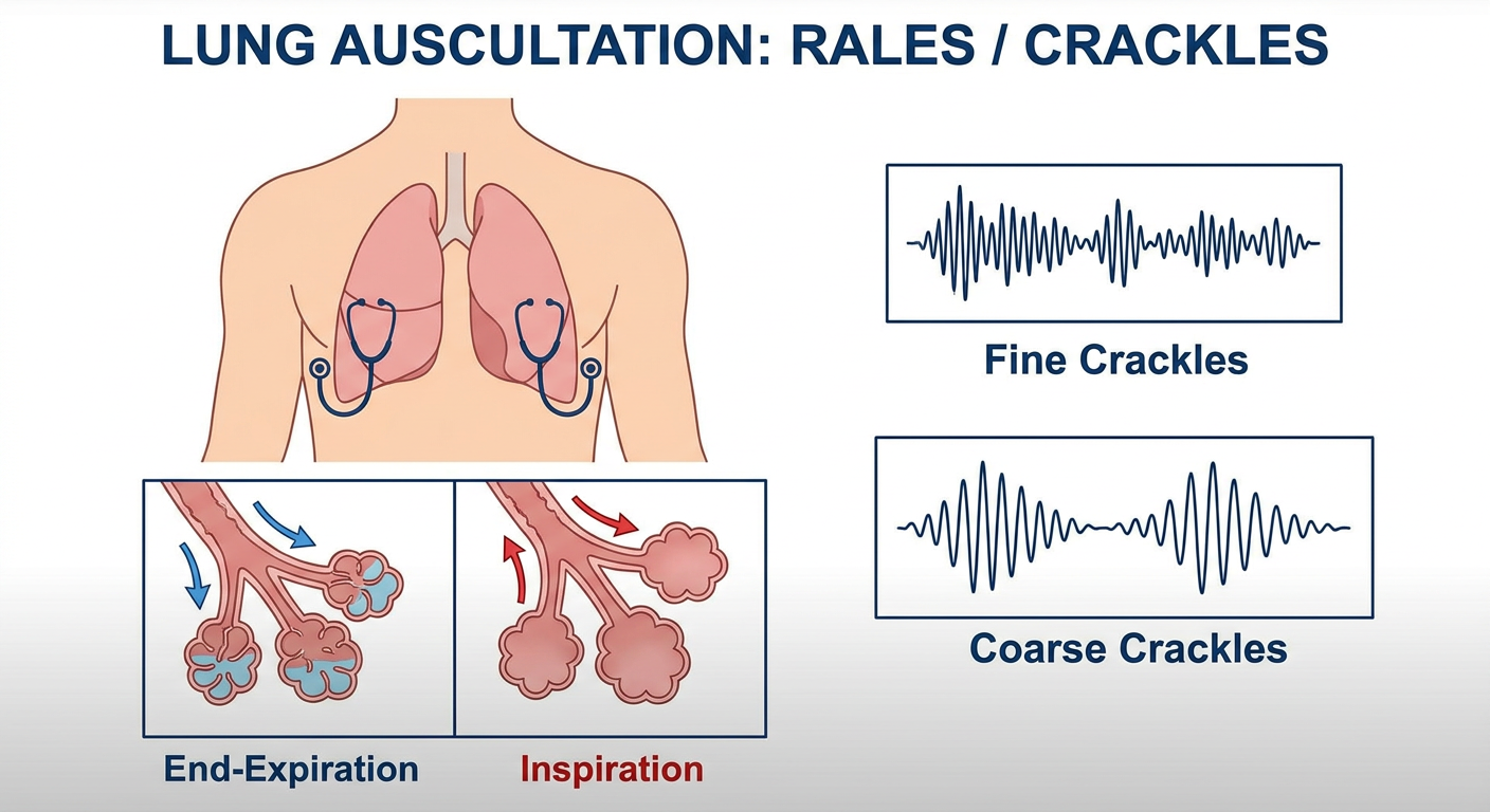
Simple medical teaching diagram for students showing rales or crackles during lung auscultation: an anterior chest with a stethoscope over both lower lung bases, small airways and alveoli that are fluid-filled or collapsed at end-expiration then pop open during inspiration, and two small sound-wave traces labelled fine crackles and coarse crackles. Clear educational labels, no patient-identifying details.
Types
| Type | Sound description | Usual timing | Common associations |
|---|---|---|---|
| Fine rales / fine crackles | Soft, high-pitched, brief. Like rubbing hair between fingers near the ear, opening Velcro, or salt on a hot pan. | Usually late inspiration, often at lung bases. | Interstitial lung disease/fibrosis, early pulmonary edema, sometimes heart failure. |
| Coarse rales / coarse crackles | Louder, lower-pitched, longer, bubbling or rattling. | Early or throughout inspiration, sometimes expiration too. | Airway secretions, pneumonia, bronchiectasis, severe pulmonary edema. May change after coughing. |
Basic mechanism
At end-expiration:
small airway/alveolus is closed or contains fluid
             ↓
During inspiration:
air enters and it suddenly pops open
             ↓
heard as a crackle (rale)
- Fine basal late-inspiratory crackles can be described as “Velcro-like” in pulmonary fibrosis.
- Bilateral basal crackles with breathlessness and leg swelling may suggest pulmonary edema/heart failure, but are not diagnostic alone.
- Rales are heard by auscultation, not percussion.