
Branches of the internal iliac artery.
internal iliac artery branches diagram anatomy
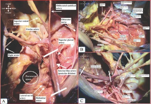
This surgical clinical photograph series illustrates the detailed anatomy of the right internal iliac artery and its branches through three perspectives: superior (A), medial (B), and lateral (C) views. The dissection highlights the pelvic vasculature essential for gynecological and pelvic surgery. Key arterial structures shown include the posterior division of the internal iliac artery giving rise to the superior gluteal, iliolumbar, and lateral sacral arteries. The anterior division branches are clearly visible, including the uterine artery, superior vesical artery, and the obliterated umbilical artery. Also demonstrated are the obturator nerve, the right ureter, and the pararectal space. View B specifically identifies venous components like the superficial and deep uterine veins, and the common trunk of the internal pudendal and inferior gluteal arteries. This educational material is intended for advanced medical students and surgical residents to understand the complex spatial relationships within the female pelvis, emphasizing vascular preservation and identification during retroperitoneal dissection.
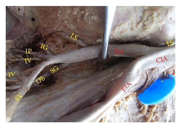
Anatomical dissection of the right hemipelvis demonstrating the arterial supply and branching patterns of the internal iliac artery (IIA). The image shows the common iliac artery (CIA) bifurcating into the external iliac artery (EIA) and the internal iliac artery. This specific specimen exhibits a high variation in IIA length, categorized as a long internal iliac artery. Key vascular branches are labeled, including the superior gluteal artery (SG) originating proximally, and a distal bifurcation forming the inferior gluteal (IG) and internal pudendal (IP) arteries. The anterior division branches, specifically the superior vesical (SV) and inferior vesical (IV) arteries, are visible coursing toward the pelvic viscera. Associated neural and vascular structures such as the lumbosacral trunk (LS) and obturator artery (Ob) are shown in their characteristic anatomical positions relative to the IIA. This clinical photograph is used in medical education to illustrate pelvic surgical anatomy and common vascular variations critical for procedures like arterial ligation or pelvic lymphadenectomy.
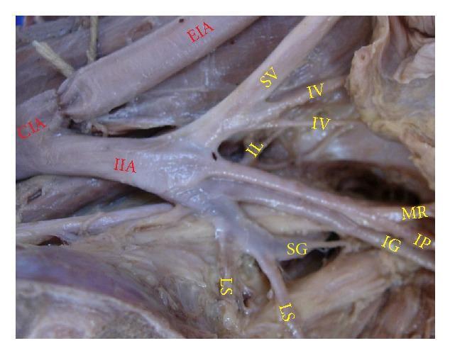
Anatomical dissection photograph of a left pelvic half demonstrating the branching morphology of the internal iliac artery (IIA). The image shows the common iliac artery (CIA) bifurcating into the external iliac artery (EIA) and the IIA. This specific specimen exhibits a direct branching pattern where the IIA terminates into a 'spray' of multiple vessels rather than distinct anterior and posterior trunks. Key visceral and parietal branches are labeled, including the superior vesical (SV), inferior vesical (IV), lateral sacral (LS), and iliolumbar (IL) arteries. Notably, the superior gluteal artery (SG) originates independently from the IIA, while the inferior gluteal artery (IG) and internal pudendal artery (IP) share a common trunk before bifurcating, representing a Type Ia variation. Other visible structures include the middle rectal artery (MR). This visual is highly relevant for medical education in surgical anatomy, particularly for understanding pelvic vascular variations essential for gynecological, urological, and orthopedic procedures.
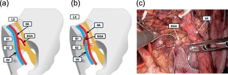
This composite image illustrates the anatomical variations in the spatial relationship between the superior gluteal artery (SGA) and the internal iliac vein (IIV). The image consists of two schematic diagrams (a and b) and one laparoscopic photograph (c). Schematic (a) depicts the SGA (red) running dorsal and lateral to the IIV (blue) as it branches from the internal iliac artery (IIA). Schematic (b) demonstrates an anatomical variation where the SGA runs dorsal but medial to the IIV. In both diagrams, the relationship to the sacral nerve plexus (L5, S1, S2) is shown, with the SGA typically passing between L5 and S1. Panel (c) is a clinical photograph from a right-sided laparoscopic lateral lymph node dissection, confirming the medial SGA variation shown in diagram (b). Key structures are labeled, including the IIA, SGA, and the IIV (marked with an asterisk and dashed white outlines). This material serves as a surgical anatomy reference for identifying neurovascular landmarks in the pelvic cavity.
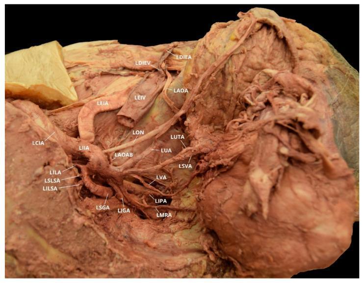
This clinical photograph of a gross anatomical dissection showcases the intricate vascular anatomy of the left female pelvis, with a focus on variations of the obturator artery. The image depicts the left common iliac artery (LCIA) bifurcating into the external (LEIA) and internal (LIIA) iliac arteries. The LIIA is shown branching into its posterior and anterior divisions. Notable posterior branches include the iliolumbar (LILA), superior lateral sacral (LSLSA), inferior lateral sacral (LILSA), and superior gluteal (LSGA) arteries. Anterior division branches visible include the umbilical (LUA), uterine (LUTA), superior vesical (LSVA), vaginal (LVA), middle rectal (LMRA), and internal pudendal (LIPA) arteries. A significant anatomical variation is highlighted: a left aberrant obturator artery (LAOA) originating from the left deep inferior epigastric artery (LDIEA), rather than the internal iliac system. Additionally, a left accessory obturator artery branch (LAOAB) is seen arising from the posterior division of the LIIA. The spatial relationship of these vessels to the left external iliac vein (LEIV) and left obturator nerve (LON) is clearly labeled.
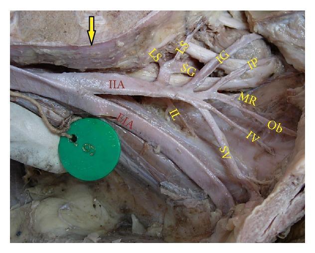
This clinical photograph displays a cadaveric dissection of the right human pelvic wall, focusing on the arterial anatomy. The image demonstrates the bifurcation of the common iliac artery at the level of the lumbosacral joint (indicated by a yellow arrow). The external iliac artery (EIA) is seen continuing toward the lower limb, while the internal iliac artery (IIA) branches extensively to supply pelvic viscera and muscles. Key vascular branches are labeled with yellow text: the superior gluteal (SG), inferior gluteal (IG), and internal pudendal (IP) arteries; the superior vesical (SV), inferior vesical (IV), and middle rectal (MR) arteries; and the obturator (Ob), lateral sacral (LS), and iliolumbar (IL) arteries. This visual serves as an educational anatomical reference for surgical training and radiological correlation, illustrating the complex branching patterns and spatial relationships of the internal iliac arterial system within the pelvic cavity.
Internal Iliac Artery - Branches
POSTERIOR TRUNK (3 branches)
| Branch | Course & Supply |
|---|---|
| Iliolumbar artery | Ascends laterally out of the pelvis. Divides into: (1) lumbar branch - supplies psoas, quadratus lumborum, and cauda equina via a spinal branch through the L5/S1 foramen; (2) iliac branch - passes into the iliac fossa to supply muscle and bone. |
| Lateral sacral arteries (usually 2) | Course medially and inferiorly along the posterior pelvic wall. Send branches into the anterior sacral foramina supplying bone, soft tissue, sacral canal structures, and overlying skin/muscle. |
| Superior gluteal artery | Largest branch of the IIA and terminal continuation of the posterior trunk. Passes posteriorly between the lumbosacral trunk and S1 anterior ramus, exits through the greater sciatic foramen above piriformis (suprapiriform foramen). Supplies gluteal muscles and skin via superficial and deep branches. |
ANTERIOR TRUNK (8-9 branches)
| Branch | Course & Supply |
|---|---|
| Umbilical artery | First branch; gives off the superior vesical artery and courses to the umbilicus. After birth, the distal part obliterates to form the medial umbilical ligament. |
| Superior vesical artery | Arises from the root of the umbilical artery; supplies the superior bladder and distal ureter. In males, may also supply the ductus deferens. |
| Inferior vesical artery (males) / Vaginal artery (females) | In males: supplies the bladder base, ureter, seminal vesicle, and prostate. In females: the vaginal artery (may occur as 2-3 vessels) replaces it, supplying the vagina. |
| Uterine artery (females) | Corresponds to the artery to the ductus deferens in males. Supplies the uterus with branches to the vagina, ovary, and uterine tube. |
| Middle rectal artery | Variable vessel; runs along the pelvic floor to supply the rectal musculature. |
| Obturator artery | Passes anteriorly along the lateral pelvic wall, exits through the obturator canal. Gives a pubic branch (anastomoses with the inferior epigastric artery - clinically important "corona mortis"), an acetabular branch (through the ligament of the head of femur), and a posterior branch (deep hip muscles). |
| Internal pudendal artery | Exits through the greater sciatic foramen below piriformis (infrapiriform foramen), hooks around the ischial spine, re-enters the perineum via the lesser sciatic foramen into the pudendal (Alcock's) canal. Supplies perineal structures including the penis/clitoris, scrotum/labia, external anal sphincter, and external urethral sphincter. |
| Inferior gluteal artery | Exits through the greater sciatic foramen below piriformis (infrapiriform foramen). Supplies surrounding gluteal muscles and gives rise to the artery to the sciatic nerve (accompanies the sciatic nerve). Phylogenetically it was the principal artery of the lower limb. |
Memory Aid
"I love sex, so I give Victoria my undying passion internally" (Inferior vesical, lateral sacral, Superior vesical, Inferior gluteal, internal pudendal, Vaginal/vesical, Middle rectal, Umbilical, Deferent artery, Pudendal, Internal gluteal)
- Posterior trunk = ILS - Iliolumbar, Lateral sacral, Superior gluteal
- Anterior trunk = visceral branches + obturator + internal pudendal + inferior gluteal
Cadaveric Dissection Images
Clinical Relevance
- IIA ligation is performed to control pelvic hemorrhage (e.g., post-partum bleeding). It reduces pulse pressure rather than completely cutting off blood supply, since collateral anastomoses re-establish flow.
- The corona mortis (anastomosis between the obturator artery's pubic branch and the inferior epigastric artery) is at risk during retropubic/inguinal surgery.
- Uterine artery embolization targets the anterior trunk branches for fibroids/adenomyosis.
- The superior gluteal artery must be identified during total hip replacement, as it is vulnerable during posterior approaches.