
Essay (25 marks) 1. AUB- causes, classification, investigation, management 2. CA cervix Short notes (5 marks) 1. Physiology of ovulation and menstruation 2. MTP 3. Bartholin gland 4. Histology of endometrium 5. Urogenital diaphragm
carcinoma cervix staging invasive squamous cell
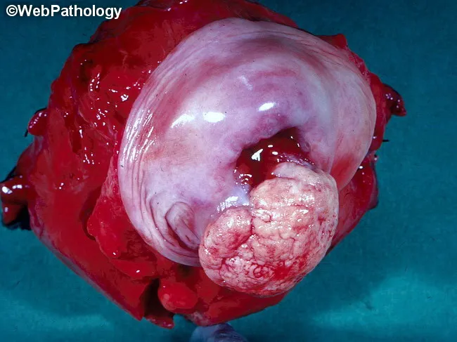
Gross pathology photograph of a cervical tissue specimen showing invasive squamous cell carcinoma of the cervix. The cervix is enlarged with a bulky, exophytic mass that presents a pink-tan, friable surface and irregular contour. Surface necrosis and hemorrhage are evident, and the lesion distorts the transformation zone with partial obstruction of the endocervical canal. The surrounding cervical stroma shows invasion, and the lesion disrupts normal architecture of the cervix; parametrial involvement is not definitively demonstrated in this single view. This macroscopic appearance is typical of HPV-related cervical squamous carcinoma, although HPV-independent cases exist. HPV infection, most commonly HPV16 and HPV18, drives oncogenic E6 and E7 oncoproteins that inactivate p53 and Rb, promoting cellular proliferation and malignant transformation. Gross features correlate with histopathology, where nests of atypical squamous cells with keratinization and intercellular bridges would be confirmed on microscopic examination. Clinically, this image underscores the importance of HPV testing, histopathologic confirmation, and accurate FIGO staging to guide treatment planning, which may include radical hysterectomy, radiotherapy, and lymphadenectomy. This photograph is valuable for education, surgical pathology review, and study of cervical cancer gross morphology in HPV-related disease. Useful for teaching and exams.
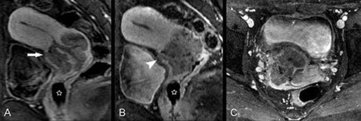
Diagnostic images (MRI) showing squamous cell carcinoma of the cervix in a 28-year-old patient. The series includes a directly acquired contrast-enhanced sagittal image (A), a sagittal reconstruction from an isotropic sequence (B), and an axial isotropic image (C). A large, heterogeneously low-signal tumor is centered in the cervix. In the sagittal views (A and B), the tumor clearly interrupts the hypointense anterior cervical stroma (indicated by arrow and arrowhead), suggesting local invasion towards the bladder. The isotropic reconstructions (B and C) provide enhanced visualization of the stromal breach and tumor margins compared to conventional sequences. An OB tampon is noted as a well-defined, low-signal void within the vaginal canal (marked with a star). These images demonstrate clinical staging features of cervical cancer, specifically the assessment of cervical stromal integrity and potential parametrial or vaginal extension.
endometrium histology proliferative secretory phase
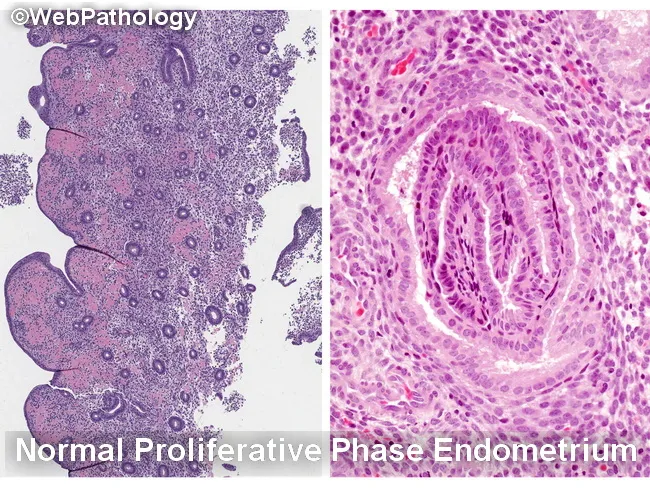
Histology image illustrating the endometrium in two complementary views. The left panel depicts normal proliferative phase endometrium with relatively sparse glands embedded in a cellular stroma, producing a low gland-to-stroma ratio and open gland spacing. The right panel shows characteristic epithelial morphology from proliferative endometrium with elongated, back-to-back glands and prominent, basally oriented nuclei at high power. The specimen emphasizes normal architectural patterns and avoids overcalling hyperplasia when glands are well separated or when fragmentation and telescoping create artifactual crowding. Key differential considerations include endometrial hyperplasia (EH) with or without atypia, disordered proliferative endometrium, reparative or regenerative changes, and endocervical/metaplastic proliferations. Within EH, nonatypical EH must be distinguished from atypical endometrial hyperplasia/endometrioid intraepithelial neoplasia (AEH/EIN), and AEH/EIN must be differentiated from endometrioid carcinoma and from endocervical lesions. Normal proliferative glands show minimal cytologic atypia, whereas AEH/EIN demonstrates architectural complexity (crumpled, crowded glands) and cytologic atypia. Artifacts such as glandular fragmentation, fragmentation- or telescoping-related crowding, and sample artifact can mimic AEH/EIN if the specimen is not intact. This image supports educational differentiation for gynecologic pathology, endometrial biopsy interpretation, and clinical correlation with bleeding patterns and imaging findings. Keywords: endometrium, proliferative phase, hyperplasia, AEH, EIN, histology, biopsy, gland-stroma ratio, artifacts, differential diagnosis teaching.
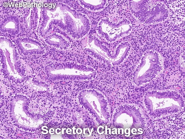
Endometrial histology in secretory phase demonstrated by light microscopy on a hematoxylin and eosin stained endometrial biopsy specimen. Glandular architecture shows irregular serration with gland crowding in focal areas, while glands are lined by tall, columnar secretory epithelium. Nuclei are enlarged and may display mild pleomorphism and loss of polarity, consistent with secretory transformation. The background stroma is edematous and commonly predecidualized, with cytoplasmic vacuolization of stromal cells. Together, these findings reflect physiologic secretory changes rather than overt endometrial hyperplasia. Notably, secretory endometrium can mimic endometrial hyperplasia/endometrioid intraepithelial neoplasia (EIN) in some cases, but the absence of marked architectural crowding, complex glandular patterns, or cytologic atypia argues against a neoplastic process. Differential considerations include secretory change, simple endometrial hyperplasia without atypia, and EIN in a background of secretory endometrium; careful architectural assessment and clinical correlation are essential. Diagnostic significance lies in distinguishing benign secretory transformation from premalignant or malignant entities to avoid overtreatment. This image is educational for pathology training, differential diagnosis exercises, and research on endometrial cycle pathology. Correlate with patient menstrual history, pregnancy status, and cycle timing; repeat sampling or adjunct studies may be warranted when discordance is suspected. Interpret interpretation should integrate histology with clinical data and follow-up.
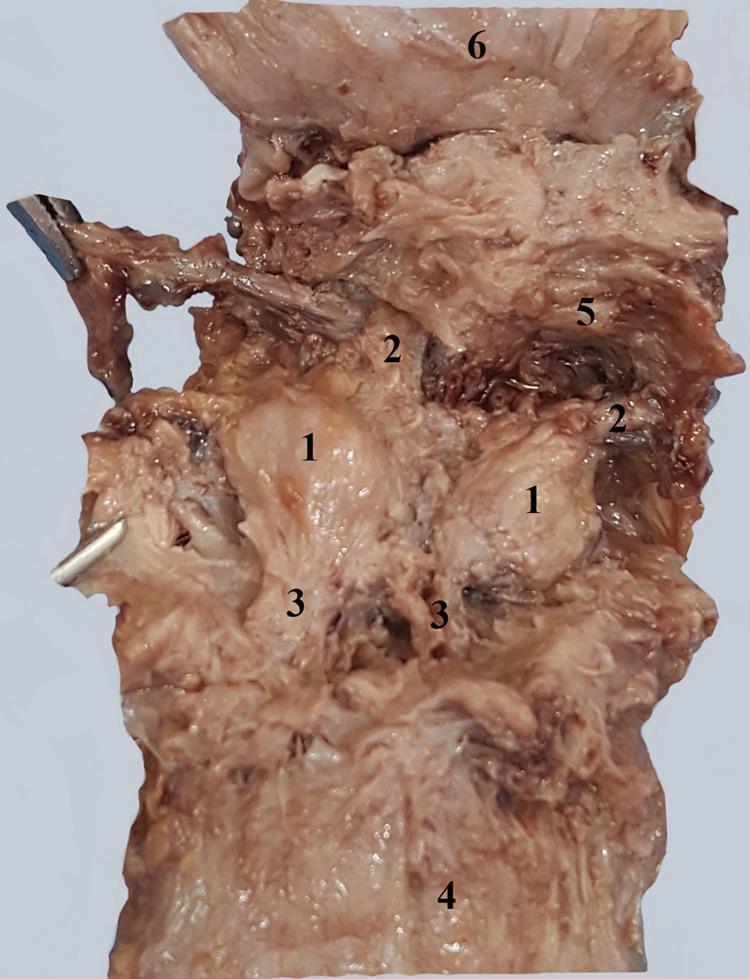
ESSAY ANSWERS
ESSAY 1: Abnormal Uterine Bleeding (AUB) — 25 Marks
Definition
Classification: The PALM-COEIN System (FIGO, 2011)
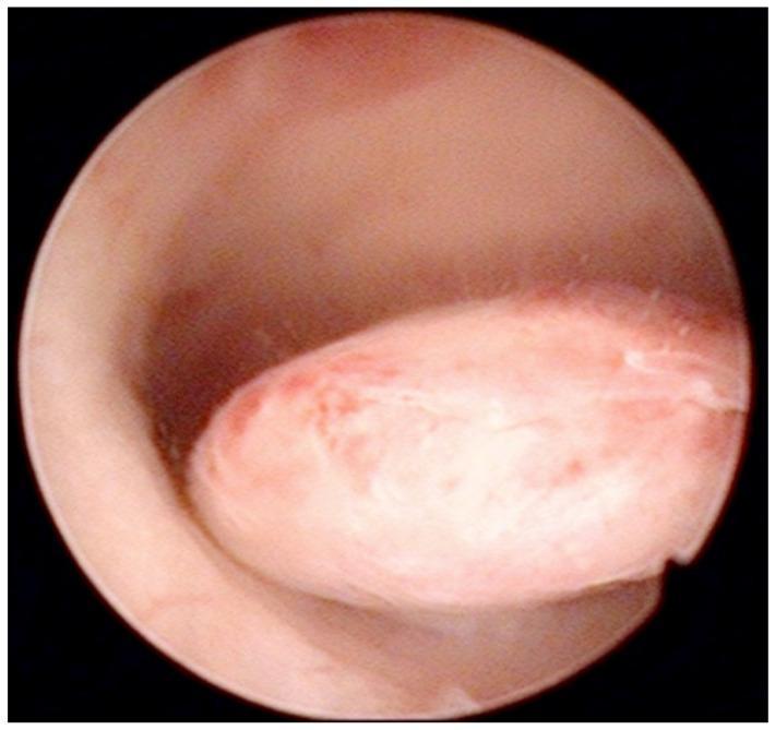
| Letter | Cause |
|---|---|
| P | Polyp (AUB-P) |
| A | Adenomyosis (AUB-A) |
| L | Leiomyoma (AUB-L) - subclassified: submucosal (AUB-LSM) vs other (AUB-LO) |
| M | Malignancy and hyperplasia (AUB-M) |
| Letter | Cause |
|---|---|
| C | Coagulopathy (AUB-C) - e.g., von Willebrand disease, ITP |
| O | Ovulatory dysfunction (AUB-O) - most common cause; anovulation |
| E | Endometrial causes (AUB-E) - primary endometrial disorder |
| I | Iatrogenic (AUB-I) - OCP, anticoagulants, IUCDs |
| N | Not yet classified (AUB-N) |
Causes
| Age Group | Common Causes |
|---|---|
| Prepuberty | Precocious puberty (hypothalamic, pituitary, or ovarian origin) |
| Adolescence | Anovulatory cycles, coagulation disorders (von Willebrand disease) |
| Reproductive age | Pregnancy complications (abortion, ectopic, trophoblastic disease), PCOS, fibroids, polyps, coagulopathy, thyroid disease, OCP use |
| Perimenopausal | Ovarian failure, anovulation, endometrial hyperplasia |
| Postmenopausal | Endometrial atrophy (most common), endometrial carcinoma, polyps, exogenous estrogen |
- The most common mechanism is estrogen excess unopposed by progesterone
- Endometrium proliferates, becomes thick and unstable, then sheds irregularly
- Common at menarche (HPO axis immaturity) and perimenopause (incipient ovarian failure)
- Also caused by: pituitary prolactinoma (reduces GnRH/LH/FSH), polycystic ovarian syndrome, granulosa cell tumors, obesity, malnutrition, chronic systemic illness
Investigations
- Detailed menstrual history (pattern, duration, amount, clots)
- Prospective bleeding diary
- Signs of anemia, thyroid disease, PCOS, coagulopathy
- Pelvic examination + Pap smear
- Complete blood count + platelet count
- Coagulation studies (PT, APTT, bleeding time) - especially if heavy bleeding since menarche, family history of coagulopathy
- Von Willebrand factor assay
- Thyroid function tests (hypothyroidism is a common cause)
- Serum prolactin
- Fasting blood glucose
- Beta-hCG (rule out pregnancy)
- Cervical cancer screening if not up to date
- STI screening
- Pelvic ultrasound: first-line - evaluates endometrial thickness, fibroids, polyps, ovarian pathology
- Saline infusion sonohysterography: better detection of intracavitary lesions
- MRI pelvis: best for adenomyosis, fibroid mapping
- All women ≥ 45 years with AUB
- Women < 45 years with: unopposed estrogen exposure (obesity, PCOS), persistent/refractory AUB, elevated familial cancer risk (Lynch syndrome)
- All postmenopausal women with bleeding
- Gold standard for intracavitary pathology
- Note: D&C alone is NOT an effective treatment for AUB
Management
- Combined oral contraceptive pills (OCP): First-line for most reproductive-age women with anovulatory bleeding; regulate cycle and reduce blood loss
- Progestins: Medroxyprogesterone acetate (MPA) 5-10 mg orally for 10-14 days per cycle; for those in whom estrogen is contraindicated
- Acute profuse bleeding: Conjugated estrogens IV 25 mg every 4 hours (up to 3 doses) to stabilize the endometrium, followed simultaneously by progestin (MPA 5-10 mg); after withdrawal bleed, start OCP for at least 3 cycles
- NSAIDs (e.g., mefenamic acid, tranexamic acid): reduce prostaglandin-mediated bleeding by 20-50%
- Tranexamic acid: antifibrinolytic, effective for heavy menstrual bleeding
- Levonorgestrel-releasing IUCD (Mirena): highly effective for menorrhagia, also useful in PCOS
- GnRH agonists (leuprolide): for fibroids - induce temporary amenorrhea, pre-surgical use
- Newer oral GnRH antagonists: relugolix 40 mg daily, elagolix 300 mg twice daily for fibroids
- Endometrial ablation: Destroys endometrium; options include thermal balloon, rollerball, NovaSure; ~70-80% success; NOT 100% effective (29% eventually require hysterectomy at 5 years)
- Hysteroscopic polypectomy / myomectomy: For polyps and submucous fibroids
- Uterine artery embolization (UAE): Minimally invasive, for uterine fibroids; ~31% require hysterectomy at follow-up
- Hysterectomy: Definitive treatment; reserved for refractory cases, failed medical therapy, malignancy; should not be done without prior endometrial sampling
ESSAY 2: Carcinoma of the Cervix (CA Cervix) — 25 Marks
Introduction/Epidemiology
Etiology and Risk Factors
- HPV E6 oncoprotein inactivates p53 (pro-apoptotic tumor suppressor)
- HPV E7 oncoprotein inactivates Rb (cell cycle checkpoint)
- Together, they promote uncontrolled proliferation and malignant transformation
- Early age of first intercourse
- Multiple sexual partners
- High-risk male partner
- Immunosuppression (HIV infection - 5x risk)
- Smoking (synergistic with HPV)
- Long-term OCP use
- High parity
- Lower socioeconomic status
- Lack of Pap smear screening
- Previous history of STIs (HSV, Chlamydia as cofactors)
Pathogenesis and Precursor Lesions
- CIN 1 (LSIL): Mild dysplasia, involves lower 1/3 of epithelium; may spontaneously regress (60%)
- CIN 2 (HSIL): Moderate dysplasia, involves lower 2/3 of epithelium
- CIN 3 (HSIL/CIS): Severe dysplasia/carcinoma in situ; full-thickness dysplasia
Histological Types (Robbins & Kumar):
| Type | Frequency |
|---|---|
| Squamous cell carcinoma | 80% |
| Adenocarcinoma + mixed adenosquamous | 15% |
| Small cell neuroendocrine carcinoma | <5% |
Morphology
- Develops in the transformation zone
- Macroscopically: exophytic or endophytic (infiltrating) growth
- Microscopically: tongues/nests of squamous cells with desmoplastic stromal response
- Well-differentiated tumors form keratin pearls
- Barrel cervix: tumor encircling and infiltrating cervical stroma
- Small cell neuroendocrine carcinoma resembles small cell lung carcinoma morphologically - very poor prognosis
- Risk < 1% for tumors < 3 mm invasion
- Risk > 10% when invasion exceeds 3 mm
- Spread to: parametrium → pelvic side wall; pelvic lymph nodes; bladder/rectum in advanced disease; distant metastases (lung, liver, bone)
Clinical Features
- Often asymptomatic in early stages (detected on screening)
- Abnormal vaginal bleeding - especially post-coital or intermenstrual
- Watery, blood-stained or offensive vaginal discharge (leukorrhea)
- Dyspareunia (painful coitus)
- Dysuria, hematuria (bladder involvement)
- Rectal bleeding, constipation (rectal involvement)
- Pelvic or back pain (parametrial/nerve involvement)
- Lower limb edema (lymphatic obstruction)
Investigations
- Pap (Papanicolaou) smear: cytology-based; detects SIL
- HPV DNA testing: more sensitive than Pap alone
- Co-testing (Pap + HPV DNA): recommended in women ≥ 30 years
- Colposcopy with directed biopsy: for abnormal Pap smear / visible lesion
- LEEP (Loop Electrosurgical Excision Procedure) or cone biopsy: for CIN 2/3
- Cervical biopsy + endocervical curettage: histological confirmation
- Pelvic examination under anesthesia
- Chest X-ray
- Intravenous urography (IVU)
- Cystoscopy and proctoscopy (for advanced disease)
- MRI pelvis: best modality for local staging, parametrial invasion
- CT chest/abdomen/pelvis: lymph node and distant metastasis assessment
- PET-CT: most accurate for lymph node involvement
FIGO Staging (2018):
| Stage | Description |
|---|---|
| I | Confined to cervix |
| IA | Microscopic; IA1: stromal invasion ≤3 mm; IA2: 3-5 mm |
| IB | Visible lesion; IB1 <2 cm; IB2 2-4 cm; IB3 ≥4 cm |
| II | Beyond cervix but not pelvic wall/lower 1/3 vagina |
| IIA | No parametrial involvement; IIA1 <4 cm; IIA2 ≥4 cm |
| IIB | Parametrial involvement |
| III | To pelvic wall / lower vagina / hydronephrosis |
| IIIA | Lower 1/3 vagina |
| IIIB | Pelvic wall or hydronephrosis |
| IIIC | Lymph node metastasis |
| IV | Bladder/rectal mucosa (IVA) or distant (IVB) |
Management
- External beam radiotherapy (EBRT) + brachytherapy + weekly cisplatin Stage IVB (distant metastasis): Palliative chemotherapy; pembrolizumab (immunotherapy) for PD-L1 positive tumors
- HPV vaccine (Gardasil 9): most effective when given before sexual debut; covers HPV 6, 11, 16, 18, 31, 33, 45, 52, 58
- Regular Pap smear screening
- Safe sex practices
SHORT NOTES (5 Marks Each)
SHORT NOTE 1: Physiology of Ovulation and Menstruation
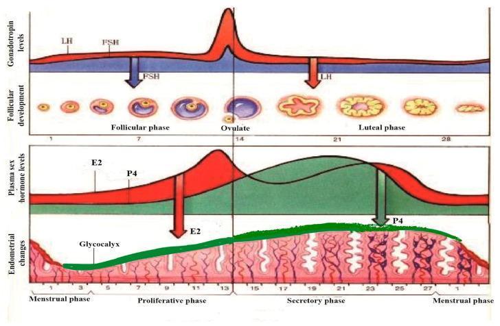
Phases of the Menstrual Cycle (Costanzo Physiology 7th Edition):
- FSH and LH secreted by anterior pituitary stimulate ovarian follicle development
- A dominant (Graafian) follicle is selected; other follicles undergo atresia
- Dominant follicle secretes increasing amounts of 17β-estradiol
- Estradiol causes endometrial proliferation (glands and stroma grow)
- Rising estradiol initially exerts negative feedback on GnRH/FSH/LH
- A burst (surge) of estradiol at the end of the follicular phase switches to positive feedback on the hypothalamus and anterior pituitary
- This triggers the LH surge (and a smaller FSH surge)
- The LH surge causes rupture of the Graafian follicle and release of the mature oocyte (ovulation)
- At ovulation, cervical mucus becomes abundant, watery, and "fern-like" (favorable for sperm penetration)
- Regardless of cycle length, ovulation occurs 14 days BEFORE menses (the variable part is the follicular phase)
- The ruptured follicle transforms into the corpus luteum
- Corpus luteum secretes both estradiol and progesterone
- Progesterone: converts endometrium from proliferative to secretory phase (tortuous glands, glycogen-rich cells, increased vascularity); prepares uterus to receive fertilized ovum
- Progesterone raises basal body temperature (basis of rhythm method)
- Cervical mucus becomes thick and less penetrable (hostile to sperm)
- If fertilization does not occur, corpus luteum regresses at day 25-26 (luteolysis)
- Estradiol and progesterone levels fall abruptly
- Abrupt withdrawal of estradiol and progesterone causes vasoconstriction of spiral arteries (due to prostaglandins - mainly PGF2α)
- Ischemic necrosis of the endometrium occurs
- The functionalis layer is shed as menstrual blood, mucus, and tissue
- Normally lasts 4-5 days with blood loss of 20-80 mL
- Primordial follicles for the next cycle are simultaneously recruited
SHORT NOTE 2: MTP (Medical Termination of Pregnancy)
Introduction
Grounds for Termination (Section 3):
- Risk to the life of the pregnant woman
- Grave physical or mental injury to the woman
- Substantial risk of fetal abnormality (incompatible with life or serious morbidity)
- Pregnancy due to rape or incest (mental trauma constitutes serious injury)
- Failure of contraception - in married or unmarried women (per 2021 Amendment)
Permissible Time Limits:
| Gestational Age | Conditions |
|---|---|
| Up to 20 weeks | Opinion of 1 RMP; based on above grounds |
| 20-24 weeks | Opinion of 2 RMPs; only for special categories: rape survivors, minors, women with disabilities, multiple pregnancies, contraceptive failure |
| Beyond 24 weeks | Medical Board approval only; for severe fetal anomalies detected at any gestational age |
Medical Board (Post 2021 Amendment):
Consent:
- Adult woman (≥18 years): Only her own written consent required
- Minor (<18 years) or mentally ill woman: Guardian consent required
- Husband's consent: NOT required legally
Approved Places (Section 4):
- Government hospitals
- Private clinics/hospitals approved by the government
- Must have facilities for: aseptic surgery, emergency resuscitation, blood transfusion, post-procedure care
Key Features of 2021 Amendment:
- Upper limit extended from 20 to 24 weeks for special categories
- Unmarried women included in contraceptive failure clause
- No upper gestational limit for severe fetal anomalies (with Medical Board approval)
- Privacy clause: No RMP shall reveal the identity of the woman - violation is punishable
Methods of MTP by Gestational Age:
- Up to 7 weeks: Medical abortion - Mifepristone 200 mg + Misoprostol 800 mcg (WHO protocol)
- 7-12 weeks: Suction evacuation (MVA - Manual Vacuum Aspiration) or EVA
- 12-20 weeks: Dilation and evacuation (D&E) or prostaglandin (misoprostol/gemeprost) induction
- >20 weeks (Medical Board approved): Labor induction with prostaglandins
SHORT NOTE 3: Bartholin Gland
Anatomy
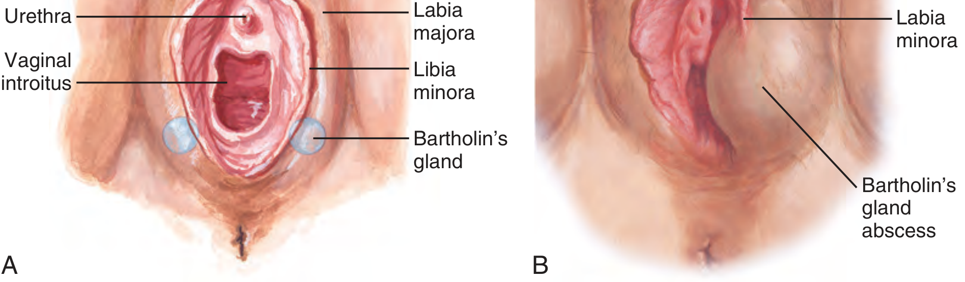
- Bartholin glands (Greater vestibular glands) are a pair of pea-sized mucous glands located at the 5 o'clock and 7 o'clock positions at the posterior part of the vestibule (vaginal introitus)
- They are the female homologue of the bulbourethral (Cowper's) glands in males
- Each gland has a duct approximately 2 cm long that opens at the inner surface of the labia minora, just outside the hymenal ring
- Their secretion lubricates the vaginal introitus, especially during sexual arousal
Histology
- Alveolar glands lined by columnar mucous-secreting epithelium
- Ducts lined by transitional epithelium proximally and squamous epithelium near the opening
Clinical Conditions:
- Formed when the duct becomes obstructed (often following infection, trauma, or mucous inspissation)
- Usually painless unless infected
- Presents as a smooth, cystic swelling in the posterior labium majus
- Treatment: Marsupialization (preferred) or Word catheter placement
- Infection of the cyst (most commonly by polymicrobial organisms; Staphylococcus, E. coli, Bacteroides; also Neisseria gonorrhoeae in younger women)
- Presents as a tender, fluctuant swelling at 5 or 7 o'clock with pain, dyspareunia, difficulty walking
- Treatment: Incision and drainage (made on the MUCOSAL surface, not the skin) + Word catheter for 2-4 weeks; antibiotics if systemic features
- Marsupialization: Definitive treatment to prevent recurrence
- Rare; accounts for 2-3% of all vulvar carcinomas
- Types: Squamous cell carcinoma (most common), adenocarcinoma, transitional cell carcinoma, adenoid cystic carcinoma
- Diagnosis requires histological confirmation
- Treatment: Wide local excision ± radical vulvectomy + inguinal lymph node dissection + radiation
SHORT NOTE 4: Histology of the Endometrium
Layers of the Endometrium:
- The superficial 2/3 of the endometrium
- This layer is shed at menstruation and regenerated each cycle
- Divided into: Stratum compactum (surface) and Stratum spongiosum (middle)
- The deep 1/3, adjacent to the myometrium
- NOT shed during menstruation
- Contains gland bases and acts as a stem cell reservoir for regeneration
Histological Phases:
- Glands: Straight, narrow, tubular; lined by pseudostratified columnar epithelium; frequent mitotic figures
- Stroma: Compact, spindle-shaped cells; abundant mitoses
- Surface epithelium: Low columnar
- Spiral arteries: Developing but not prominent
- Gland-to-stroma ratio is low; glands evenly distributed
- Early secretory (Days 14-17): Sub-nuclear glycogen vacuoles appear in glandular epithelium - earliest reliable sign of ovulation (seen 36-48 hours after ovulation)
- Mid-secretory (Days 18-24): Glands become tortuous (saw-toothed), luminal secretions prominent; stroma edematous
- Late secretory (Days 25-28): Pre-decidual change in stroma (stromal cells enlarge, become round with abundant pale cytoplasm - predecidual reaction around spiral arteries); spiral arteries become prominent and coiled
- Stromal collapse and breakdown
- Nuclear dust and red blood cell infiltration
- Glandular and stromal fragmentation
- Regeneration begins from the basalis
- Thin, inactive endometrium
- Small, cystically dilated glands lined by flat/low cuboidal epithelium with little mitotic activity
- Sparse stroma
SHORT NOTE 5: Urogenital Diaphragm
Definition
Boundaries:
- Anteriorly / apex: Pubic symphysis
- Laterally: Ischiopubic rami (right and left)
- Posteriorly: A line joining the two ischial tuberosities (the transverse perineal ligament marks the posterior edge)
Structures:
| Muscle | Function |
|---|---|
| Deep transverse perineal muscle | Stabilizes the perineal body; supports pelvic floor |
| Sphincter urethrae (External urethral sphincter) | Voluntary control of micturition |
- Sphincter urethrovaginalis
- Compressor urethrae
- Inferior fascia (perineal membrane): The strongest layer; thick triangular membrane closing the urogenital triangle below. A key pelvic support structure
- Superior fascia: Thinner layer above the muscles
- Together they form a fascial "sandwich" enclosing the deep perineal space (pouch)
- Urethra (in both sexes)
- Vagina (in females)
- The perineal body is attached posteriorly
Contents of the Deep Perineal Pouch (Space):
- Deep transverse perineal muscle
- External urethral sphincter
- Internal pudendal vessels and branches
- Dorsal nerve of clitoris/penis
- Bulbourethral (Cowper's) glands in males
Clinical Significance:
- Pelvic floor support: The UGD, along with the pelvic diaphragm (levator ani + coccygeus), supports pelvic organs against increases in intra-abdominal pressure
- Urinary continence: The external urethral sphincter is the primary voluntary mechanism of continence
- Obstetric injuries: Childbirth can damage the perineal body and UGD, contributing to stress urinary incontinence and pelvic organ prolapse
- Surgical relevance: Knowledge of the UGD anatomy is essential for perineal surgeries, repair of fourth-degree perineal tears, and anti-incontinence procedures
Sources: Sabiston Textbook of Surgery; Robbins & Kumar Basic Pathology; Goldman-Cecil Medicine; Berek & Novak's Gynecology; Costanzo Physiology 7th Edition; The Essentials of Forensic Medicine and Toxicology (36th ed); Roberts & Hedges' Clinical Procedures in Emergency Medicine; Gray's Anatomy for Students