
Give me a 10 marks answer on focus assessed trans thoracic echocardiography(FATE) from standard textbooks for AIIMS md anaesthesia exams
Reading File
Now let me search the medical library for FATE content in parallel.
Finding Sources
Finding Sources
Reading File
Finding Sources
Good - found content in Miller's Anesthesia and Morgan & Mikhail. Let me read all the relevant sections in parallel.
Reading File
Reading File
Excellent content found. Let me continue reading the Miller's section and also search for more FATE-specific content.
Reading File
Finding Sources
Reading File
Finding Sources
Finding Sources
Reading File
I found the dedicated FATE section in Morgan & Mikhail. Let me read more of it and also fetch the key images.
Reading File
Loading Image
Reading File
Loading Image
I now have comprehensive content from both Morgan & Mikhail's Clinical Anesthesiology (7e) and Miller's Anesthesia (10e). Let me fetch a couple more key images and compile the answer.
Loading Image
Loading Image
I now have all the material needed to write a comprehensive, exam-ready 10-mark answer. Let me compose it.
Focus Assessed Transthoracic Echocardiography (FATE)
Sources: Morgan & Mikhail's Clinical Anesthesiology, 7e (Ch. 5); Miller's Anesthesia, 10e (Ch. 33)
Definition and Concept
FATE (Focus-Assessed Transthoracic Echocardiography) is a bedside, limited-scope, point-of-care TTE protocol designed to rapidly assist in hemodynamic diagnosis in critically ill or perioperative patients. It is goal-directed, problem-oriented, time-sensitive, and uses qualitative or semiquantitative assessment rather than formal comprehensive measurements. FATE is performed by the treating clinician (e.g., anaesthesiologist, intensivist) and is intended to answer specific clinical questions - not to replace a formal echocardiographic examination.
(Morgan & Mikhail's Clinical Anesthesiology, 7e, p. 201)
Rationale and Indications
TTE has the advantage of being completely noninvasive and essentially risk-free. Bedside TTE exams such as FATE can readily assist in diagnosing the source of haemodynamic instability when a patient is unexpectedly hypotensive or haemodynamically compromised.
Key clinical questions FATE addresses:
- Is the heart adequately filled? (preload/hypovolaemia)
- Is the myocardium contracting adequately? (systolic function)
- Is there external cardiac compression? (tamponade)
- Are there grossly obvious structural defects?
- Is there significant pleural pathology?
(Morgan & Mikhail's Clinical Anesthesiology, 7e, p. 201)
The Four FATE Scanning Positions
The FATE examination involves scanning through four positions in the most favourable sequence:
Fig. 5-27: The FATE examination - four probe positions on the chest
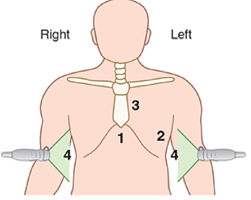
(Morgan & Mikhail's Clinical Anesthesiology, 7e, Fig. 5-27)
| Position | View | Structures Assessed |
|---|---|---|
| Pos 1 | Subcostal 4-chamber | All four chambers, pericardial space, IVC |
| Pos 2 | Apical 4-chamber | All four chambers, mitral & tricuspid valves, relative sizes |
| Pos 3 | Parasternal long axis + short axis | LV, RV, aortic root, LA, mitral valve; LV function and wall motion |
| Pos 4 | Pleural scanning (bilateral) | Pleural effusion, pneumothorax |
Subcostal 4-chamber (Position 1): Probe placed subxiphoid, indicator pointing toward patient's left. Visualises all four chambers and the pericardium. Excellent for detecting pericardial effusion and comparing right vs left chamber sizes.
Apical 4-chamber (Position 2): Probe at the cardiac apex, indicator pointing to patient's right. All four chambers and AV valves seen. Best view for comparing LV/RV size ratio and assessing wall motion.
Parasternal long axis (Position 3): Probe at the left parasternal border (2nd-4th intercostal space), indicator pointing to patient's right shoulder. Shows LV, aortic root, LA, mitral valve, and descending aorta. The short axis view (probe rotated 90°) reveals a cross-section of LV at the papillary muscle level - useful for regional wall motion and LV systolic function.
Pleural scanning (Position 4): Bilateral lateral chest scan to identify pleural effusion or pneumothorax.
Normal Apical Four-Chamber View
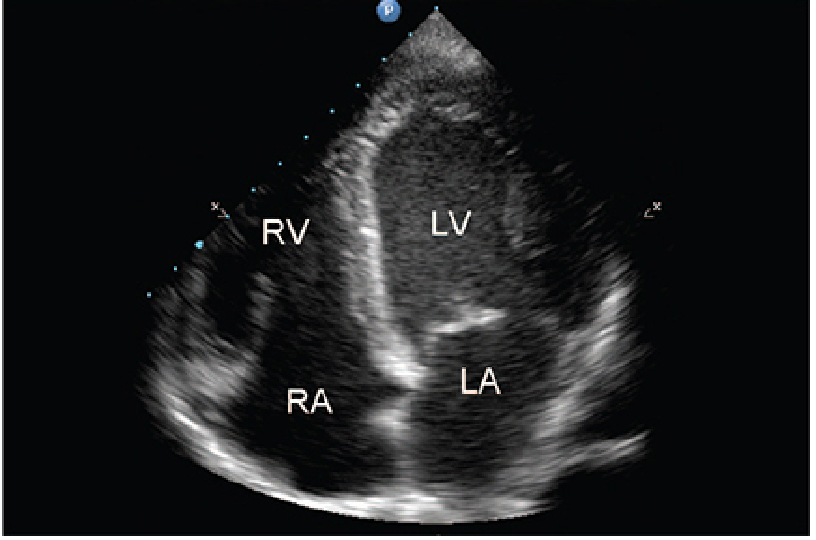
Normal apical four-chamber view. LV = left ventricle, RV = right ventricle, LA = left atrium, RA = right atrium. (Morgan & Mikhail's Clinical Anesthesiology, 7e, Fig. 5-26)
Pathological Patterns Identified by FATE
The FATE protocol enables recognition of important pathologies by pattern recognition:
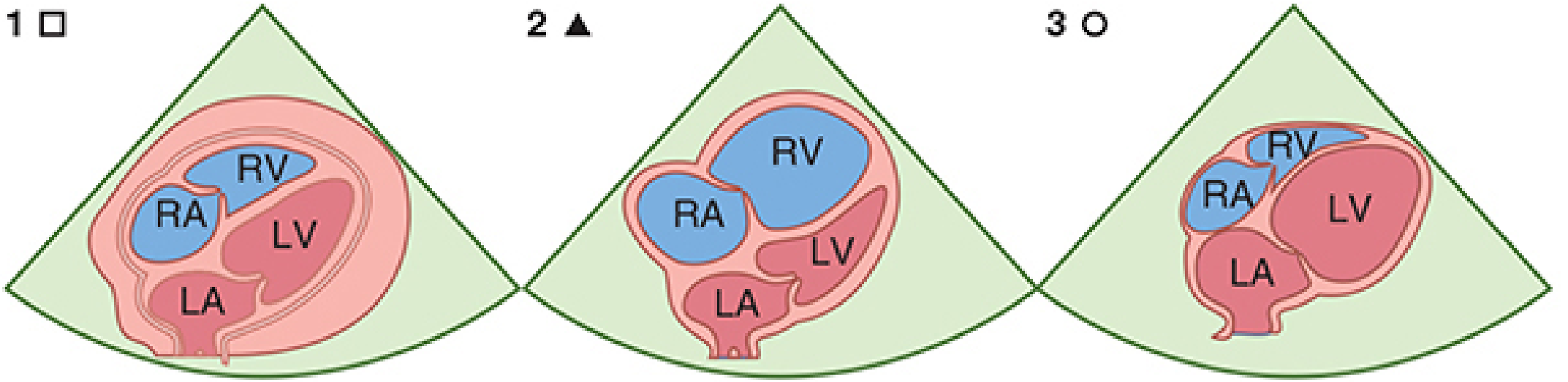
Fig. 5-28: Important pathological conditions identified with FATE - pericardial effusion (pattern 1/square), dilated RA+RV (pattern 2/triangle), dilated LA+LV (pattern 3/circle). (Morgan & Mikhail's Clinical Anesthesiology, 7e)
1. Pericardial Effusion / Cardiac Tamponade
- Echo-free space surrounding the heart visible in positions 1, 2, and 3
- Clinical context: post-cardiac surgery, post-catheterisation, trauma, uraemia, infection
- Causes haemodynamic compromise by external cardiac compression
2. Dilated Right Atrium + Right Ventricle (RV Dominance)
- RV appears enlarged relative to LV (normally LV > RV)
- RV:LV ratio >1 in apical 4-chamber view suggests acute cor pulmonale
- Clinical context: pulmonary embolus, RV infarction, pulmonary hypertension, volume overload
3. Dilated Left Atrium + Left Ventricle (LV Failure)
- LV appears globally dilated and poorly contracting
- Clinical context: ischaemic heart disease, dilated cardiomyopathy, septic cardiomyopathy, volume overload, aortic insufficiency
4. LV Hypertrophy with Dilated LA
- Thick LV walls, small cavity, dilated LA
- Clinical context: aortic stenosis, arterial hypertension, LV outflow tract obstruction (e.g., HOCM), hypertrophic cardiomyopathy, myocardial deposit diseases (amyloid)
5. Hypovolaemia
- "Kissing walls" phenomenon - near-complete LV cavity obliteration at end-systole on parasternal short axis
- IVC diameter <2 cm with >50% inspiratory collapse suggests low CVP/preload
6. Pleural Pathology (Position 4)
- Pleural effusion: anechoic space between lung and chest wall
- Pneumothorax: absence of lung sliding
(Morgan & Mikhail's Clinical Anesthesiology, 7e, p. 203-204)
Features of Focused Cardiac Ultrasound (FoCUS / FATE)
As defined by the American Society of Echocardiography (Via et al., 2014) and summarised in Miller's Anesthesia, Box 33.2:
| Feature | Description |
|---|---|
| Goal-directed | Answers a specific haemodynamic question |
| Problem-oriented | Triggered by a clinical problem |
| Limited in scope | Does not replace comprehensive echo |
| Simplified | Uses qualitative/semiquantitative assessment |
| Time-sensitive | Performed rapidly at the bedside |
| Repeatable | Can be done serially to track response |
| Point-of-care | Done by the treating clinician |
(Miller's Anesthesia, 10e, Box 33.2, p. 4981)
FATE vs Comprehensive TTE
| Parameter | FATE / FoCUS | Comprehensive TTE |
|---|---|---|
| Views | 4-5 key views | 12+ views (parasternal, apical, subcostal, suprasternal) |
| Performed by | Treating clinician | Dedicated echocardiographer |
| Purpose | Haemodynamic diagnosis | Formal structural/functional assessment |
| Quantification | Qualitative / semiquantitative | Full quantitative measurements |
| Time | Minutes | 30-60 minutes |
| Certification | Basic level | Advanced (ASE/NBE/EchoScot) |
If FATE does not reveal pathology but there is clinical suspicion of cardiac disease, formal comprehensive echocardiography must be pursued.
(Miller's Anesthesia, 10e, p. 4981)
Advantages and Limitations
Advantages:
- Completely noninvasive, risk-free
- Rapid bedside assessment
- No need for formal echocardiography training at the basic level
- Guides immediate therapeutic decisions (fluid resuscitation, vasopressors, pericardiocentesis)
- Useful intraoperatively and in the ICU
- Repeatable - can monitor response to treatment
Limitations:
- Acquiring adequate windows can be difficult (obese patients, post-surgical wounds, subcutaneous emphysema, mechanical ventilation with high PEEP)
- Qualitative only - cannot provide precise quantitative measurements
- Operator-dependent
- Cannot reliably exclude all pathology
- Does not replace formal echocardiography for valvular disease assessment or complex structural diagnoses
(Morgan & Mikhail's Clinical Anesthesiology, 7e, p. 201)
Role of FATE in Perioperative Anaesthesia
Echocardiography, including FATE, has the following perioperative uses:
- Diagnosis of the source of haemodynamic instability - myocardial ischaemia, systolic and diastolic heart failure, valvular abnormalities, hypovolaemia, pericardial tamponade
- Estimation of haemodynamic parameters (stroke volume, cardiac output, intracavitary pressures)
- Diagnosis of structural diseases
- Guiding surgical and therapeutic interventions
- Differential diagnosis of unexplained intraoperative hypotension or cardiac arrest
In the ICU and operating room, FATE and the related FAST (Focused Assessment with Sonography in Trauma) protocol have become standard adjuncts to traditional haemodynamic monitoring.
(Morgan & Mikhail's Clinical Anesthesiology, 7e, p. 201)
Summary
FATE is a 4-position bedside TTE protocol (subcostal 4-chamber, apical 4-chamber, parasternal long/short axis, and bilateral pleural scanning) designed for rapid, goal-directed haemodynamic assessment by the anaesthesiologist or intensivist. It enables rapid pattern recognition of five key pathological states: pericardial effusion/tamponade, right heart failure/acute cor pulmonale, left heart failure/cardiomyopathy, LV hypertrophy, and hypovolaemia - in addition to pleural pathology. It is qualitative in nature, complementary to (not a replacement for) comprehensive echocardiography, and is now a standard skill expected of anaesthesiologists managing critically ill patients.
References:
- Morgan & Mikhail's Clinical Anesthesiology, 7e - Chapter 5 (Noninvasive Monitoring), pp. 199-204
- Miller's Anesthesia, 10e - Chapter 33 (Transesophageal and Transthoracic Echocardiography), Box 33.2 and Fig. 33.8, pp. 4981-4983
- Via G, et al. International evidence-based recommendations for focused cardiac ultrasound. J Am Soc Echocardiogr. 2014;27(7):683
This is a shared conversation. Sign in to Orris to start your own chat.