
Seminar on nural tude defect
"neural tube defects"[MeSH Terms] AND prevention
myelomeningocele neural tube defect spina bifida
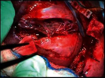
This clinical photograph captures an intraoperative view of a fetal surgical repair for a myelomeningocele (spina bifida). The image displays a large 5 x 5 cm neural tube defect located in the lumbosacral region, characterized by exposed neural placode and surrounding meningeal tissue. Associated with the defect is a visible kyphotic spinal deformity. Surgical instruments are seen manipulating the area where the repair is being performed. The procedure demonstrates the closure technique using a skin patch sewn over the open defect to protect the spinal cord and prevent further damage from amniotic fluid exposure. The surgical field includes medical drapes, sutures, and specialized instrumentation utilized in maternal-fetal surgery. This visual illustrates key concepts in pediatric neurosurgery and fetal intervention, specifically the management of congenital central nervous system malformations and the techniques required for anatomical closure of myelomeningocele.
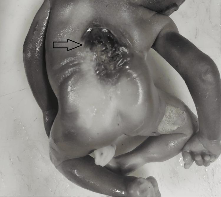
This clinical photograph shows a posterior view of a human fetus presenting with an open neural tube defect (NTD), specifically spina bifida aperta (myelomeningocele). The primary abnormality is located in the mid-to-lower thoracic spine, indicated by a black arrow. The defect appears as a large, midline ulcerated lesion where the skin and underlying vertebral arches have failed to fuse, exposing the neural placode and underlying meningeal tissues. The lesion shows evidence of hemorrhage and irregular borders. Surrounding the spinal defect, the skin appears thinned and tense. Additionally, the lower extremities and overall body structure are visible; there is evidence of skin maceration or epidermal desquamation on the right thigh, which may be secondary to intrauterine positioning or underlying pathology. This image illustrates a severe congenital central nervous system malformation, serving as a primary example of spinal dysraphism for medical education in embryology, neonatology, and fetal pathology.
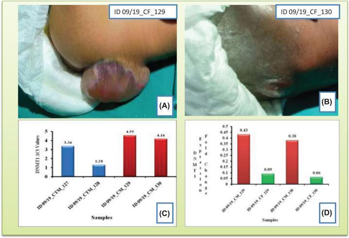
This composite educational image illustrates Neural Tube Defects (NTDs) and related genetic expression data. Panels (A) and (B) are clinical photographs of newborns showing manifestations of spina bifida. Image A demonstrates a cystic spina bifida (myelomeningocele or meningocele) presented as a prominent, spherical, fluid-filled sac protruding from the lumbosacral region, covered by thin, translucent skin with pink and purplish discoloration. Image B shows a different case with a less pronounced protrusion, possibly indicating spina bifida occulta or a smaller closed neural tube defect with associated skin dimpling or scarring. Panels (C) and (D) are quantitative bar charts related to DNMT1 gene expression. Chart C compares ΔCt values between control mothers (CTM) and case mothers (CM), showing higher ΔCt (lower expression) in mothers who gave birth to NTD babies. Chart D shows the fold change (2−ΔΔCt) comparing case mothers to their respective fetuses (CF), illustrating a significant reduction in DNMT1 expression in the affected newborns. This material is designed for medical genetics and pediatrics, demonstrating the phenotypic presentation of NTDs alongside molecular epigenetic findings.
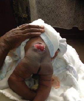
This clinical photograph shows a neonate in a prone position, demonstrating a prominent neural tube defect in the lumbar spine. The lesion is consistent with spina bifida cystica (myelomeningocele). There is a visible midline protrusion with an open, erythematous neural plaque (placode) where the spinal cord and meningeal tissues are exposed through a defect in the skin and vertebral arches. The surrounding skin appears thin at the margins of the sac. The lower extremities are visible, appearing somewhat flaccid with internal rotation of the thighs, which is clinically significant for potential neurological impairment associated with the spinal lesion. The image is a primary resource for medical education regarding congenital central nervous system malformations and neonatal physical examination findings.
anencephaly encephalocele neural tube defect
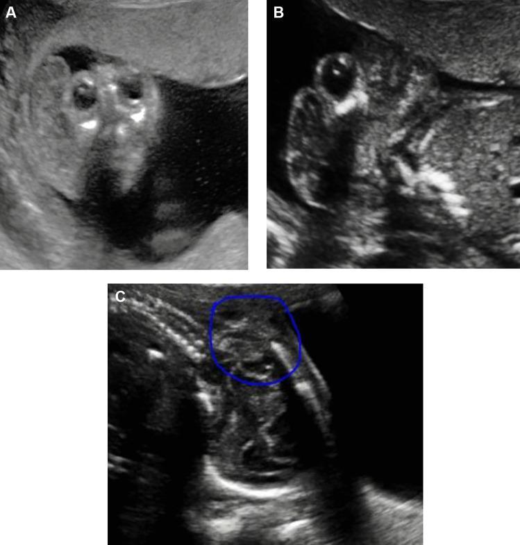
This composite diagnostic image features three grayscale fetal ultrasound panels (A, B, and C) illustrating different types of neural tube defects (NTDs) in the second trimester. Panel A shows anencephaly, characterized by the absence of the cranial vault and cerebral hemispheres; the protruding orbits and residual neural tissue create the classic 'Mickey Mouse' sign in a coronal or transverse view. Panel B demonstrates an encephalocele, specifically an occipital myelomeningocele, visualized as a fluid-filled, sac-like protrusion through a defect in the occipital bone. Panel C displays a longitudinal view of the fetal spine with a thoracolumbar myelomeningocele (highlighted by a blue circle). This view reveals a disruption in the spinal line with a cystic mass protruding from the posterior aspect of the vertebrae, indicative of spina bifida aperta where the spinal cord and meninges are exposed. The collection serves as an educational reference for prenatal screening and the sonographic identification of major congenital central nervous system malformations.
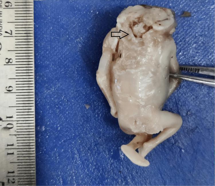
This clinical photograph shows a human fetal specimen demonstrating anencephaly, a severe open neural tube defect. The image highlights a significant malformation of the cephalic region characterized by the absence of a major portion of the brain (forebrain and cerebral hemispheres) and the overlying cranial vault (acrania). An arrow points to the area of the defect where disorganized neural tissue, often referred to as the area cerebrovasculosa, is exposed. In contrast to the severe cranial deformity, the rest of the fetal anatomy, including the torso, upper limbs, and lower limbs with visible feet, appears relatively well-developed and intact. A ruler is positioned to the left for scale, and surgical forceps are visible on the right, indicating a pathological or embryological examination. This specimen serves as a classic educational example of a failure of the neural tube to close at the cranial end during early embryonic development.
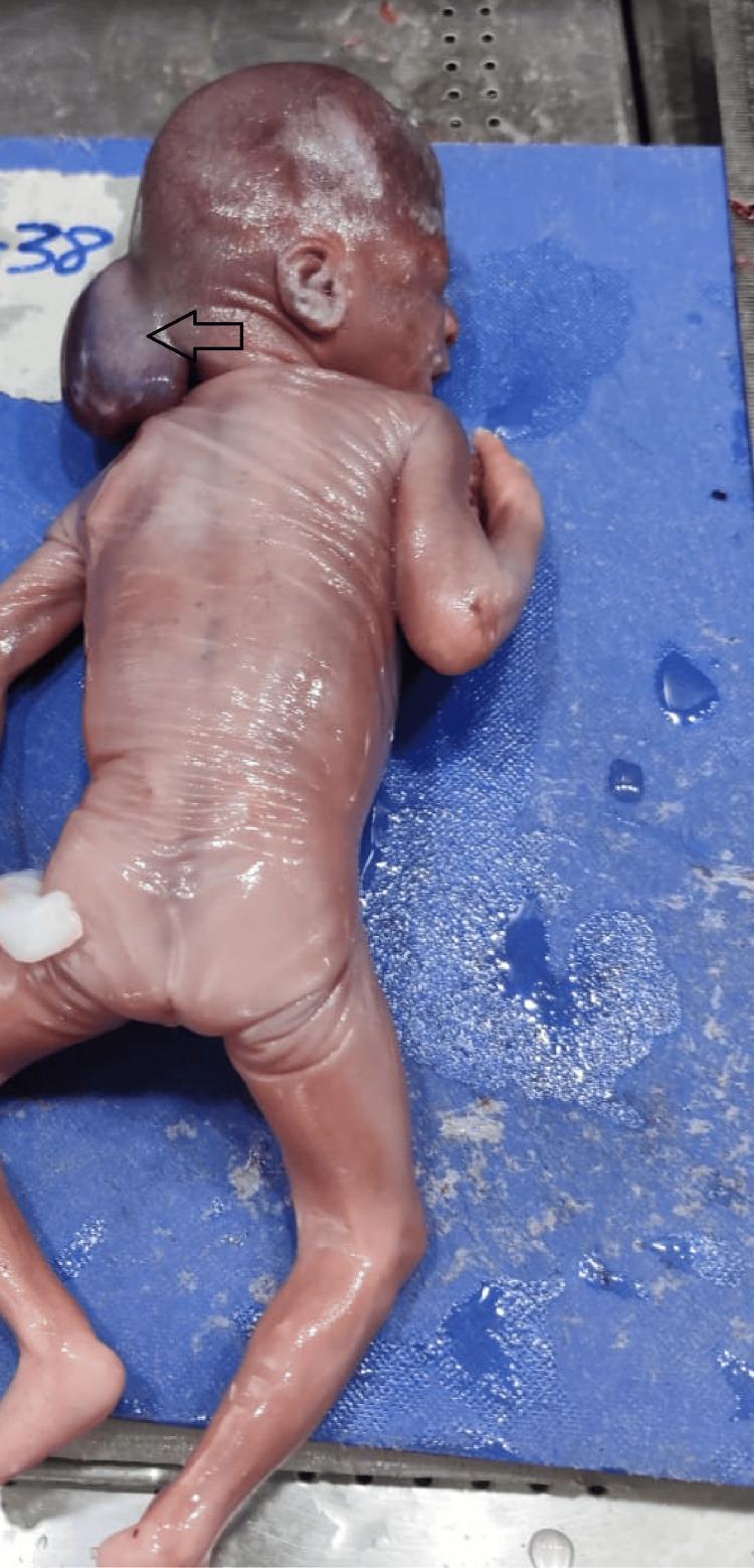
A clinical photograph of a fetus in posterior view, demonstrating a large occipital encephalocele. The primary pathology is a significant, sac-like protrusion in the occipital region of the head, indicated by a black arrow. This mass is smooth, taut, and spherical, representing the herniation of intracranial contents through a midline skull defect. The fetus exhibits thin, translucent skin with visible underlying vascular patterns, characteristic of early second-trimester development. The limbs appear well-formed but are positioned in a flexed posture. The image serves as a clear educational example of a cranial neural tube defect (NTD). It is highly relevant for medical studies in embryology, obstetrics, and pathology, specifically illustrating the morphological presentation of encephaloceles versus other cranial defects like anencephaly or exencephaly.
Seminar: Neural Tube Defects (NTDs)
1. Introduction and Definition
- Failure of neural tube closure - secondary mesenchymal tissue defects stem from aberrant skeletal modeling around the malformed tube (e.g., anencephaly, myelomeningocele)
- Primary bony defects - caused by abnormal axial mesoderm development leading to secondary CNS abnormalities (e.g., encephalocele, meningocele, spina bifida)
- Robbins & Kumar Basic Pathology, p. 1044; Robbins Cotran & Kumar Pathologic Basis of Disease, p. 1150
2. Embryology - Neurulation
- Shaping, folding, and midline fusion of the neural plate
- Normally complete by day 25 after conception
- Creasy & Resnik's Maternal-Fetal Medicine, p. 585
3. Epidemiology
| Parameter | Data |
|---|---|
| Incidence (ONTD) | ~1:1000 live births |
| Most common forms | Anencephaly and spina bifida |
| US annual burden | ~3,500 births per year |
| Most common location | Lumbosacral region (>80% of cases) |
| Aneuploidy association | 10% (trisomy 18, trisomy 13, triploidy) |
| Recurrence risk | 3%-5% in subsequent pregnancies |
| Perinatal mortality (isolated ONTD) | ~10%-15% |
- Goldman-Cecil Medicine, p. 430; Creasy & Resnik's MFM, p. 585
4. Classification of NTDs
A. Spina Bifida (Most Common NTD)
| Type | Description |
|---|---|
| Spina Bifida Occulta | Bony defect only; no exposed neural tissue; asymptomatic; no visible sac |
| Meningocele | Sac containing only meninges and CSF; spinal cord not herniated |
| Myelomeningocele | Sac containing spinal cord or other neural elements; most severe spinal form |
| Myeloschisis | Wide splaying of vertebral arch; no visible covering; neural tube completely exposed |
B. Anencephaly
- Malformation of the anterior end of the neural tube
- Leads to absence of most of the brain and calvarium
- Forebrain disrupted at approximately 28 days gestation
- Remnant is the area cerebrovasculosa - disorganized brain tissue with admixed ependymia, choroid plexus, and meningothelial cells
- Posterior fossa structures may be partially spared
- Incompatible with sustained life
C. Encephalocele
-
Diverticulum of malformed CNS tissue extruding through a midline defect in the cranium
-
Most often occurs in the occipital region or posterior fossa
-
Anterior variants may involve orbit, ethmoid, or cribriform plate ("nasal glial heterotopia")
-
Robbins Cotran & Kumar Pathologic Basis of Disease, p. 1150-1151
5. Clinical Images
6. Etiology and Risk Factors
| Risk Factor | Odds Ratio (OR) / Risk |
|---|---|
| Folic acid deficiency (<400 mcg/day) | OR = 3.72 (95% CI, 1.77-7.81) |
| Prior affected pregnancy | 3%-5% recurrence risk |
| Folate antagonists (valproic acid, carbamazepine) | ~1%-2% risk |
| Pregestational diabetes (type 1 or 2) | OR = 2.88 (95% CI, 1.79-4.65) |
| Periconceptional fever (≥101°F) | OR = 2.4 (95% CI, 1.5-4.0) |
| Maternal obesity (BMI ≥30) | OR = 1.79 (95% CI, 1.51-2.13) |
| MTHFR mutation (677C>T) | OR = 1.34 (95% CI, 1.17-1.54) |
- Creasy & Resnik's MFM, p. 585
7. Genetics
-
MTHFR gene polymorphism (677C>T) - impairs folate metabolism, raising NTD risk
-
Overall recurrence rate 4%-5% after one affected pregnancy
-
Differences in NTD rates among populations are partly explained by polymorphisms in enzymes involved in folic acid metabolism
-
Genomic sequencing has begun uncovering additional genetic variants
-
Emery's Elements of Medical Genetics and Genomics; Robbins Cotran & Kumar Pathologic Basis of Disease
8. Clinical Features of Myelomeningocele
- Motor and sensory deficits in the lower extremities (paraparesis/paraplegia)
- Level of deficit corresponds to level of lesion
- Neurogenic bladder and bowel
- Urinary and fecal incontinence
- Clubfeet (talipes)
- Scoliosis and kyphoscoliosis
- Hip dislocation
- Arnold-Chiari malformation type II (downward herniation of brainstem and cerebellum)
- Hydrocephalus (requires ventriculoperitoneal shunting)
- Cerebral ventriculomegaly
- Aberrant corpus callosum development (70%-90%)
- Microcephaly (HC <5th percentile, 71%)
- Creasy & Resnik's MFM, p. 586-589; Robbins & Kumar Basic Pathology, p. 1051
9. Prenatal Diagnosis
Maternal Serum Screening
- Elevated alpha-fetoprotein (AFP) in maternal serum - most ONTDs are associated with elevated MSAFP
- AFP is also elevated in amniotic fluid; acetylcholinesterase in amniotic fluid is a more specific marker
First-Trimester Screening (11-14 weeks)
- Intracranial translucency (IT): Qualitative non-visualization of the echolucent brainstem, 4th ventricle, or cisterna magna on mid-sagittal plane is suspicious for open spina bifida
- Obliterated cisterna magna and elevated BS-BSOB ratio (brainstem-to-occipital bone ratio >1:1 is abnormal; >1:2 suspicious)
- Early anatomy imaging at 15-16 weeks if first-trimester screen is abnormal
Second-Trimester Ultrasound Signs
| Sign | Finding |
|---|---|
| Lemon sign | Frontal scalloping of the cranium (bifrontal retraction) |
| Banana sign | Effacement/obliteration of the cisterna magna due to Chiari II herniation |
| Spinal defect | Sagittal: dorsal defect with overlying cystic mass; Axial: V- or U-shaped posterior vertebrae |
Amniocentesis
-
Karyotype / chromosomal microarray to assess for aneuploidy
-
Elevated amniotic fluid AFP + acetylcholinesterase confirms open NTD
-
Creasy & Resnik's MFM, p. 585-589; Tietz Textbook of Laboratory Medicine
10. Management
Antenatal Management
- Genetic counseling - discuss prognosis, recurrence risk, syndrome associations
- Amniocentesis with microarray or NIPT for karyotyping
- Fetal echocardiogram to assess cardiac structure
- Multidisciplinary counseling - involve pediatric neurosurgery, urology, orthopedics
- Fetal surgery (prenatal repair) - the landmark MOMS trial (Management of Myelomeningocele Study) showed prenatal repair significantly reduces need for VP shunting (40% vs. 82%) and improves motor outcomes vs. conventional postnatal surgery
- Must be performed at highly specialized fetal surgery centers
- Strict patient selection criteria apply
- Delivery should be at a tertiary care center
- Delivery mode: Recent meta-analysis shows no difference in motor-anatomic level between vaginal vs. cesarean delivery; vaginal delivery less likely to need a shunt. Cesarean indicated for breech or severe hydrocephalus.
- Use nonlatex gloves throughout to prevent latex sensitization
Neonatal Management
- Protect open spinal lesion from infection and desiccation - wrap with moist sterile bandages in sterile isolette
- Use nonlatex gloves
- Immediate pediatric neurosurgery consultation
- Prophylactic antibiotics pending surgery
- Postnatal surgical repair within 48 hours of birth
- Followed by ongoing management of hydrocephalus, neurogenic bladder, orthopedic complications
- Creasy & Resnik's MFM, p. 587-589
11. Prevention
Primary Prevention: Folic Acid
| Population | Folic Acid Dose | Timing |
|---|---|---|
| All women of childbearing age (low-risk) | 0.4 mg (400 mcg)/day | At least 1 month before and through first trimester |
| High-risk women (prior NTD pregnancy) | 4 mg/day | Starting 1 month before conception |
- Folate supplements reduce NTD incidence by up to 70%
- Supplemental folic acid before conception reduces recurrent ONTD risk by 70%
- The US CDC, USPSTF, and AAP all recommend folic acid supplementation
- Mandatory folic acid fortification of grain products in the US since 1998 has significantly reduced NTD prevalence
- Average American folic acid intake is only ~half the recommended amount
Recent Evidence (2025-2026)
- Neuroscience: Exploring the Brain 5th ed., p. 1963-1977; Creasy & Resnik's MFM, p. 406; Robbins Cotran & Kumar, p. 1151
12. Summary Table
| Feature | Details |
|---|---|
| Neural tube closure | Complete by day 25-28 post-conception |
| Most common type | Spina bifida (spinal dysraphism) |
| Most common location | Lumbosacral (>80%) |
| Key biochemical marker | Elevated AFP (maternal serum + amniotic fluid) |
| Best prenatal sign | Lemon + banana sign on 2nd-trimester US |
| Genetic association | MTHFR 677C>T mutation |
| Key risk factor | Folic acid deficiency |
| Prevention | 0.4 mg/day folic acid periconceptionally |
| High-risk prevention | 4 mg/day folic acid |
| Risk reduction with folate | Up to 70% |
| Prenatal surgery | MOMS trial - proven benefit for myelomeningocele |
| Recurrence risk | 3%-5% |
13. Key Teaching Points
- NTDs result from failure of neural tube closure, normally complete by day 25-28 post-conception
- The two main mechanisms are failed neural tube closure (anencephaly, myelomeningocele) and primary bony defects (spina bifida, encephalocele)
- Myelomeningocele is the most clinically significant survivable NTD; anencephaly is lethal
- The "lemon sign" + "banana sign" on second-trimester ultrasound are hallmark prenatal markers
- Elevated maternal serum AFP should always prompt careful sonographic evaluation of the spine
- Periconceptional folic acid (0.4 mg/day) reduces NTD risk by up to 70% - this is the single most important preventive measure
- High-risk women (prior affected pregnancy) should receive 4 mg/day starting one month before conception
- Prenatal surgical repair (MOMS trial) significantly improves outcomes compared to postnatal-only repair
- MTHFR gene polymorphisms and multifactorial inheritance are the genetic basis
- Emerging evidence highlights vitamin B12 as an additional important nutritional factor beyond folate
Summery conclusion bibloigraphy
Summary
Conclusion
Bibliography
-
Creasy RK, Resnik R, et al. Creasy and Resnik's Maternal-Fetal Medicine: Principles and Practice. 8th ed. Philadelphia: Elsevier; 2023. Chapter 26: Neural Tube Defects, pp. 584-589.
-
Kumar V, Abbas AK, Aster JC. Robbins and Cotran Pathologic Basis of Disease. 10th ed. Philadelphia: Elsevier; 2021. Chapter 28: Neural Tube Defects, pp. 1150-1151.
-
Kumar V, Abbas AK, Aster JC. Robbins and Kumar's Basic Pathology. 11th ed. Philadelphia: Elsevier; 2023. Chapter 21: Neural Tube Defects, pp. 1044-1052.
-
Goldman L, Cooney KA. Goldman-Cecil Medicine. 27th ed. Philadelphia: Elsevier; 2024. Neural Tube Defects, pp. 430-432.
-
Turnpenny PD, Ellard S, Cleaver R. Emery's Elements of Medical Genetics and Genomics. 16th ed. Philadelphia: Elsevier; 2022. Multifactorial Inheritance - Neural Tube Defects, pp. 4327-4331.
-
Bear MF, Connors BW, Paradiso MA. Neuroscience: Exploring the Brain. 5th ed. Burlington: Jones & Bartlett Learning; 2020. Nutrition and the Neural Tube, pp. 1963-1977.
-
Katzung BG. Basic and Clinical Pharmacology. 16th ed. New York: McGraw-Hill; 2023. Folic Acid - Prevention of Neural Tube Defects, p. 956.
-
Rifai N, Chiu RWK, Young I, et al. Tietz Textbook of Laboratory Medicine. 7th ed. Philadelphia: Elsevier; 2023. Maternal Serum AFP Screening and Neural Tube Defects.
-
Park K. Park's Textbook of Preventive and Social Medicine. 26th ed. Jabalpur: Banarsidas Bhanot; 2023. Defects at Birth: Neural Tube Defects, p. 2732.
-
Adzick NS, Thom EA, Spong CY, et al. (MOMS Investigators). A randomized trial of prenatal versus postnatal repair of myelomeningocele. N Engl J Med. 2011;364(11):993-1004. PMID: 21306277.
-
Sinha A, P P, Chakraborty H. Burden of neural tube defects in India: a systematic review and meta-analysis. Childs Nerv Syst. 2024 Dec. PMID: 39365302
-
Nie L, Liu X, Li X. Beyond Folate: The Emerging Role of Maternal Vitamin B12 in Neural Tube Development. Nutrients. 2025 Jun 19. PMID: 40573151
-
Wu X, Bian X, Zheng Q. Influencing factors of neural tube malformation: a systematic review and meta-analysis. Afr Health Sci. 2025 Mar. PMID: 41640554
-
Centers for Disease Control and Prevention (CDC). Use of folic acid for the prevention of spina bifida and other neural tube defects. MMWR. 1991;40(30):513-516.
-
US Preventive Services Task Force. Folic acid supplementation for the prevention of neural tube defects: USPSTF recommendation statement. JAMA. 2017;317(2):183-189.