
topography of internal thoracic artery
Reading File
The internal thoracic artery (ITA), also called the internal mammary artery, arises from the subclavian artery and runs down the anterior chest wall just lateral to the sternum. It gives branches to the pericardium, mediastinum, and intercostal spaces, and terminates as the musculophrenic and superior epigastric arteries at the level of the 6th intercostal space.
Searching sources...
Finding Sources
Finding Sources
Good - I have several relevant sources. Let me read the primary anatomy sections now.
Reading File
Reading File
Reading File
I have a diagram image URL. Let me fetch it and also read more content from THIEME Atlas for complete branch details.
Loading Image
Reading File
Finding Sources
Excellent - I have rich content from multiple authoritative anatomy textbooks and a detailed diagram. Let me also fetch the lateral view diagram for completeness and search for relation details (transversus thoracis, etc.).
Loading Image
Finding Sources
Excellent - now let me read the detailed sections from Gray's Anatomy for Students on anterior intercostal arteries and the internal thoracic artery course, plus the Fischer's Mastery of Surgery section for additional surgical anatomy detail.
Reading File
Reading File
I now have comprehensive information from multiple authoritative sources. Let me also fetch the surgical anatomy diagram from Fischer's.
Loading Image
I now have comprehensive, well-illustrated information from multiple authoritative textbooks. Here is the complete answer:
Topography of the Internal Thoracic Artery
Also called the internal mammary artery (IMA), the internal thoracic artery (ITA) is one of the most important vessels of the anterior thoracic wall, widely harvested as a coronary bypass conduit.
Origin
- Arises from the inferior (concave) surface of the first part of the subclavian artery, just medial to the anterior scalene muscle.
- On the right side, the subclavian itself arises from the brachiocephalic trunk; on the left, directly from the aortic arch.
- Variant (10%): arises from the thyrocervical trunk; in 8% the thyrocervical trunk itself is formed by the ITA plus inferior thyroid and suprascapular arteries.
- Color Atlas of Human Anatomy Vol. 2, p. 103; THIEME Atlas of General Anatomy, p. 211
Course
The ITA follows a strictly vertical, paramedian course through four segments:
| Segment | Description |
|---|---|
| Cervical | Passes posterior to the clavicle and the large veins (subclavian, brachiocephalic). Crosses over the cervical dome of the pleura. |
| Superior thoracic aperture | Passes anteriorly through the superior thoracic aperture posterior to the 1st costal cartilage. |
| Thoracic (main) | Descends along the posterior surface of costal cartilages 1-6, lying ~1 cm lateral to the lateral border of the sternum. It lies anterior to the transversus thoracis muscle and posterior to the costal cartilages and internal intercostal muscles. |
| Bifurcation | At the level of the 6th intercostal space / 6th costal cartilage, divides into its two terminal branches. |
- Gray's Anatomy for Students, p. 192; Fischer's Mastery of Surgery, p. 2149
Relations
| Direction | Structure |
|---|---|
| Anterior | Costal cartilages of ribs 1-6, internal intercostal muscles |
| Posterior | Transversus thoracis muscle (lower part), pleura (upper part) |
| Medial | Sternum (~1 cm away) |
| Lateral | Internal intercostal spaces |
The internal thoracic vein accompanies the artery (actually two veins - venae comitantes - in the lower part, which join into one superiorly), lying medial to it and draining into the brachiocephalic vein.
Branches
1. Pericardiacophrenic Artery
- Arises high up, accompanies the phrenic nerve all the way to the diaphragm.
- Supplies the pericardium and diaphragm.
2. Anterior Intercostal Arteries
- Two branches per intercostal space (one along the inferior margin of the upper rib, one along the superior margin of the lower rib) - these anastomose with the posterior intercostal arteries from the aorta.
- Supply upper 6 intercostal spaces directly from the ITA.
- The 7th-9th anterior intercostals arise from the musculophrenic artery.
3. Perforating Branches
- One per intercostal space, pass forward through the costal cartilages to supply the skin and anterior chest wall.
- Carry the medial mammary branches - a major arterial source to the medial breast.
4. Sternal Branches
- Multiple small branches to the posterior surface of the sternum and sternal periosteum.
5. Mediastinal Branches
- Small branches to the thymus, mediastinal fat, and areolar tissue.
Terminal Branches (at 6th costal cartilage)
| Branch | Course | Supply |
|---|---|---|
| Superior epigastric artery | Passes through the diaphragm between its sternal and costal heads; enters the rectus sheath; anastomoses with the inferior epigastric artery (from external iliac) at the level of the umbilicus | Anterior abdominal wall, rectus abdominis |
| Musculophrenic artery | Curves along the costal margin behind the costal arch; perforates the diaphragm; ends near the last intercostal space | Diaphragm, lower intercostal spaces (7th-9th anterior intercostals arise from it) |
- Gray's Anatomy for Students, p. 192; Color Atlas of Human Anatomy, p. 103
Diagrams
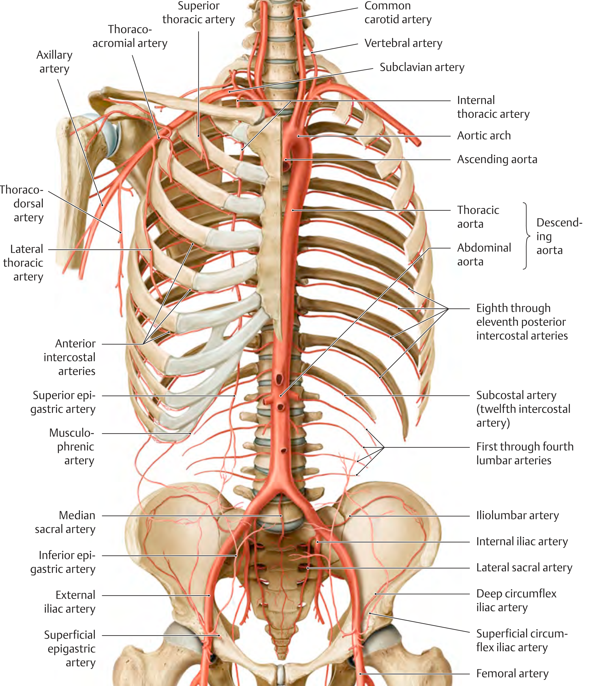
Arteries of the trunk wall (anterior view) - THIEME Atlas:
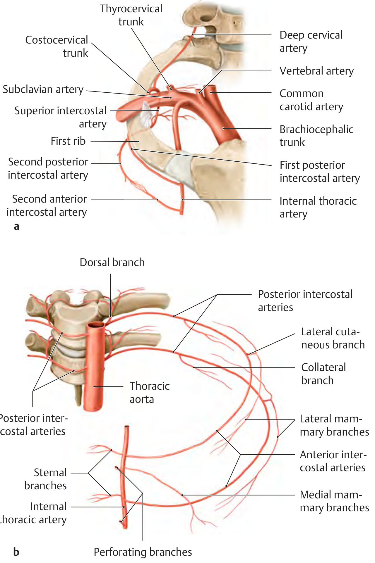
Origin and branches of the intercostal arteries - THIEME Atlas:
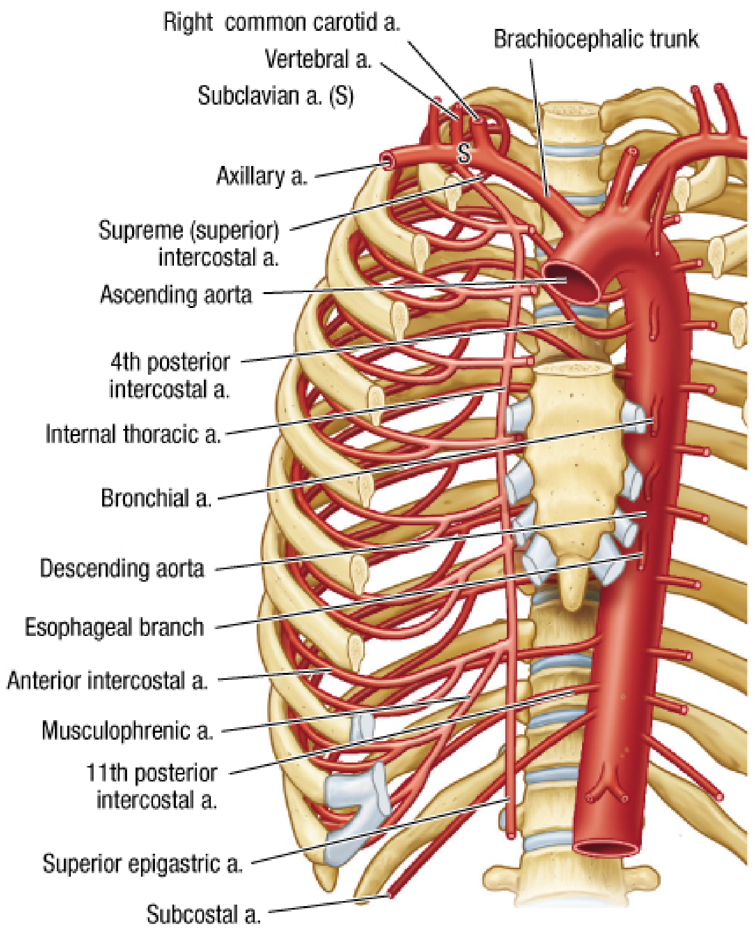
Surgical view of thoracic arterial supply - Fischer's Mastery of Surgery:
Anastomoses
The ITA is a key component of several anastomotic chains:
- Superior epigastric (ITA) ↔ inferior epigastric (external iliac) - clinically important collateral pathway in aortoiliac occlusion.
- Anterior intercostals (ITA) ↔ posterior intercostals (aorta) - chest wall collaterals in coarctation of the aorta.
- Pericardiacophrenic ↔ inferior phrenic (abdominal aorta) - diaphragmatic anastomosis.
Clinical Significance
- The left ITA (LITA) is the gold-standard conduit for coronary artery bypass grafting (CABG), particularly to the left anterior descending (LAD) artery, due to its superior long-term patency compared to saphenous vein grafts.
- The right ITA can be used as a free graft or in situ conduit, or as a composite arterial graft off the LITA.
- The vessel is harvested either as a pedicled or skeletonized graft during cardiac surgery.
Sources: Gray's Anatomy for Students; THIEME Atlas of General Anatomy and Musculoskeletal System; Color Atlas of Human Anatomy Vol. 2 (Sobotta/Schiebler); Fischer's Mastery of Surgery, 8th ed.; Goldman-Cecil Medicine
This is a shared conversation. Sign in to Orris to start your own chat.