
Analyse this report and provide prognosis
cd "/tmp/workspace/attachments/ac09fc87-a44f-4c6b-a043-d29566724766/" && python3 -c " import pdfplumber with pdfplumber.open('GOPAL SINGH DANGI 82YM.pdf') as pdf: print(f'Pages: {len(pdf.pages)}') for i, page in enumerate(pdf.pages): print(f'--- PAGE {i+1} ---') text = page.extract_text() print(text) tables = page.extract_tables() if tables: for t in tables: print('TABLE:', t) "
bladder cancer muscle invasive elderly prognosis
transitional cell carcinoma bladder muscle invasion prognosis elderly CT staging VUJ infiltration
"bladder cancer" AND "vesicoureteric junction" AND invasion prognosis
CT Urography Report Analysis - Gopal Singh Dangi, 82 Y/M
Summary of Findings
| Structure | Finding |
|---|---|
| Left bladder wall | Heterogeneously enhancing sessile polypoidal mass, ~3.7 x 2.5 x 3.5 cm |
| Mass location | Left posterolateral wall, infiltrating left vesico-ureteric junction (VUJ) |
| Muscle invasion | Suspected (?invasion of muscularis propria) |
| Extravesical spread | None detected on CT |
| Left urinary tract | Mild hydro-uretero-nephrosis (upstream obstruction from VUJ infiltration) |
| Both kidneys | Bosniak I cysts (benign), tiny concretions (1-1.5 mm, clinically minor) |
| Prostate | Enlarged - 50.6 cc, Grade II prostatomegaly |
| Bladder wall | Diffuse circumferential mural thickening + coarsened trabeculae (chronic cystitis) |
Radiological Diagnosis
- Sessile (non-pedunculated) morphology - higher risk of muscle invasion than papillary tumors
- Heterogeneous enhancement - hallmark of high-grade, aggressive urothelial carcinoma
- Size ~3.7 cm - large tumor, associated with worse prognosis
- Left VUJ infiltration with resultant upstream hydronephrosis - indicates locally advanced involvement
- Suspected muscle invasion (radiologist's qualifier "?invasion of muscles") - places this in the MIBC category pending histological confirmation
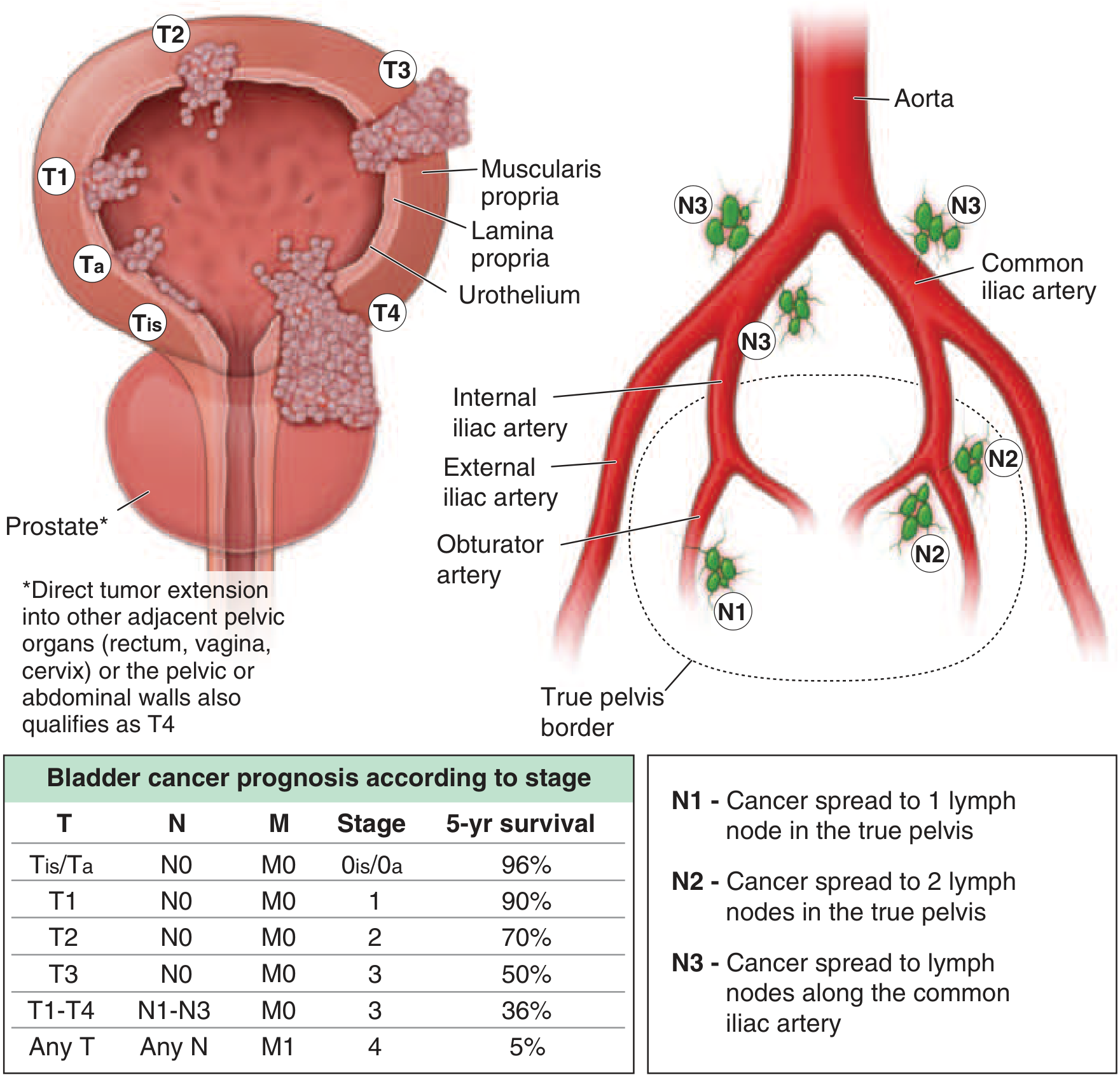
TNM Staging (CT-Based, Provisional)
| Parameter | Assessment |
|---|---|
| T stage | Likely T2 (muscle-invasive) or T3 (perivesical spread - cannot exclude on CT) |
| N stage | N0 (no nodal disease reported) |
| M stage | M0 (no distant metastasis detected) |
| Clinical Stage | Stage II (T2N0M0) to Stage III (T3N0M0) |
Important caveat: CT has well-known limitations in distinguishing T2 from T3 disease, and in detecting microscopic extravesical extension. MRI (multiparametric) is more accurate for local staging. The radiologist's use of "?" for muscle invasion means cystoscopic biopsy with TURBT is mandatory to confirm pathological staging - which has been correctly recommended in the report.
Prognosis
Stage-Based Survival (Harrison's Principles of Internal Medicine, 22nd Ed.)
| Stage | 5-Year Survival |
|---|---|
| Stage 0is/0a (Tis/Ta) | 96% |
| Stage I (T1) | 90% |
| Stage II (T2N0M0) | ~70% |
| Stage III (T3N0M0) | ~50% |
| Stage III (T1-T4, N+) | 36% |
| Stage IV (any M1) | 5% |
Age-Specific Prognostic Modifiers (82 years)
-
Reduced surgical candidacy: Among SEER database patients aged 80-84, only 16% underwent radical cystectomy (vs. 40-55% in those under 75) due to operative risk and comorbidities. The perioperative mortality for radical cystectomy (RC) is 3-7% in mixed-age populations and higher still in octogenarians.
-
Higher comorbidity burden: Elderly patients with muscle-invasive bladder cancer have higher prevalence of cardiac disease, prior cancers, chronic anemia, and poor ASA classification - all of which increase treatment-related complications.
-
VUJ obstruction: The infiltration of the left vesico-ureteric junction causing hydro-uretero-nephrosis indicates the tumor has already compromised ureteral drainage. This is a marker of locally advanced disease and, if bilateral in future progression, poses a risk of obstructive nephropathy and renal failure.
-
Grade II prostatomegaly (50.6 cc): This is a concurrent finding and contributes to the presenting symptom of voiding difficulty. It does not directly worsen the cancer prognosis but complicates urological management and is an independent surgical risk factor.
-
Chronic cystitis: Diffuse mural thickening and trabeculation suggests long-standing lower urinary tract dysfunction, consistent with the chronic obstructive picture from BPH.
Next Steps (Clinically Indicated)
- Cystoscopic biopsy + TURBT (correctly advised by the radiologist) - to confirm TCC histology, determine grade (high vs. low), establish pathological T stage, and assess for carcinoma in situ (CIS)
- MRI pelvis - for superior soft tissue staging, particularly to assess true muscle vs. perivesical invasion
- Urology/Uro-oncology MDT discussion - treatment planning individualized to age and fitness
- Fitness assessment: ECOG performance status, ASA grading, renal function (creatinine, GFR - relevant given hydronephrosis), cardiac workup
- Possible treatment pathways:
- If fit: radical cystectomy (gold standard for MIBC) ± neoadjuvant cisplatin-based chemotherapy
- If unfit for surgery: bladder-preserving trimodality therapy (TURBT + radiotherapy + concurrent chemotherapy) - evidence in elderly patients shows disease-specific survival comparable to surgery in selected cases (5-year DSS ~71%)
- Palliative/conservative approach: if frail/unfit - TURBT for local control, intravesical agents, symptom management
Summary for the Clinician
Disclaimer: This analysis is based on radiological findings only. Final diagnosis and management must be made by a qualified clinician after clinical examination, histopathological confirmation, and complete workup.
Analyse this report and provide prognosis
CT Urography Report Analysis - Gopal Singh Dangi, 82 Y/M
Summary of Findings
| Structure | Finding |
|---|---|
| Left bladder wall | Heterogeneously enhancing sessile polypoidal mass, ~3.7 x 2.5 x 3.5 cm |
| Mass location | Left posterolateral wall, infiltrating left vesico-ureteric junction (VUJ) |
| Muscle invasion | Suspected (?invasion of muscularis propria) |
| Extravesical spread | None detected on CT |
| Left urinary tract | Mild hydro-uretero-nephrosis (upstream obstruction from VUJ infiltration) |
| Both kidneys | Bosniak I cysts (benign), tiny concretions (1-1.5 mm, clinically minor) |
| Prostate | Enlarged - 50.6 cc, Grade II prostatomegaly |
| Bladder wall | Diffuse circumferential mural thickening + coarsened trabeculae (chronic cystitis) |
Radiological Diagnosis
- Sessile (non-pedunculated) morphology - higher risk of muscle invasion than papillary/pedunculated tumors
- Heterogeneous enhancement - hallmark of high-grade, aggressive urothelial carcinoma
- Size ~3.7 cm - large tumor; size >3 cm independently associated with worse prognosis
- Left VUJ infiltration with upstream hydronephrosis - indicates locally advanced disease
- Suspected muscle invasion - places this in the Muscle-Invasive Bladder Cancer (MIBC) category pending histological confirmation
TNM Staging (CT-Based, Provisional)
| Parameter | Assessment |
|---|---|
| T stage | Likely T2 (muscle-invasive) or T3 (cannot exclude perivesical spread on CT alone) |
| N stage | N0 (no nodal disease reported) |
| M stage | M0 (no distant metastasis detected) |
| Clinical Stage | Stage II (T2N0M0) to Stage III (T3N0M0) |
Important caveat: CT has recognized limitations in distinguishing T2 from T3 disease. MRI (multiparametric) provides superior soft-tissue characterization. The radiologist's use of "?" for muscle invasion means cystoscopic biopsy + TURBT is mandatory for pathological staging - correctly recommended in the report.
Prognosis
Stage-Based 5-Year Survival (Harrison's Principles of Internal Medicine, 22nd Ed., 2025)
| Stage | 5-Year Overall Survival |
|---|---|
| Stage 0 (Tis/Ta) | 96% |
| Stage I (T1) | 90% |
| Stage II (T2, N0M0) | ~70% |
| Stage III (T3, N0M0) | ~50% |
| Stage III (any N+) | 36% |
| Stage IV (M1) | 5% |
Age-Specific Prognostic Modifiers (Age 82)
-
Reduced surgical candidacy: SEER data shows only 16% of patients aged 80-84 with MIBC undergo radical cystectomy (vs. 40-55% in those under 75). Perioperative mortality for radical cystectomy is 3-7% in mixed-age populations, and significantly higher in octogenarians.
-
Higher comorbidity burden: Elderly MIBC patients carry a higher prevalence of cardiac disease, chronic anaemia, prior cancers, and poor ASA classification - all of which compound operative and chemotherapy risk.
-
VUJ obstruction and hydronephrosis: The tumor's infiltration of the left vesico-ureteric junction is a marker of locally advanced disease. If bilateral obstruction develops, it poses a direct risk of progressive obstructive nephropathy and renal failure - which would further limit systemic treatment options (cisplatin requires adequate GFR).
-
Grade II BPH (prostate 50.6 cc): Contributes to the presenting voiding difficulty. Does not worsen cancer prognosis directly but adds surgical complexity and is an independent anesthetic/operative risk factor.
-
Diffuse chronic cystitis: Circumferential bladder wall thickening reflects long-standing bladder outlet dysfunction, likely secondary to BPH. This background inflammatory change may complicate pathological interpretation of biopsy margins.
Recommended Next Steps
- Cystoscopy + TURBT (mandatory, as correctly advised) - to confirm TCC histology, establish grade (high vs. low), determine true pathological T stage, and evaluate for carcinoma in situ (CIS)
- MRI pelvis (mpMRI) - superior to CT for locoregional staging, especially to differentiate T2 vs. T3 invasion
- Renal function panel (serum creatinine, eGFR) - critical given left-sided hydronephrosis; determines cisplatin eligibility
- Multidisciplinary team (Urology + Uro-Oncology + Radiation Oncology) discussion
- Fitness assessment: ECOG performance status, ASA grade, cardiac evaluation
Possible Treatment Pathways Based on Fitness
| Fitness | Approach |
|---|---|
| Fit for surgery | Radical cystectomy ± neoadjuvant cisplatin-based chemotherapy (gold standard for MIBC) |
| Unfit for surgery but fit for systemic therapy | Trimodality therapy: maximal TURBT + concurrent chemoradiotherapy - evidence shows 5-year disease-specific survival ~71% in elderly, comparable to surgery in selected patients |
| Frail / poor performance status | Palliative TURBT for local control + best supportive care + symptom management |
Clinical Summary
Disclaimer: This is an AI-assisted analysis based on radiological findings only. Final diagnosis, staging, and management decisions must be made by a qualified clinician following cystoscopic biopsy, complete workup, and clinical examination.
Search for recent clinical guidelines on elderly MIBC management and trimodality therapy outcomes
"bladder cancer" AND "trimodality" AND elderly
"muscle-invasive bladder cancer" AND guideline
MIBC elderly trimodality therapy guidelines 2024 2025 EAU AUA bladder preservation chemoradiation
PMID: 37187202
PMID: 39986911
PMID: 38661067
PMID: 40118736
PMID: 41955932
PMID: 39934792
PMID: 38754471
https://www.renalandurologynews.com/features/muscle-invasive…
Recent Guidelines & Evidence: Elderly MIBC Management and Trimodality Therapy
1. Current Major Guidelines
EAU 2025 Guidelines on MIBC (Most Current)
- NEW - Trimodality therapy (TMT) formally recognized: New recommendations now mandate management of all TMT candidates in a multidisciplinary team (MDT) setting using shared decision-making - no longer a fallback, but a structured parallel pathway.
- NEW - Salvage cystectomy after TMT: Explicit recommendations now exist for salvage radical cystectomy if TMT fails, underlining that bladder preservation does not foreclose surgical options.
- Adjuvant nivolumab updated: Recommended for pT3/4 and/or pN+ patients who are not eligible for, or declined, adjuvant cisplatin-based chemotherapy - directly applicable to elderly/cisplatin-ineligible patients like this one post-operatively.
- MRI before TURBT: New recommendation to perform MRI before TURBT (not after) for optimal local staging.
- NEW - Erdafitinib: Patients with susceptible FGFR3 alterations in unresectable/metastatic disease should receive erdafitinib (strong recommendation) - a molecularly targeted oral option.
- NIAGARA trial added: Perioperative cisplatin/gemcitabine + durvalumab now EMA and FDA approved (July 2025) as neoadjuvant/adjuvant therapy in eligible surgical candidates.
AUA/ASCO/SUO 2024 Guideline on Non-Metastatic MIBC
- Radical cystectomy remains the guideline standard for non-metastatic MIBC in fit patients.
- Multimodal bladder-preserving therapy (TMT) explicitly incorporated as a guideline option for patients who are unfit for or decline cystectomy, or as a patient preference alternative.
- Neoadjuvant cisplatin-based chemotherapy (prior to RC) carries a strong recommendation (Grade A evidence).
- Pelvic lymphadenectomy extent updated based on new evidence (standard template recommended).
NCCN Bladder Cancer Guidelines, Version 3.2024
- Aligns with EAU/AUA: TMT is explicitly listed as an alternative to radical cystectomy for selected MIBC patients.
- Updated recommendations for BCG-unresponsive NMIBC and systemic therapy in advanced/metastatic disease.
- Emphasizes patient eligibility criteria and comorbidity assessment before treatment assignment.
2. Landmark Clinical Evidence: RC vs. TMT Head-to-Head
The Zlotta Trial (Lancet Oncology 2023) - Most Cited Comparative Study
| Outcome | Radical Cystectomy (5-yr) | Trimodality Therapy (5-yr) | p-value |
|---|---|---|---|
| Metastasis-free survival | 74% | 75% | 0.40 (NS) |
| Cancer-specific survival | 83% | 85% | 0.071 (NS) |
| Disease-free survival | 76% | 76% | NS |
| Overall survival | Comparable | Comparable | NS |
- No statistically significant difference in any survival outcome between RC and TMT in propensity-matched analysis.
- The 2025 EAU guidelines discussion explicitly cites this as the most robust comparative data available.
- This data was also highlighted at EAU Congress 2025: "Disease-free survival was the same in both groups... overall survival was even better in patients treated with trimodality treatment."
3. Systematic Review & Meta-Analysis: TMT vs. RC
Fong et al. 2025 - IPD Meta-Analysis (Highest Evidence)
| Outcome | TMT | Radical Cystectomy |
|---|---|---|
| OS at 5 years | 47% | 57% |
| OS at 10 years | 18% | 22% |
| CSS at 5 years | 62% | 72% |
| CSS at 10 years | 29% | 29% |
- 11 studies, 6,780 patients analyzed via individual patient data (IPD) meta-analysis.
- RC still confers a statistically significant OS advantage (HR 1.14, 95% CI 1.08-1.21) and CSS advantage (HR 1.09, 95% CI 1.01-1.18) over TMT.
- Conclusion: RC should remain the standard of care for fit patients; TMT is a "valid alternative for carefully selected and informed patients."
- Important caveat for elderly patients: Most studies in this meta-analysis enrolled younger, fitter patients - the OS gap may narrow considerably in older cohorts where RC carries higher operative mortality.
4. Elderly-Specific Evidence
Sakaguchi et al. 2025 - Systematic Review of Radiotherapy in Elderly MIBC
| Outcome | Elderly vs. Young MIBC Patients |
|---|---|
| Overall survival (OS) | Worse in elderly (as expected from competing comorbidities) |
| Progression-free survival (PFS) | No significant difference |
| Cancer-specific survival (CSS) | No significant difference |
| Grade 3+ toxicities | No significant difference |
5. Novel Option: Radiation + Immunotherapy for Cisplatin-Ineligible Elderly Patients
Mouw et al. 2026 - Phase II Trial (Avelumab + Radiation)
| Result | Value |
|---|---|
| Complete response (cCR) at 3 months | 64% |
| Median overall survival | 30.9 months |
| Grade 3 toxicity | 3/14 patients (21%) |
| Grade 4/5 toxicity | None |
| Radiation completion rate | 100% |
- This trial was specifically designed for MIBC patients who cannot receive cisplatin - the most relevant scenario for an 82-year-old with hydronephrosis and possible renal impairment.
- Avelumab (PD-L1 checkpoint inhibitor) + radical-dose bladder radiation is a safe and active regimen in this very elderly population.
- Provides a viable alternative to standard cisplatin-based TMT for this patient if renal function is compromised.
6. Practical Application to Gopal Singh Dangi (82 Y/M)
| Factor | Implication |
|---|---|
| Age 82, likely multiple comorbidities | RC carries very high perioperative risk; only 16% of 80-84 yr olds undergo surgery (SEER data) |
| Hydronephrosis from VUJ involvement | Likely reduced eGFR - may render cisplatin ineligible |
| No extravesical spread on CT | Favorable for TMT eligibility (solitary tumor, no extensive CIS suggested) |
| EAU 2025 recommendation | MDT + shared decision-making for all TMT candidates - mandatory |
| If cisplatin-eligible | Standard TMT: maximal TURBT + concurrent cisplatin/5-FU or gemcitabine + RT |
| If cisplatin-ineligible (likely) | Radiation + avelumab (Mouw 2026) OR radiation + carboplatin/paclitaxel OR radiation alone |
| If RC is chosen despite age | Standard template lymphadenectomy; perioperative durvalumab (NIAGARA, EMA approved July 2025) |
| Post-cystectomy, pT3/4, cisplatin-ineligible | Adjuvant nivolumab (EAU 2025 updated recommendation) |
Evidence Summary Table
| Paper | Type | Year | Key Finding |
|---|---|---|---|
| EAU MIBC Guidelines 2025 | Practice Guideline | 2025 | TMT mandatory via MDT; adjuvant nivolumab for cisplatin-ineligible; salvage cystectomy defined |
| AUA/ASCO/SUO Guideline 2024 | Practice Guideline | 2024 | RC gold standard; TMT guideline option for unfit/preference |
| NCCN v3.2024 | Practice Guideline | 2024 | TMT listed as alternative; updated systemic therapy |
| Zlotta et al. Lancet Oncol | Propensity-matched analysis | 2023 | RC vs TMT - no significant survival difference in matched cohorts |
| Fong et al. IPD Meta-analysis | Systematic Review + Meta-analysis | 2025 | RC has marginal OS advantage; TMT valid for selected patients |
| Sakaguchi et al. | Systematic Review | 2025 | In elderly: PFS/CSS/toxicity equivalent between age groups with chemoRT |
| Mouw et al. | Phase II RCT | 2026 | Median age 83 yrs: Radiation + avelumab safe and active; 64% cCR, no grade 4-5 toxicity |
Disclaimer: This literature summary is for clinical reference purposes. All treatment decisions must be made by a qualified multidisciplinary team after full clinical assessment.
What is the cause of such carcinoma
transitional cell carcinoma bladder causes risk factors etiology smoking occupational carcinogens 2024
https://pmc.ncbi.nlm.nih.gov/articles/PMC9411696
Causes of Urothelial (Transitional Cell) Carcinoma of the Bladder
The Core Mechanism: "Field Cancerization"
- Oncogenes (e.g., FGFR3, HRAS, PIK3CA) - promoting uncontrolled cell proliferation
- Tumor suppressor genes (e.g., TP53, RB1, PTEN) - disabling normal cell cycle checkpoints
Causes by Category
1. Tobacco Smoking - The Single Largest Cause (30-40% of all cases)
- The key carcinogens in tobacco smoke are aromatic amines (beta-naphthylamine, 4-aminobiphenyl) and polycyclic aromatic hydrocarbons (PAHs)
- These are absorbed, circulate systemically, then are filtered and concentrated in urine, bathing the urothelium
- Cigarettes, pipe tobacco, and cigars all carry risk - it is not limited to cigarettes
- Risk decreases with cessation: relative risk drops from 2.6 (after 1-3 years of quitting) to 1.1 (after >15 years of quitting)
- ~20% of bladder cancer patients are still smoking at diagnosis, worsening prognosis
2. Occupational Carcinogen Exposure - Second Largest Cause (5-20% of cases)
| Chemical | Industry/Occupation |
|---|---|
| 2-Naphthylamine, Benzidine, 4-Aminobiphenyl | Dye, rubber, textile manufacturing |
| Polycyclic aromatic hydrocarbons (PAHs) | Chimney sweeps, coal/tar/asphalt workers |
| Industrial solvents | Petrochemical, paint, leather workers |
| Aromatic amines | Hairdressers (hair dyes), printers |
- Average 7 years of exposure needed for tumor development; latency up to 20 years after exposure stops - Grainger & Allison, p. 2503
- This is directly relevant to an elderly man in his 80s: occupational exposure from his working years decades ago could be the causative factor today
- Benzidine and β-naphthylamine were banned in most industrialized countries in the 1960s, but those exposed before the bans are only now presenting with cancer
3. Advanced Age and Male Sex
- Bladder cancer incidence rises sharply with age - peak incidence is in patients in their 70s and 80s
- Men are 3-4 times more likely to develop bladder cancer than women, largely because of higher historical rates of smoking and occupational carcinogen exposure
- Age itself is not a direct cause, but reflects the cumulative DNA damage accumulated over a lifetime of exposures
4. Chronic Bladder Irritation and Inflammation
- Bladder stones - repeated mucosal trauma
- Chronic catheterization - mechanical irritation
- Recurrent urinary tract infections (UTIs) - inflammatory cytokines promote mutagenesis
- Bilharzia (Schistosoma haematobium) - a parasitic infection endemic in Africa and the Middle East; strongly linked to squamous cell carcinoma (not TCC) of the bladder
- Bladder outlet obstruction (BPH) - as seen in this patient - chronic urinary stasis and recurrent infections may contribute. The CT report already shows chronic cystitic changes, indicating long-standing bladder wall inflammation
5. Drug and Chemical Exposures
| Agent | Mechanism | Risk |
|---|---|---|
| Cyclophosphamide (chemotherapy) | Mutagenic metabolite (phosphoramide mustard) | 4.5x increased risk; dose-dependent |
| Pioglitazone (diabetes drug, thiazolidinedione) | Direct carcinogenic effect on urothelium | Modest increased risk |
| Phenacetin (old painkiller, withdrawn 1970s) | Metabolites damage upper tract urothelium | Historical; rarely seen now |
| Arsenic (contaminated drinking water) | Directly mutagenic to urothelium | Significant in endemic regions |
| Radiation therapy (prior pelvic RT) | DNA damage to surrounding bladder wall | Relevant for prior prostate/cervical cancer treatment |
6. Genetic and Hereditary Factors (~7% of cases)
- First-degree relatives of bladder cancer patients have a 2-fold increased risk
- Lynch syndrome (HNPCC) - caused by mismatch repair gene mutations (especially MSH2) - associated with elevated urothelial cancer risk. Should be suspected in patients under 60 or with a family history of colorectal/uterine cancers
- NAT2 and GSTM1 polymorphisms - genes encoding carcinogen-detoxifying enzymes; slow-metabolizer variants increase susceptibility to aromatic amines and tobacco smoke
- No classic Mendelian inheritance pattern; risk is multifactorial (gene-environment interaction) - Campbell-Walsh-Wein Urology, p. 4462
7. Aristolochic Acid (Herbal Medicine / Endemic Nephropathy)
- Aristolochic acid found in certain Chinese herbal medicines (Aristolochia species) is a potent urothelial carcinogen
- Also underlies Balkan Endemic Nephropathy - a geographic cluster of urothelial carcinoma in Croatia, Bosnia, Bulgaria, Romania, Serbia
- Creates a characteristic DNA mutational signature (A:T → T:A transversions) highly specific to aristolochic acid exposure
- Increasingly recognized worldwide due to use of herbal remedies containing Aristolochia species - Grainger & Allison, p. 2505
Summary: Causes Most Relevant to This Patient (82-Year-Old Male)
| Risk Factor | Relevance to This Case |
|---|---|
| Tobacco smoking | Most likely primary cause - 30-40% of all TCC; 82-year-old male with decades of potential exposure |
| Occupational exposure | Possible - decades ago as a working-age adult; latency up to 20 years |
| Age (82 years) | Peak incidence decade for bladder cancer; cumulative DNA damage over a lifetime |
| Male sex | 3-4x higher incidence than women |
| Chronic BPH + bladder obstruction | Chronic cystitis shown on CT - long-standing inflammation; contributing factor |
| Chronic UTIs/cystitic changes | Directly visible on CT as mural thickening and trabeculation |
Patient has been heavy smoker and marijuana user
"marijuana" OR "cannabis" AND "bladder cancer" urothelial carcinoma
marijuana cannabis bladder cancer TCC urothelial carcinoma risk evidence 2023 2024
cannabis marijuana bladder cancer risk urothelial
https://www.cannabisclinicians.org/wp-content/uploads/2023/0…
cannabis dependence urologic cancer bladder risk population study
PMID: 40484779
Tobacco + Marijuana Use as Causative Factors in This Patient's TCC
1. Heavy Tobacco Smoking - The Established Primary Cause
How tobacco causes bladder TCC:
- Aromatic amines (2-naphthylamine, 4-aminobiphenyl, benzidine)
- Polycyclic aromatic hydrocarbons (PAHs)
- FGFR3 (activating - drives papillary low-grade tumors)
- TP53, RB1, PTEN (inactivating - drives high-grade, muscle-invasive tumors like this one)
Dose-Response Relationship (Pack-Years Matter):
| Smoking Status | Relative Risk of Bladder Cancer |
|---|---|
| Never smoker | 1.0 (baseline) |
| Former smoker (quit <1-3 yrs) | 2.6x |
| Former smoker (quit >15 yrs) | 1.1x |
| Current light smoker | 2-3x |
| Heavy smoker (current/decades) | 3-6x |
- Heavy, long-duration smokers have the highest risk category
- ~20% of bladder cancer patients are still smoking at diagnosis - continued smoking in MIBC is associated with increased risk of bladder cancer death
- Even after diagnosis, smoking worsens NMIBC local recurrence rates and MIBC-specific mortality
Genetic Susceptibility Modifier:
2. Marijuana (Cannabis) Use - Emerging and Significant Evidence
Why marijuana is biologically plausible as a bladder carcinogen:
- Polycyclic aromatic hydrocarbons (PAHs) - present in higher concentrations per cigarette than tobacco
- Ammonia - ~20x higher in cannabis than tobacco smoke
- Hydrogen cyanide, carbon monoxide, benzene - all present
Clinical Evidence:
- 74,642 cannabis-dependent/abusing patients vs. 4.7 million controls
- Propensity-score matched for tobacco use, nicotine dependence, demographics
- Cannabis dependence was associated with a 4.21x increased risk of bladder cancer (0.14% vs. 0.03%, RR = 4.21, 95% CI 2.70-6.57)
- Also: 3.7x kidney cancer risk, 2.8x prostate cancer risk
- This is the most robust study to date, and specifically controlled for tobacco as a confounder
- 88.5% of urothelial carcinoma patients reported habitual cannabis use vs. 69.2% of matched controls (p = 0.008)
- Cannabis use correlated significantly with higher tumor stage, higher grade, and more recurrences - suggesting it not only causes TCC but may drive more aggressive disease
- Limitation: small sample (n = 124), could not fully separate tobacco confounding
- Found a slightly reduced risk of bladder cancer in cannabis users (HR 0.66) but this became statistically non-significant after adjusting for tobacco (HR 0.77, 95% CI 0.58-1.02)
- Illustrates the importance of controlling for tobacco smoking - the two are heavily co-used
| Study | Design | Finding |
|---|---|---|
| Davis et al. 2025 (n=74,642) | Propensity-matched cohort | 4.2x increased bladder cancer risk in cannabis dependence |
| Chacko et al. 2006 (n=124) | Case-control | Higher TCC stage, grade, recurrence with cannabis use |
| Huang et al. 2022 (n=151,945) | Large cohort | Risk attenuated but non-significant after tobacco adjustment |
| Narrative Review, Thomson et al. 2023 | Literature review | Evidence "mixed" but biological plausibility strong |
3. The Synergistic Double Exposure in This Patient
Combined Carcinogen Load
Deeper Inhalation Pattern
Prolonged Urinary Carcinogen Exposure
- Nicotine metabolites: excreted over ~12 hours
- THC metabolites: excreted over up to 60 hours
- Combined: the bladder mucosa is exposed to active carcinogens almost continuously in a heavy dual user
Tumor Aggressiveness
4. Implications for Management
| Clinical Implication | Detail |
|---|---|
| Cessation is still advised | Even at diagnosis, continued smoking increases MIBC-specific mortality. Cessation should be formally recommended |
| Cannabis cessation | Should also be advised; no protective evidence outweighs the 4x bladder cancer risk signal in dependent users |
| Urothelial field effect | Entire urinary tract is at risk - upper tracts (renal pelvis, ureters) need surveillance alongside the bladder |
| Recurrence risk elevated | Active smokers with NMIBC have higher local recurrence; with MIBC higher disease-specific mortality |
| NAT2 genotyping | Optional but can confirm smoking as direct cause - useful for medicolegal and genetic counselling |
| Second primary cancer vigilance | Heavy smokers are at risk for concurrent lung, oropharyngeal, and esophageal cancers - systemic oncological review is prudent |
Summary
Disclaimer: This analysis is for clinical reference purposes. All management decisions should be made by a qualified multidisciplinary team.
What is mural invasive
bladder wall layers anatomy urothelium lamina propria muscularis propria invasion staging diagram
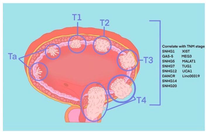
Anatomical diagram of a transverse section of the human urinary bladder illustrating the TNM staging system for bladder cancer (BLCA). The illustration depicts the progression of tumor invasion through the bladder wall layers. Ta stages are shown as small, papillary, finger-like projections confined to the inner urothelium. T1 represents a localized nodular mass invading the lamina propria. T2 shows deeper invasion into the muscularis propria (detrusor muscle). T3 demonstrates extension through the muscle layer into the surrounding perivesical adipose tissue. T4 illustrates advanced invasion into adjacent pelvic organs or the pelvic wall. To the right of the diagram, a list of molecular markers including various Small Nucleolar RNA Host Genes (SNHGs) and long intergenic non-coding RNAs (lincRNAs) such as SNHG1, SNHG14, SNHG20, MALAT1, and Linc00319 are provided, indicating their correlation with these pathological stages. The diagram serves as an educational tool for differentiating non-muscle-invasive bladder cancer (NMIBC) from muscle-invasive bladder cancer (MIBC).
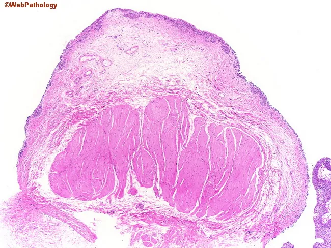
Low-magnification light microscopic image of a section through the urinary bladder wall stained with Hematoxylin and Eosin. The mucosal surface is out of focus at the top, revealing urothelium transitioning into the lamina propria and then into the muscularis propria. The detrusor muscle, comprising thick, densely packed smooth muscle bundles, dominates the deeper aspect of the wall. In contrast, the muscularis mucosae is represented by wispy, discontinuous fascicles more superficially, making the distinction between muscularis propria and mucosa identifiable yet challenging in small specimens. The detrusor fibers are arranged in interlacing and parallel orientations with brisk eosinophilic cytoplasm and elongated, spindle-shaped nuclei; connective tissue septa are sparse in this field. Variation in thickness and organization is evident, with the region near the bladder neck showing coarser, more tightly packed bundles compared with more distal bladder segments. Although this image does not show neoplastic invasion, understanding the architecture is critical for accurate bladder cancer staging. In practice, invasion of the muscularis propria (detrusor) would denote stage T2 disease, driving definitive treatment such as radical cystectomy or radiation therapy. This slide illustrates normal histologic landmarks that underlie diagnostic decision-making, sampling adequacy, and staging accuracy in urothelial carcinoma. For clinical and research use.
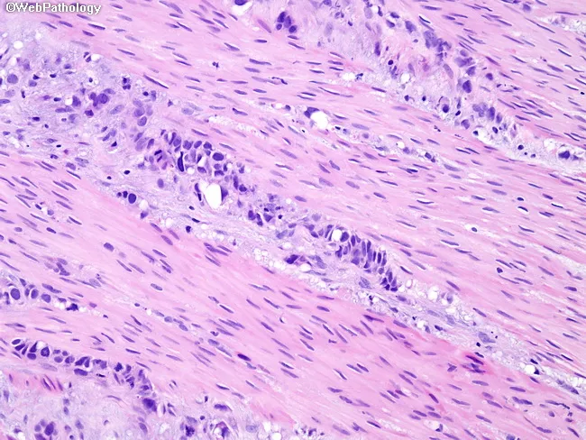
This is a histopathology image of a bladder wall biopsy or cystectomy specimen prepared with Hematoxylin and Eosin staining. The tissue shows detrusor muscle fibers (detrusor = muscularis propria) interspersed with nests and single cells of urothelial carcinoma invading into the muscularis propria. The malignant cells exhibit high-grade cytology: marked nuclear pleomorphism, hyperchromasia, and conspicuous nucleoli, with frequent mitotic figures. The interface between urothelial tumor and smooth muscle is irregular, with tumor infiltration extending among the smooth muscle fascicles. In some regions, muscularis propria appears disrupted by tumor cords, while surrounding stroma shows a desmoplastic response. The overlying urothelium demonstrates variable atypia; there is no definitive invasion of mucosal layers evident in this field, but the critical observation is deep invasion into the detrusor muscle, consistent with muscle-invasive bladder cancer. Clinically this finding has major implications: staging as pT2, guiding definitive therapy decisions such as radical cystectomy, systemic chemotherapy, or radiotherapy, and prognosis. In equivocal sections, pathologists reserve judgment and recommend resampling or re-cutting to confirm muscularis propria invasion. This image supports the diagnoses of muscle-invasive urothelial carcinoma and underscores the necessity for precise depth of invasion reporting and multidisciplinary clinical correlation.
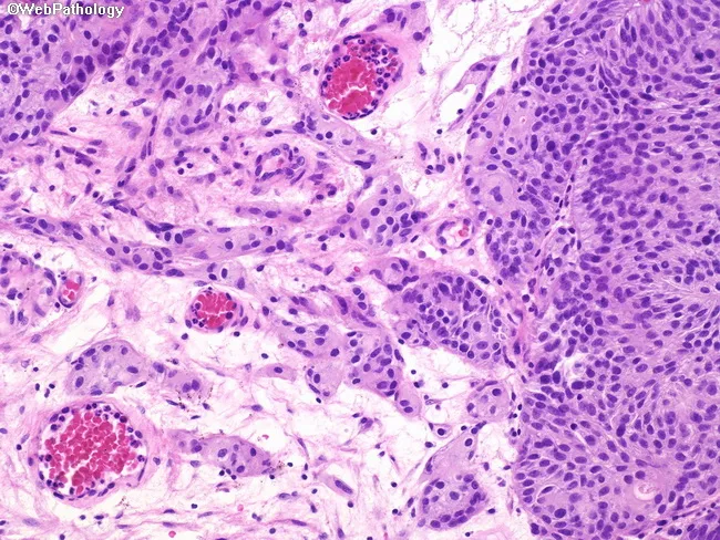
Imaging modality: Brightfield light microscopy of a hematoxylin and eosin (H&E) stained bladder mucosa section. Anatomical context: urinary bladder urothelium with invasion into the lamina propria; muscularis propria is not involved. The histology shows urothelial carcinoma arranged in nests, cords, and clusters at the base of the lesion, with invasion into the subepithelial lamina propria. Individual tumor cells and small clusters are embedded within desmoplastic stroma and peritumoral inflammatory infiltrate. Cytology is predominantly high-grade with marked nuclear pleomorphism and hyperchromasia, though occasional fields may exhibit lower-grade cytology. The lesion lacks invasion of the muscularis propria (pathologic stage T1); the surrounding muscularis shows no tumor cell infiltration. Pathologic interpretation: urothelial carcinoma with lamina propria invasion (T1), a non–muscle-invasive but higher-risk subset, often associated with recurrence and progression risk. Management decisions include intravesical therapies (e.g., BCG) and surveillance, with cystectomy reserved for select higher-grade or unresponsive cases. Diagnostic significance and staging context emphasize lamina propria invasion as a key prognostic factor. Differential considerations include Ta urothelial carcinoma without invasion and carcinoma in situ when deeper invasion is not demonstrated. This image is valuable for educational and research purposes in bladder cancer histopathology, staging, and pathology-to-clinical-management correlations.
What is "Muscle-Invasive" (Mural Invasive)?
The Bladder Wall: Layer by Layer
| Layer | What It Is | Cancer Stage if Involved |
|---|---|---|
| Urothelium | The innermost lining (transitional epithelium, 5-7 cells thick) - directly exposed to urine | Ta, Tis |
| Lamina propria (submucosa) | Loose connective tissue just below the urothelium; contains blood vessels, nerves, thin muscularis mucosae | T1 |
| Muscularis propria (detrusor muscle) | Thick, powerful smooth muscle - the actual "wall" of the bladder that contracts during urination | T2 = muscle-invasive |
| Perivesical fat | Fatty tissue surrounding the outside of the bladder | T3 |
| Adjacent organs | Prostate, uterus, rectum, pelvic wall | T4 |
The Critical Dividing Line
-
Above the line (T1 and below) = Non-Muscle-Invasive Bladder Cancer (NMIBC)
- Cancer is limited to the inner lining or lamina propria
- Manageable with TURBT (endoscopic removal) + intravesical therapy (BCG)
- 5-year survival: 90%+
-
At or beyond the line (T2 and above) = Muscle-Invasive Bladder Cancer (MIBC)
- Cancer has penetrated the detrusor muscle
- TURBT alone is not curative - requires radical cystectomy or trimodality therapy
- 5-year survival drops to 50-70%
Visual: Tumor Invasion Through Bladder Wall Layers
- Ta - papillary fronds only in the urothelium, not yet invading below
- T1 - pushed through urothelium into lamina propria, still above the muscle
- T2 - breached the muscularis propria (detrusor) - this is muscle-invasive
- T3 - penetrated through the muscle into surrounding fat
- T4 - invaded adjacent organs (prostate, rectum, pelvic wall)
What It Looks Like Under the Microscope
Applied to This Patient (Gopal Singh Dangi)
"Heterogeneously enhancing sessile polypoidal mass... with ?invasion of muscles..."
- The mass is sessile (flat-based), not pedunculated - flat-based tumors are far more likely to invade the muscle wall than papillary, finger-like ones
- It is large (3.7 cm) - size correlates with depth of invasion
- There is heterogeneous enhancement - irregular blood supply suggests aggressive, infiltrating growth
- It is causing hydronephrosis via VUJ infiltration - the tumor is growing into neighboring structures, not just sitting on the surface
In Simple Terms
Can the bladder be removed surgically in this patient
radical cystectomy octogenarian elderly outcomes morbidity mortality bladder cancer
radical cystectomy 80 year old octogenarian safety outcomes MIBC bladder cancer surgical risk 2023 2024
PMID: 41617218
PMID: 36933124
PMID: 33591898
https://pmc.ncbi.nlm.nih.gov/articles/PMC12134441
Can the Bladder Be Surgically Removed in This Patient?
The Short Answer: Technically yes - but the risk is very high, and alternatives are equally effective with far less danger.
What the Operation Involves: Radical Cystectomy (RC)
- The entire bladder is removed along with surrounding lymph nodes
- In men: prostate and seminal vesicles are also removed
- A urinary diversion must be created to redirect urine - either:
- Ileal conduit (urostomy bag, most common in elderly)
- Neobladder (new bladder formed from bowel - rarely appropriate in this age group)
- Operating time: 4-8 hours
- Hospital stay: 9-14 days
- Recovery time: 6-12 weeks
The Evidence: What Happens When 80+ Year Olds Undergo RC?
1. Systematic Review and Meta-Analysis of RC Mortality in Older Patients (PMC 2025)
| Age Group | 90-Day Post-Operative Mortality | vs. Younger Patients |
|---|---|---|
| Age ≥75 years | 7% | OR 2.48 (2.5x higher) |
| Age ≥80 years | 11% | OR 3.42 (3.4x higher) |
| Age <75 years | 3% | Baseline |
| Age <80 years | 2% | Baseline |
- 1 in 9 patients aged 80+ dies within 90 days of radical cystectomy
- Higher comorbidity burden is the strongest predictor of 90-day mortality in the elderly
- Minor complications are more common in older patients; major complications are similar
2. Savin et al. 2021 - RC in Octogenarians vs. Younger Patients (n=119)
| Outcome | Octogenarians (≥80) | Younger (<80) |
|---|---|---|
| 30-day post-op mortality | Not significantly different | - |
| Major complications (Clavien ≥3) | Not significantly different | - |
| 2-year cancer-specific survival | 40% | 75% |
| 2-year overall survival | 30% | 69% |
| Received perioperative chemotherapy | Only 13% | 34% (p=0.03) |
- Short-term surgical safety is comparable - but long-term survival after RC is markedly worse in octogenarians
- Most receive no chemotherapy with surgery, removing a key survival benefit
3. Ray et al. 2023 - Open RC vs. Robot-Assisted RC in Octogenarians (n=2,527)
- Robot-assisted RC (RARC) significantly reduces 30-day mortality vs. open surgery in patients ≥80 (HR 0.40, p=0.004)
- Shorter hospital stay: 9.3 days (RARC) vs. 10.3 days (open)
- RARC use in this age group is growing rapidly (12% in 2010 → 28% in 2016)
- If surgery is chosen, robotic approach is strongly preferred in elderly patients
4. Morikawa et al. 2026 - RARC Specifically in Patients ≥80 (n=24)
- Complication profile was comparable to younger patients with robotic technique
- Ileus (bowel obstruction) was significantly more common in the elderly group - the most relevant post-op complication
- Hospital stay was longer in elderly patients
- Conclusion: "RARC is a safe treatment option for elderly patients, but requires cautious perioperative management"
Factors Specific to This Patient That Increase Surgical Risk
| Risk Factor | This Patient's Status | Impact |
|---|---|---|
| Age 82 | Confirmed | 11% 90-day mortality baseline; 3.4x higher than younger patients |
| Heavy smoker | Confirmed | Increases cardiopulmonary risk (COPD, cardiac disease likely); directly raises anaesthetic risk |
| Grade II BPH (prostate 50.6cc) | Confirmed | Prostate must also be removed with RC in men; adds to operative complexity |
| Left hydronephrosis | Confirmed | Suggests renal compromise; may affect GFR → makes neoadjuvant cisplatin high-risk |
| Chronic cystitis / bladder wall changes | Confirmed | Indicates long-standing pelvic inflammation; adds surgical difficulty |
| Marijuana use history | Confirmed | Associated with increased respiratory complications under anaesthesia |
| Comorbidities unknown | Not stated | Likely has cardiac, pulmonary disease from decades of heavy smoking |
Key EAU 2025 Guideline Statement:
"Radical cystectomy is associated with the greatest risk reduction in disease-related and non-disease-related death in patients aged <80 years. Chronological age is less important than frailty." - EAU MIBC Guidelines 2025
SEER Population Data: How Often Do 80-84 Year Olds Actually Get RC?
| Age Group | Rate of Undergoing RC |
|---|---|
| Under 75 years | 40-55% |
| 75-79 years | 24% |
| 80-84 years | 16% |
What Needs to Be Assessed Before Deciding
| Assessment | Why It Matters |
|---|---|
| Frailty score (e.g. Clinical Frailty Scale, G8 screening tool) | More predictive of outcome than chronological age alone; frail patients have dramatically higher operative mortality |
| Cardiopulmonary workup (ECG, echo, PFTs) | Heavy smoker - COPD and cardiac disease likely; determines anaesthetic fitness |
| eGFR / renal function | Hydronephrosis = likely reduced GFR; if GFR <60, cisplatin ineligible; if severely impaired, affects diversion choice |
| Nutritional status (albumin, BMI) | Malnutrition predicts post-op complications after RC |
| Performance status (ECOG 0-4) | ECOG ≥2 strongly favours non-surgical management |
| Patient's own preference | EAU 2025 mandates shared decision-making - patient must be fully informed of all options |
Decision Framework: Surgery vs. No Surgery
Is the patient fit for major surgery?
│
├── Frail / ECOG ≥2 / multiple comorbidities
│   └── ➜ TRIMODALITY THERAPY (TURBT + Chemoradiation)
│           or Radiation + Immunotherapy (if cisplatin-ineligible)
│
├── Borderline fit / ECOG 1
│   └── ➜ MDT discussion; RARC preferred if surgery chosen
│           Strong consideration of TMT as equivalent alternative
│
└── Fit / ECOG 0 / low frailty score
    └── ➜ RC (robotic preferred) ± neoadjuvant chemotherapy
            (though chemotherapy uptake is low at this age)
Practical Summary for This Patient
- The 90-day mortality is ~11% for patients ≥80 undergoing RC - meaning roughly 1 in 9 does not survive past 3 months post-surgery
- 2-year overall survival after RC in octogenarians is only ~30% - worse than trimodality therapy outcomes
- Frailty matters more than age - formal frailty assessment is essential before deciding
- Heavy smoking history (COPD, cardiac risk) and renal compromise (hydronephrosis) likely push him toward cisplatin-ineligible and high operative risk categories
- EAU 2025 guidelines state RC gives the greatest net survival benefit only in patients aged under 80
- Trimodality therapy (TMT) or radiation + avelumab (shown safe and active in patients with median age 83) offer comparable cancer control with far lower procedural mortality
Disclaimer: This is a clinical reference summary. All surgical decisions must be made by the treating multidisciplinary team after comprehensive assessment of this individual patient.
Reload again