
Types of paronychia
paronychia types acute chronic nail fold infection
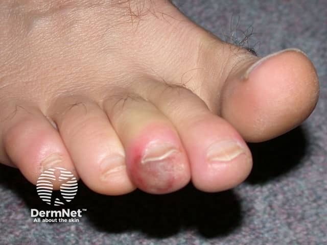
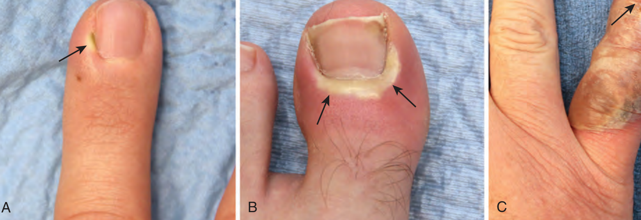
Clinical photography of a toenail unit demonstrating acute paronychia. The image shows a dorsal close‑up of the great toe with surrounding periungual soft tissue erythema and marginal swelling at the proximal nail fold. The nail plate remains present and intact, but the adjacent tissue is inflamed, with a reddish-purple hue and mild edema extending from the cuticle onto the lateral nail folds. No well‑formed abscess is clearly visible in this view, though localized tenderness and warmth are suggested by the tissue characteristics. This dermatologic finding is most consistent with acute paronychia or periungual cellulitis, often secondary to trauma, nail biting, moisture exposure, or minor infection. Differential diagnoses include chronic paronychia, fungal nail infection (onychomycosis), irritant/contact dermatitis, or less likely herpetic infection. Clinically, the image supports decision‑making regarding initial management: warm soaks, analgesia, antiseptic care, and consideration of oral antibiotics directed at common pathogens such as Staphylococcus aureus; incision and drainage if an abscess forms. This photograph serves educational and search-retrieval purposes for dermatology, infectious disease, and primary care, enabling keyword‑rich indexing: paronychia, periungual, nail fold inflammation, toe infection, dorsal toe view, nail unit, soft tissue edema, erythema, swelling, acute, bacterial. Images like this guide clinicians in triaging therapy and patient education.
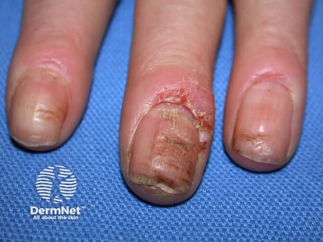
Clinical photography of the periungual digits demonstrating acute paronychia with inflamed, erythematous nail folds. The central and ring fingers show pronounced edema surrounding the proximal nail folds, with fissuring of the cuticle and crusted debris along the eponychial region. The adjacent distal nail plates appear slightly deformed and tender, consistent with early nail-fold infection. No penetrating foreign body is evident. Skin over the dorsal fingertip is uninjured beyond the periungual zones, while surrounding epidermis shows mild desquamation. The left-handed orientation presents a close-up, magnified view of three digits; the middle finger bears the most conspicuous swelling, whereas the index and ring digits display milder involvement. The overall picture is suggestive of acute bacterial paronychia, most commonly caused by Staphylococcus aureus, with potential moisture-related colonization or trauma-induced microfissures providing entry for pathogens. Clinically, this finding warrants supportive care, topical antisepsis, possible incision and drainage if abscess forms, and consideration of oral antibiotics in extensive edema or systemic symptoms. Diagnosis should be distinguished from herpetic whitlow and chronic paronychia by history, distribution, and presence of vesicles or chronic changes. Documentation of nail dystrophy, secondary infection, and risk factors such as diabetes or immunosuppression is essential for management and prognosis. Clinical relevance acknowledged.
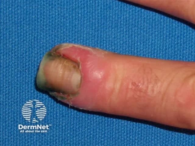
Comprehensive Description: A high-magnification clinical photograph of a single fingernail demonstrates chronic paronychia affecting the periungual region. The image captures the dorsal-lateral aspect of the distal finger, highlighting erythema and edema of the lateral nail fold with fissuring and crusting at the cuticle. The nail plate is partially displaced by surrounding inflammation, and there may be mild hypertrophy of the proximal nail fold. These findings reflect a protracted inflammatory process often linked to repeated moisture exposure, trauma from manicure practices, irritant contact, or candidal colonization. The visual features include localized redness, tenderness, soft tissue swelling, and occasional crusted debris at the cuticle; the adjacent skin appears thickened in places. Clinically, this presentation suggests chronic paronychia rather than an acute infection, and a clinician would consider secondary fungal or bacterial involvement, eczema, or psoriasis of the nail unit in the differential. The diagnostic significance lies in guiding management toward topical or systemic anti-inflammatory therapy, barrier protection, meticulous nail hygiene, and targeted antimicrobial or antifungal treatment when indicated. This image is valuable for dermatology education, clinical case assessment, and comparative studies of nail-fold inflammation or chronic paronychia progression. Early recognition improves outcomes through targeted therapy and preventive hand care for patients daily.
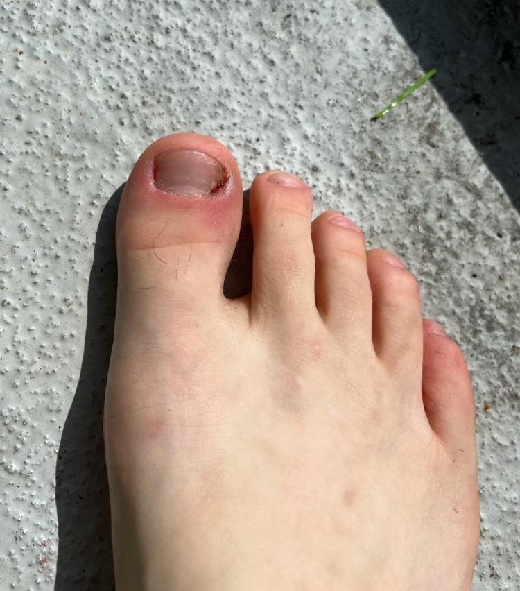
Clinical photograph of a right foot demonstrating skin and nail abnormalities associated with onychotillomania and chronic excoriation. The primary focus is the first toe (hallux), which displays significant nail plate shortening and distal-lateral onycholysis. The lateral nail fold exhibits acute-on-chronic paronychia, characterized by focal erythema, edema, and crusting, indicative of localized inflammation and secondary infection from repetitive trauma. While the medial portion of the nail shows signs of recent regrowth and less severe plate damage, the overall nail morphology remains irregular. The adjacent toes (second through fifth) appear largely uninvolved with normal skin tone and intact nail units. This image serves as a clinical example of self-induced dermatological injury (factitious disorder) and its infectious complications like paronychia, relevant for dermatology and psychiatry education regarding impulse control disorders and body-focused repetitive behaviors.
Types of Paronychia
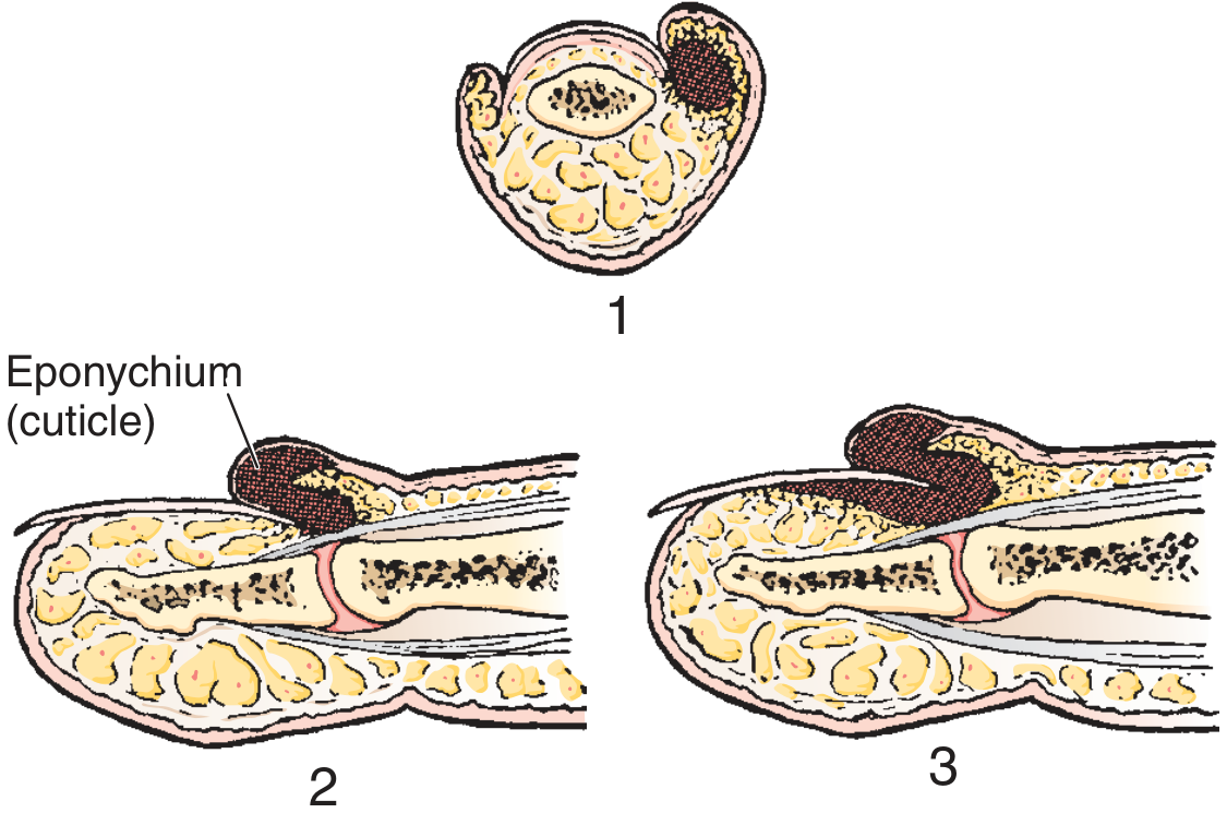
1. Acute Paronychia
- Trauma - nail biting, manicures, artificial nails, hangnails, finger sucking
- Minor cuts or abrasions of the cuticle
- CA-MRSA has been reported
- Staphylococcus aureus is the most common organism
- In children: often anaerobes (linked to finger/thumb sucking)
- Group A beta-hemolytic streptococcus (in children with pharyngitis who suck their thumb)
- Mixed aerobic + anaerobic (50% of cases in one series), pure anaerobic (25%), pure aerobic (25%)
- MRSA involvement is increasingly recognized
- Painful swelling, erythema, and tenderness at one corner of the proximal nail fold, spreading to the lateral fold
- May progress to frank abscess with fluctuance and pus
- If the nail bed is mobile, infection has spread under the nail (subungual abscess)
- Localized abscess at one side of the nail
- Spreads around the nail base, raising the eponychium but not under the nail
- End-stage: subeponychial AND subungual abscess
- Early (cellulitic, no fluctuance): warm soaks 3x/day ± oral antistaphylococcal antibiotic (cephalosporin, semisynthetic penicillin)
- Abscess formed: drainage by lifting the eponychial fold; formal incision rarely required
- Subungual extension: remove proximal third of nail with gauze wick
2. Chronic Paronychia
- Repeated/prolonged moisture exposure (water, cleaning agents, detergents)
- Contact with irritants or allergens
- Occupational: food handlers, bartenders, hairdressers, meat handlers
- Damaged cuticle allows penetration of dirt, bacteria, and particles under the proximal nail fold
- Edema and redness of the proximal nail fold
- Absence or loss of the cuticle (key sign)
- Beau's lines (transverse grooves) on the nail plate
- Nail plate surface abnormalities and dystrophy
- Gradual onset, recurrent
- Protective measures: cotton-lined rubber gloves to avoid water/irritant contact
- Topical corticosteroids (primary treatment - reduces inflammation)
- Topical antimicrobials/antifungals when indicated
- Avoid cuticle manipulation
3. Herpetic Whitlow (Viral Paronychia)
- Healthcare workers (exposure to oral secretions - nurses, dentists)
- Immunocompromised patients
- Children with primary HSV oral infection who suck their fingers
- Localized swelling with clear vesicle formation (key distinguishing feature)
- Lymphangitis and lymphadenopathy may be present
- Extremely painful
- Viral cultures from vesicle fluid
- Tzanck smear (multinucleated giant cells)
- Serum HSV antibody titers
4. Drug-Induced / EGFR-Inhibitor Paronychia
- Epidermal growth factor receptor (EGFR) inhibitors (e.g., cetuximab, erlotinib) - cause acute paronychia, often with periungual pyogenic granuloma-like lesions
- Antiretroviral therapy for HIV (particularly indinavir, lamivudine)
- Retinoids (isotretinoin)
Summary Table
| Feature | Acute (Bacterial) | Chronic | Herpetic Whitlow |
|---|---|---|---|
| Onset | Rapid (hours-days) | Slow (weeks-months) | Days |
| Cause | S. aureus, anaerobes | Moisture/irritants, Candida | HSV-1/HSV-2 |
| Key sign | Pus, fluctuance | Cuticle loss, Beau's lines | Vesicles |
| Treatment | Drainage ± antibiotics | Steroids, barrier protection | Acyclovir (NO I&D) |
| Drain? | Yes (if abscess) | No | Never |
Management of paronychia
paronychia incision drainage procedure technique nail fold
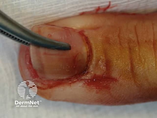
This high-resolution clinical photograph captures a close-up view of the distal finger tip focusing on the nail unit and surrounding paronychia region. The image appears to show a postoperative or laceration site along the lateral nail fold with a curvilinear incision, likely from a nail bed repair or paronychia procedure. The nail plate is partially or fully in place, with the surrounding periungual skin reddened and mildly edematous. Blood-tinged tissue and minor superficial bleeding are present at the incision margins, consistent with recent surgical manipulation or acute injury. A pair of surgical graspers can be seen near the wound, indicating active tissue handling. The skin texture shows intact epidermis with subtle desquamation; no exposed bone or deep necrosis is evident. Lighting is bright and diffuse, highlighting the pinkish-tan skin tone and vascular pattern. This image demonstrates common postoperative fingertip wound appearance, periungual skin involvement, and nail-fold anatomy, including the nail plate, surrounding cuticle, and lateral gutter. Clinical relevance includes evaluation of wound edges, signs of infection, alignment of the nail bed, and healing trajectory; used for dermatology, hand surgery, and medical education to teach nail unit injuries, wound care, suture technique, and postoperative monitoring.
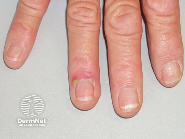
This is a clinical color photograph captured with digital close-up macro technique focusing on the nail unit of the right middle finger. The image documents the proximal nail fold region and surrounding cuticle. Visible findings include marked erythema and edema of the proximal nail fold with a small yellowish pustule/crust at the nail fold base, consistent with acute paronychia. The adjacent nail plate appears structurally normal, with no obvious subungual hematoma or distal nail dystrophy. The presentation is unilateral, localized to the medial digit, and the skin surface over the nail apparatus shows mild surface tenderness suggested by swelling. The lesion demonstrates an inflammatory process that may be secondary to minor trauma, nail-biting, manicuring injury, or moisture exposure, allowing bacterial inoculation. Differential considerations include chronic paronychia, candidal paronychia, herpetic whitlow, and ungual abscess if fluctuation develops. Clinically, the finding is important because early management with warm compresses, topical antiseptics, and antibiotic therapy or incision and drainage if an abscess is present can prevent progression. This image is relevant for dermatology education, hand surgery preoperative assessment, infectious disease case discussions, and clinical teaching on nail-fold infections and their visual differentiation from other nail disorders.
Clinical photography of a toenail unit demonstrating acute paronychia. The image shows a dorsal close‑up of the great toe with surrounding periungual soft tissue erythema and marginal swelling at the proximal nail fold. The nail plate remains present and intact, but the adjacent tissue is inflamed, with a reddish-purple hue and mild edema extending from the cuticle onto the lateral nail folds. No well‑formed abscess is clearly visible in this view, though localized tenderness and warmth are suggested by the tissue characteristics. This dermatologic finding is most consistent with acute paronychia or periungual cellulitis, often secondary to trauma, nail biting, moisture exposure, or minor infection. Differential diagnoses include chronic paronychia, fungal nail infection (onychomycosis), irritant/contact dermatitis, or less likely herpetic infection. Clinically, the image supports decision‑making regarding initial management: warm soaks, analgesia, antiseptic care, and consideration of oral antibiotics directed at common pathogens such as Staphylococcus aureus; incision and drainage if an abscess forms. This photograph serves educational and search-retrieval purposes for dermatology, infectious disease, and primary care, enabling keyword‑rich indexing: paronychia, periungual, nail fold inflammation, toe infection, dorsal toe view, nail unit, soft tissue edema, erythema, swelling, acute, bacterial. Images like this guide clinicians in triaging therapy and patient education.
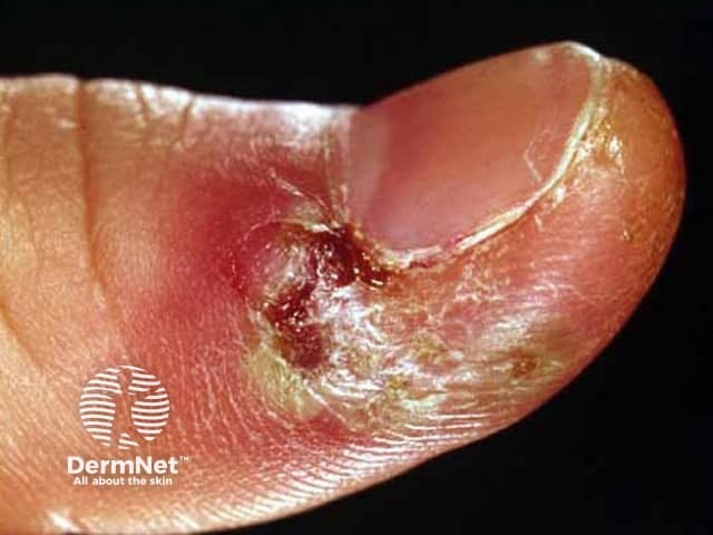
This is a high-resolution clinical photograph in visible light depicting acute herpetic paronychia of the fingernail region. Modality: Clinical photography; technique: close-up, macro view of the nail fold. Anatomical location: digits, paronychia around the nail plate of a fingertip (paronychia at the lateral/lateral nail fold). Appearance: marked erythema and edema of the periungual tissue with surrounding desquamation, crusting and whitish exudate; nearby skin may be tense; central vesicles or pustules may be present but not clearly visible in this frame; the tissue shows vesicular clusters typical of herpes simplex infection; the nail fold fissures and crusts indicate acute inflammatory response; arrangement is unilateral, localized to the lateral nail border. Pathology: HSV-induced vesiculobullous lesions targeting the proximal and lateral nail folds; differential includes bacterial paronychia; differentiation through vesicle groups on erythematous base; clinical features include painful swelling and rapid onset. Diagnostic significance: recognition of herpetic etiology guides antiviral management (acyclovir, valacyclovir) and cautions incision/drainage; may require microbiology confirmation (PCR, Tzanck smear). Clinical use: educational illustration for dermatology training, infectious disease differential, primary care assessment of paronychia; relevant for telemedicine triage; keywords: paronychia, herpes simplex virus, HSV, distal finger infection, periungual edema, vesicles, vesicular lesions, crusting, acute, infectious dermatology.
Management of Paronychia
A. Acute Paronychia
Stage 1 - Early Cellulitis (No Abscess / No Fluctuance)
- Warm soaks in warm water or chlorhexidine solution, 3 times daily
- Topical antibiotics (e.g., mupirocin/Bactroban) ± topical corticosteroids
- Short course of oral antibiotics if significant cellulitis is present:
- First-line: first-generation cephalosporin or antistaphylococcal penicillin (semisynthetic penicillin)
- If anaerobes / oral flora suspected (bite wounds, finger sucking): clindamycin or amoxicillin-clavulanate (Augmentin)
- If CA-MRSA suspected (especially in urban areas): trimethoprim-sulfamethoxazole (TMP-SMX) or clindamycin
- Incision has no role at this cellulitic stage
"If soft tissue swelling is present without fluctuation, treatment consists of frequent warm soaks (three times a day) and a short course of oral antibiotics." - Roberts & Hedges' Clinical Procedures in Emergency Medicine, p. 888
Stage 2 - Abscess Formed (Fluctuance Present)
Drainage Technique (Standard):
- Consider soaking the finger first to soften the eponychium
- Anesthesia: Digital nerve block for large paronychias; smaller ones can often be drained without anesthesia
- Instrument: No. 11 scalpel blade, scissors, or a 21-23 gauge needle (Ogunlusi technique)
- Approach: Advance the instrument parallel to the nail plate, under the eponychium, at the site of maximal swelling - lift the eponychial fold away from the nail matrix to release pus
- Fan the instrument under the eponychium to break up loculations
- Packing: Insert a small piece of gauze wick beneath the eponychium for 24 hours if significant pus is drained; remove at 24-36 hours after warm soak
"One can invariably obtain adequate drainage by simply lifting the eponychial fold away from the nail matrix to allow the pus to drain... a paronychia is not a cutaneous abscess per se, but rather a collection of pus in the potential space between the cuticle and the proximal end of the fingernail." - Roberts & Hedges', p. 889
Antibiotics Post-Drainage:
- Not essential if drainage is complete and surrounding cellulitis is minimal
- Prescribe if: significant remaining cellulitis, immunocompromised patient, or incomplete drainage
- Cultures are generally indicated given the prevalence of MRSA (Roberts & Hedges')
Tourniquet:
- A tourniquet at the base of the finger can limit bleeding and help identify the true extent of infection during drainage
"Run-around" / Both-Nail-Fold Involvement - Swiss Roll Technique:
- For severe paronychia with contiguous infection of both nail folds
- Incise both sides of the nail fold with a No. 15 scalpel (pointing away from the nail bed)
- Irrigate thoroughly with saline
- Roll the nail fold proximally over non-adherent dressing (like a "swiss roll") and suture to skin
- Remove sutures at 48 hours; unfold the nail fold back to heal by secondary intention
- Advantage: spares the nail plate (Roberts & Hedges', p. 890)
Stage 3 - Subungual Abscess (Pus Under the Nail)
- Trephination: Create one or more holes in the proximal nail with a hot paper clip or electrocautery device to allow drainage (least invasive)
- Partial nail removal (preferred): After digital block:
- Bluntly elevate the eponychium to expose the proximal nail edge
- Elevate and resect the proximal one-third of the nail with scissors
- Leave the distal two-thirds in place - acts as a physiologic dressing and reduces postoperative pain
- Place a gauze wick under the eponychium for 48 hours
- If infection is under the lateral nail edge: Gently elevate and excise longitudinally - avoid the nail matrix
"Leave the distal two thirds of the nail in place to act as a physiologic dressing and to decrease postoperative pain." - Schwartz's Principles of Surgery 11e, p. 1979
Post-Procedure Care (Acute)
- Warm tap water soaks at home after anesthesia wears off
- Antibiotic ointment periodically to the site for several days
- Remove packing at 24-36 hours with warm water soak
- 1-2 follow-up visits to assess healing
- If infection lasts > few weeks: evaluate for osteomyelitis of the distal phalanx (rare but well-known complication)
B. Chronic Paronychia
Conservative (First-Line):
- Barrier protection: Cotton-lined rubber gloves to prevent contact with water and irritants - essential and must be addressed for treatment to succeed
- Topical corticosteroids: Primary anti-inflammatory treatment
- Topical antifungals/antimicrobials: When Candida colonization is confirmed
- Avoid cuticle manipulation or manicures
Surgical (Refractory Cases):
- Marsupialization (eponychial marsupialization): Excision of the dorsal eponychium; nail plate removal with marsupialization of skin proximal to the eponychial fold allows secondary healing
- Required when chronic paronychia is unresponsive to conservative/oral therapy
"They do not respond to antibiotic treatment, and nail plate removal with marsupialization of the skin proximal to the eponychial fold will allow the wound to heal secondarily. The environmental factors leading to the chronic paronychia must also be corrected in order for treatment to be successful." - Schwartz's Principles of Surgery 11e, p. 1979
C. Herpetic Whitlow (Viral)
| Step | Action |
|---|---|
| DO NOT incise and drain | No pus - incision causes dissemination and secondary bacterial infection |
| Analgesia | Oral analgesics for pain |
| Antivirals (if < 48 hrs) | Acyclovir or valacyclovir - may shorten course |
| Recurrent herpetic whitlow | Suppressive antiviral therapy |
| Secondary bacterial infection | Then add antibiotics |
| Self-limiting | Resolves in ~2-3 weeks |
"I&D is contraindicated" for herpetic whitlow. "Antivirals may be prescribed if infection has been present for <48 hr. For recurrent herpetic whitlow, suppressive therapy with an antiviral agent may be helpful." - Roberts & Hedges', Table 57.1
Antibiotic Summary Table
| Condition | Organisms | Antibiotic |
|---|---|---|
| Acute paronychia | S. aureus, streptococci; anaerobes/GNR if oral flora exposure | 1st-gen cephalosporin or antistaphylococcal penicillin; clindamycin or amoxicillin-clavulanate if anaerobes |
| MRSA suspected | CA-MRSA | TMP-SMX or clindamycin (oral); vancomycin IV |
| Chronic paronychia | Candida albicans (fungal) | Topical antifungal; steroids for inflammation |
| Herpetic whitlow | HSV-1 / HSV-2 | Acyclovir/valacyclovir (supportive); I&D contraindicated |
Key Principles Summary
- No abscess - soaks + antibiotics; no incision
- Abscess present - lift the eponychium (needle/blade parallel to nail); simple and curative
- Subungual extension - remove proximal third of nail; leave distal two-thirds
- Both folds involved - Swiss roll technique
- Chronic - barrier protection + steroids ± antifungal; marsupialization if refractory
- Herpetic whitlow - NEVER incise; antivirals early
- Always culture (MRSA is prevalent)
- Complication watch: Osteomyelitis of distal phalanx in persistent cases