
Range of movements of wrist joint
- Flexion: ~80° (palmar flexion)
- Extension: ~70° (dorsiflexion)
- Abduction (radial deviation): ~20°
- Adduction (ulnar deviation): ~30-40°
- Circumduction: combination of the above
range of movements of wrist joint diagram
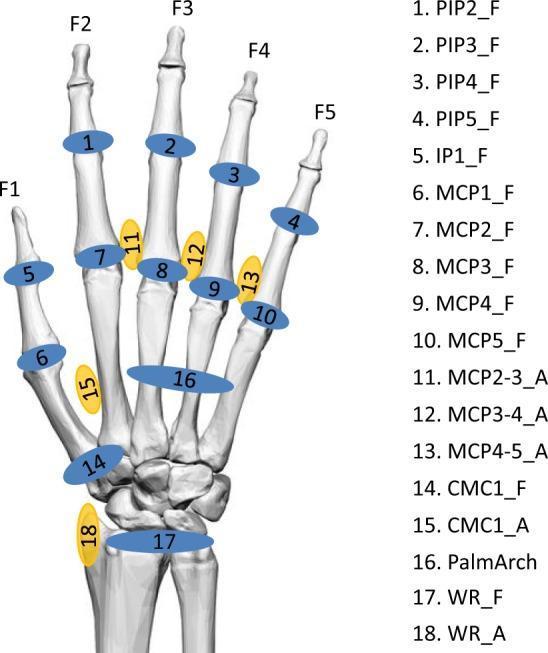
This anatomical diagram displays a skeletal model of the human hand and wrist, serving as a biomechanical reference for recorded anatomical angles. The illustration uses a color-coded numbering system to identify specific joint movements across the digits (F1-F5) and carpus. Blue markers represent flexion (_F), while yellow markers represent abduction (_A). The labeled joints include: the distal and proximal interphalangeal joints (IP1, PIP2-5), the metacarpophalangeal joints (MCP1-5), the carpometacarpal joint of the thumb (CMC1), the palmar arch (PalmArch), and the wrist (WR). Specifically, it identifies flexion for all major finger joints and abduction for the inter-metacarpal spaces (MCP2-3, 3-4, 4-5), the thumb (CMC1_A), and the wrist (WR_A). This diagram is intended for clinical study and biomechanical research, providing a standardized nomenclature for range of motion (ROM) tracking and orthopedic assessment of hand kinematics.
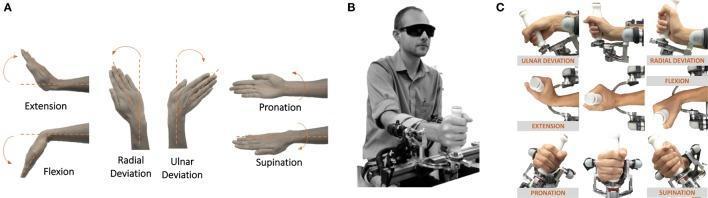
This composite educational image illustrates the three degrees of freedom (DoFs) of the human wrist and a corresponding robotic rehabilitative/experimental device. Section A is an anatomical diagram depicting the range of motion for flexion and extension in the sagittal plane, radial and ulnar deviation in the coronal plane, and pronation and supination along the longitudinal axis of the forearm. Section B shows a clinical photograph of a blindfolded participant utilizing a high-precision robotic wrist manipulandum, with the forearm strapped to a mechanical support to ensure joint alignment and repeatability. Section C provides a detailed series of clinical images showing the kinematics of the robotic device as it facilitates specific wrist movements: ulnar and radial deviation, flexion and extension, and pronation and supination. This visual material is intended for medical training in orthopedics, physical therapy, and robotic rehabilitation, demonstrating the interface between biomechanical joint movement and haptic haptic feedback technology.
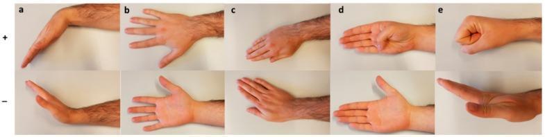
This clinical photograph series demonstrates the range of motion (ROM) assessment for the human hand and wrist, categorized into five distinct movements (a-e). The top row (+) represents positive directional movements, while the bottom row (-) represents negative directional movements. (a) Wrist Flexion (+) and Extension (-): illustrating palmar and dorsal angulation of the radiocarpal joint. (b) Radial (+) and Ulnar (-) Deviation: showing lateral movement of the hand toward the radius and ulna respectively. (c) Pronation (+) and Supination (-): demonstrating rotation of the forearm resulting in palm-down and palm-up orientations. (d) Thumb Flexion (+) and Extension (-): focusing on the movement of the metacarpophalangeal (MCP) and interphalangeal (IP) joints of the thumb. (e) Finger Flexion (+) and Extension (-): showing the transition from a closed fist to fully extended fingers, involving the MCP, proximal interphalangeal (PIP), and distal interphalangeal (DIP) joints. This visual serves as a standardized reference for clinical orthopedic examination and kinesiologic evaluation.
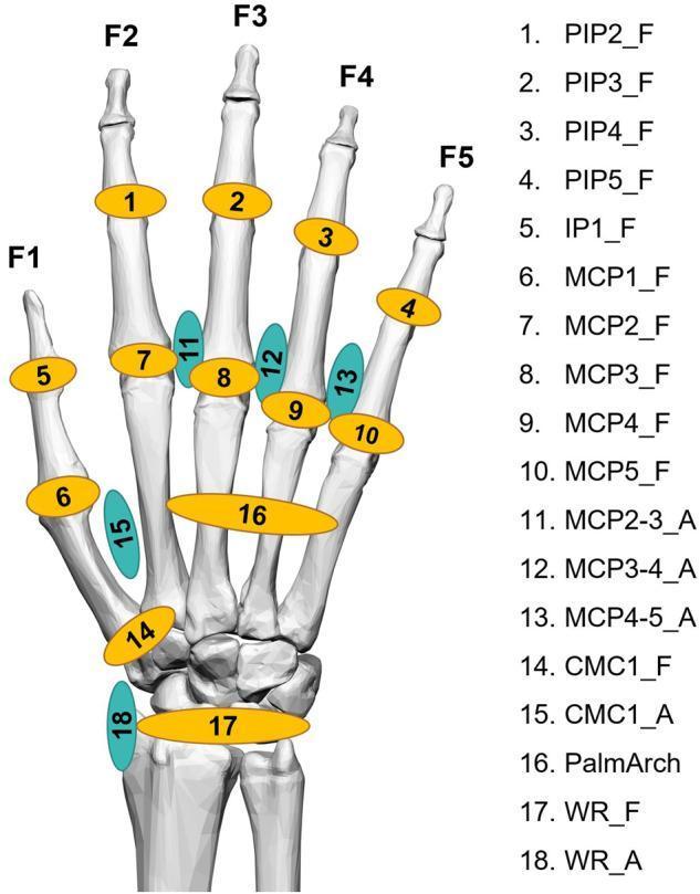
This anatomical diagram illustrates the 18 key kinematic angles of the human hand and wrist used for biomechanical analysis and data glove calibration. The visual depicts a skeletal model of the hand with numbered markers corresponding to specific joint movements. Markers 1-10 and 14 represent flexion (indicated by yellow ovals and the suffix '_F'), including the interphalangeal (IP), proximal interphalangeal (PIP), metacarpophalangeal (MCP), and carpometacarpal (CMC) joints for digits F1 through F5. Turquoise ovals (suffix '_A') denote abduction/adduction movements, located between the MCP joints (11-13), at the CMC1 joint (15), and the wrist (18). Marker 16 highlights the palmar arch, representing the complex movement of the ring and little finger CMC joints. Markers 17 and 18 represent wrist flexion/extension and abduction/adduction, respectively. This diagram serves as a reference for hand kinesiology, orthopedic assessment, and the development of wearable motion-capture technologies in rehabilitation and ergonomics.
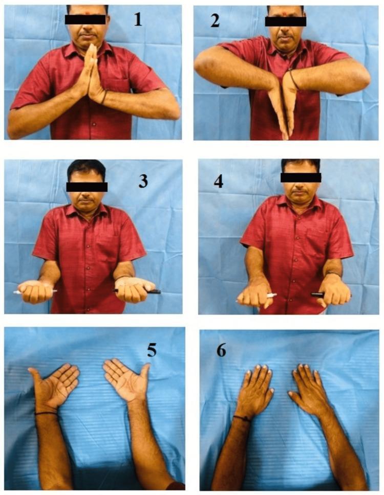
This composite of clinical photographs illustrates the standard functional range of motion (ROM) assessment for the wrist and forearm. The six panels depict a patient performing specific bilateral movements: 1. Dorsiflexion (extension) with palms pressed together; 2. Palmar flexion with the dorsal surfaces of the hands together; 3. Forearm supination with palms facing upward while holding pens to visualize rotational axis; 4. Forearm pronation with palms facing downward; 5. Ulnar deviation showing the hands angled away from the midline; and 6. Radial deviation showing the hands angled toward the midline. The images serve as an educational guide for orthopedic and physical therapy examinations, demonstrating the clinical evaluation of the radiocarpal and distal radioulnar joints. These maneuvers are essential for assessing joint mobility following distal radius fractures or other musculoskeletal injuries.
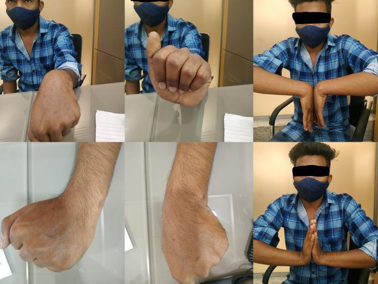
A composite of six clinical photographs demonstrating the range of motion (ROM) of the wrist and hand in a patient six months post-surgery for a wrist injury. The top row shows the patient performing active wrist movements: the first frame displays a neutral wrist position with a closed fist; the second shows a fist with thumb extension; and the third demonstrates bilateral wrist palmar flexion in a 'reverse Phalen' maneuver. The bottom row provides detailed views: the left and middle frames are close-up lateral and dorsal photographs of the wrist during active palmar flexion and dorsiflexion, showing healthy skin integrity with minimal scarring and no significant edema. The final frame depicts bilateral wrist extension with palms pressed together (Prayer sign). This series evaluates postoperative functional recovery, specifically volar tilt, radial inclination, and the modified Mayo wrist score, illustrating successful restoration of joint mobility and grip strength following surgical intervention.
Range of Movements of the Wrist Joint
1. Flexion (Palmar Flexion)
- Normal range: 80-90°
- Functional range: ~10°
- Produced mainly by: flexor carpi radialis, flexor carpi ulnaris, palmaris longus (assisted by the long digital flexors)
- ~2/3 of wrist flexion-extension occurs at the radiocarpal joint; ~1/3 at the intercarpal (midcarpal) joint
2. Extension (Dorsiflexion)
- Normal range: 70-80°
- Functional range: ~35°
- Produced by: extensor carpi radialis longus & brevis, extensor carpi ulnaris
- Loss of extension is the most incapacitating functional impairment of wrist motion
3. Radial Deviation (Abduction)
- Normal range: 15-20° (up to 30°)
- Functional range: ~10°
- Primarily an intercarpal movement
- Produced by: flexor carpi radialis + extensor carpi radialis (acting together)
4. Ulnar Deviation (Adduction)
- Normal range: 35-50°
- Functional range: ~15°
- Relies on both radiocarpal and intercarpal motion
- Produced by: flexor carpi ulnaris + extensor carpi ulnaris (acting together)
- Greater range than radial deviation (ulna's styloid process is shorter than the radius's)
5. Circumduction
- A combination of the four movements above (not a pure rotation)
- The hand traces a cone shape
Note: Pronation and supination of the forearm are not wrist movements - they occur at the proximal and distal radioulnar joints.
Summary Table
| Movement | Normal ROM | Functional ROM |
|---|---|---|
| Flexion (palmar) | 80-90° | ~10° |
| Extension (dorsal) | 70-80° | ~35° |
| Radial deviation | 15-20° | ~10° |
| Ulnar deviation | 35-50° | ~15° |
| Circumduction | Combined | - |
Biomechanical Notes
- The instant center of rotation is usually the head of the capitate, but it varies
- The most functionally important motion in daily activities is "dart-thrower's motion" - a coupled out-of-plane movement from radial extension to ulnar flexion (e.g., throwing, hammering, drinking). Nearly all of this occurs at the midcarpal joint
- The wrist acts as an intercalated link system: radius → lunate → capitate
- The scaphoid links the two carpal rows and coordinates their motion
Clinical Reference Image

- Firestein & Kelley's Textbook of Rheumatology - Wrist and Carpal Joints section (p. 4787-4789)
- Miller's Review of Orthopaedics, 9th Ed. - Wrist and Hand Biomechanics (p. 128): Normal = 65° flexion, 55° extension, 15° radial deviation, 35° ulnar deviation
- Imaging Anatomy, Vol. 3 - Biomechanics (p. 605)