
Clear definition or visual pathway
What topic would you like a clear definition or visual pathway for?
Please type the specific medical or clinical concept you want a clear definition or visual pathway for (e.g., 'coagulation cascade', 'action potential', 'renin-angiotensin-aldosterone system', etc.):
All about visual pathway, definition parts and all
visual pathway diagram optic nerve chiasm tract cortex
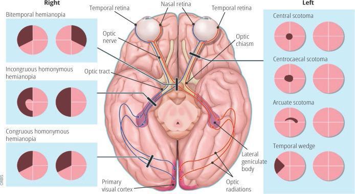
Anatomical diagram and educational infographic detailing the human visual pathway and associated visual field defects. The central illustration shows a superior (dorsal) view of the brain, highlighting the optic nerve, optic chiasm (decussation point), optic tract, lateral geniculate body, optic radiations, and the primary visual cortex in the occipital lobes. Pathways are color-coded (red and blue) to demonstrate the transition from nasal and temporal retinal fibers to the contralateral and ipsilateral hemispheres. Black lesion bars indicate specific sites of damage along this pathway. Flanking the central diagram are clinical representations of visual field loss patterns. The 'Right' panel displays bilateral defects including bitemporal hemianopia, incongruous homonymous hemianopia, and congruous homonymous hemianopia. The 'Left' panel displays unilateral or localized defects including central scotoma, centrocaecal scotoma, arcuate scotoma, and temporal wedge. This diagram serves as a neuro-ophthalmological guide for localizing lesions within the central nervous system based on perimetry findings.
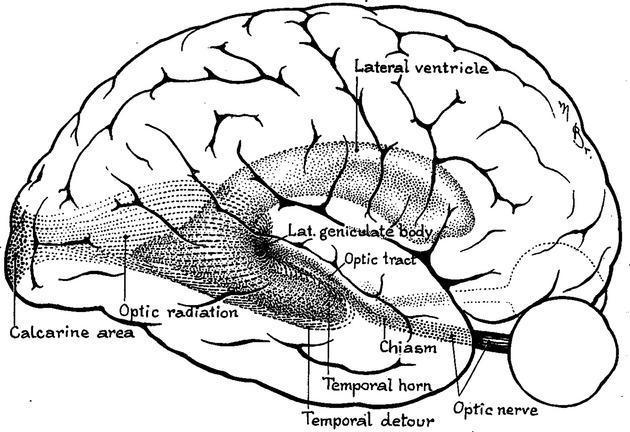
This anatomical diagram provides a lateral view of the human brain, specifically illustrating the visual pathway and the spatial distribution of the optic radiation. Based on Harvey Cushing's 1921 drawing, the illustration highlights the geniculocalcarine tract from its origin at the lateral geniculate body (LGB) to its termination in the primary visual cortex (calcarine area). A key educational focus is the 'temporal detour' or Meyer’s loop, where the ventral fibers of the optic radiation arc anteriorly around the temporal horn of the lateral ventricle before heading posteriorly toward the occipital lobe. Other labeled anatomical landmarks include the optic nerve, optic chiasm, optic tract, and the main body of the lateral ventricle. This diagram is significant for understanding neuroanatomy related to visual field deficits (e.g., quadrantanopia) that may arise from lesions or neurosurgical procedures in the temporal lobe, such as epilepsy surgery or tumor resection.
The Visual Pathway - Complete Guide
Definition
Master Diagram
Step-by-Step Pathway
1. Retina - The Starting Point
- Parasol cells (Magnocellular / M pathway) - large receptive fields, detect motion and gross features
- Midget cells (Parvocellular / P pathway) - small receptive fields, detect fine detail and color
- Neuroanatomy through Clinical Cases, 3rd Ed.
2. Optic Nerve (CN II)
- Exits the orbit through the optic canal of the sphenoid bone
- Unique among cranial nerves: its axons are myelinated by oligodendrocytes (not Schwann cells), and it is covered by cranial meninges - making it a true CNS structure
- Carries information from one eye only (monocular)
- Gray's Anatomy for Students
3. Optic Chiasm
- Located on the ventral surface of the brain, just anterior to the pituitary gland (sits ~10 mm above the pituitary fossa)
- Named after the Greek letter chi (χ) but in life resembles the letter H
- The critical crossing point: 50% of fibers decussate
- Nasal (medial) retinal fibers → cross to the contralateral optic tract (these carry temporal visual field information)
- Temporal (lateral) retinal fibers → remain ipsilateral (these carry nasal visual field information)
- Scott-Brown's Otorhinolaryngology, Vol. 1 | Neuroanatomy through Clinical Cases
4. Optic Tract
- Runs from the chiasm, wrapping around the midbrain laterally, to reach the LGN
- Carries fibers from the contralateral visual field of both eyes
- A small branch diverts to the pretectal area and superior colliculus - this mediates the pupillary light reflex (not conscious vision)
5. Lateral Geniculate Nucleus (LGN) of the Thalamus
- Layers 1 & 2 = Magnocellular (receive input from parasol cells; motion/coarse vision) → project to cortical layer 4Cα
- Layers 3-6 = Parvocellular (receive input from midget cells; fine detail/color) → project to cortical layer 4Cβ
- Inputs from the two eyes are kept separate in alternating layers
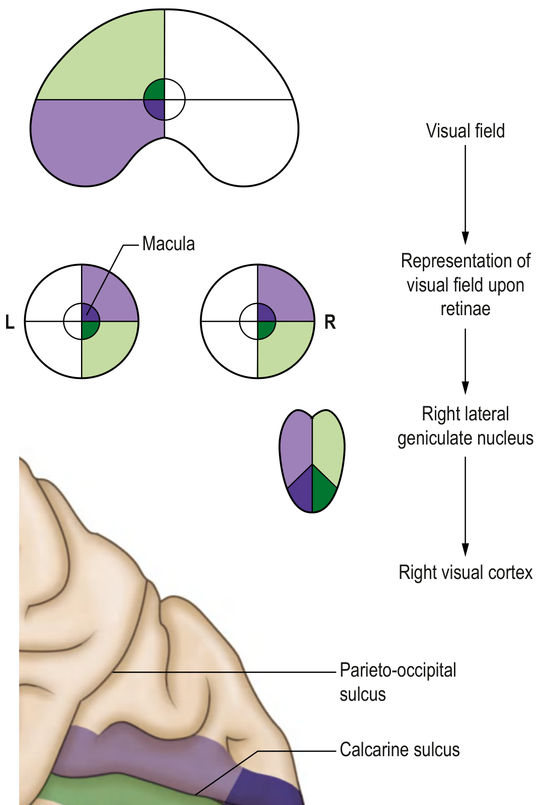
6. Optic Radiations (Geniculocalcarine Tract)
| Division | Route | Carries | Lesion Effect |
|---|---|---|---|
| Inferior fibers (Meyer's loop) | Arc forward into the temporal lobe | Superior visual field | Contralateral superior quadrantanopia - "pie in the sky" |
| Superior fibers | Pass under the parietal lobe | Inferior visual field | Contralateral inferior quadrantanopia - "pie on the floor" |
7. Primary Visual Cortex (V1 / Area 17 / Striate Cortex)
- Located in the occipital lobe, along the calcarine sulcus
- Organized retinotopically - each point in the visual field has a corresponding cortical representation
- The macula (central vision) has a disproportionately large cortical representation (macular magnification)
- Upper visual field → maps to cortex below the calcarine sulcus
- Lower visual field → maps to cortex above the calcarine sulcus
Beyond V1 - Higher Visual Processing
| Stream | Route | Function |
|---|---|---|
| Dorsal ("Where?" stream) | V1 → Parieto-occipital cortex | Spatial location, motion, visuospatial navigation |
| Ventral ("What?" stream) | V1 → Occipitotemporal cortex | Object recognition, color, faces, reading |
- Neuroanatomy through Clinical Cases, 3rd Ed.
Summary of Lesions and Visual Field Defects
| Lesion Site | Visual Field Defect |
|---|---|
| Optic nerve (before chiasm) | Monocular blindness (one eye only) |
| Optic chiasm (central) | Bitemporal hemianopia |
| Optic tract | Incongruous contralateral homonymous hemianopia |
| Meyer's loop (temporal lobe) | Contralateral superior quadrantanopia ("pie in the sky") |
| Parietal optic radiations | Contralateral inferior quadrantanopia ("pie on the floor") |
| Complete optic radiations | Congruous contralateral homonymous hemianopia |
| Occipital cortex (V1) | Congruous homonymous hemianopia ± macular sparing |
Key Concepts to Remember
- Before the chiasm = monocular defect (one eye affected)
- At the chiasm = bitemporal defect (nasal fibers from both eyes cross)
- After the chiasm = homonymous defect (both eyes lose the same visual hemifield)
- The more posterior (closer to V1) the lesion, the more congruous (symmetrical) the defect
- Meyer's loop makes the temporal lobe vulnerable to causing superior visual field loss
- Macular sparing in occipital lesions is because the macular cortex receives dual blood supply