
Vasoclusive crisis
Reading File
Vaso-occlusive crisis (VOC) is the hallmark complication of sickle cell disease. It occurs when sickle-shaped RBCs obstruct small blood vessels, causing ischemia and severe pain - typically in bones, chest, abdomen, and joints. It is triggered by dehydration, infection, cold exposure, hypoxia, or stress.
Searching sources...
Finding Sources
Searching PubMed
"vaso-occlusive crisis" AND "sickle cell disease"
Finding Sources
Finding Sources
Reading File
Reading File
Reading File
Reading File
Reading File
Reading File
Loading Image
Here is a comprehensive overview of Vaso-Occlusive Crisis (VOC) in sickle cell disease, synthesized from Tintinalli's Emergency Medicine and related textbooks.
Vaso-Occlusive Crisis (VOC) in Sickle Cell Disease
Pathophysiology
Sickle cell anemia (SCA) results from a single amino acid substitution - valine replaces glutamic acid at the sixth position of the beta-globin chain - creating abnormal hemoglobin S (HbS). When HbS is deoxygenated, it undergoes noncovalent polymerization, forming chains that distort the RBC membrane into the characteristic sickle shape.
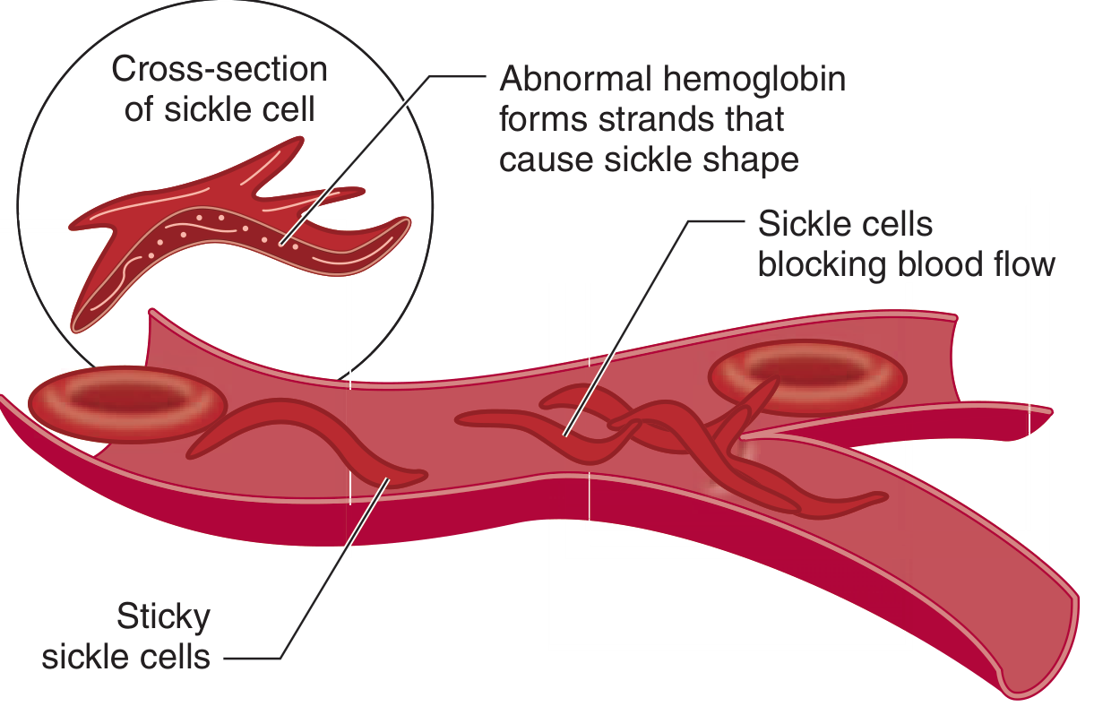
The sickled, rigid cells:
- Adhere to the vascular endothelium (activated endothelium plays a key role)
- Increase blood viscosity
- Obstruct small vessels (arterioles and capillaries)
- Cause tissue ischemia and ischemia-reperfusion injury
This creates a vicious cycle: obstruction → hypoxia and acidosis → further sickling → more obstruction. From 5% to 50% of circulating erythrocytes in HbSS patients can be irreversibly sickled, with a markedly shortened RBC lifespan (~20 days vs. 120 days normal).
The disease does not manifest until fetal hemoglobin (HbF) is replaced by HbS, typically after 4 months of age.
Types of Sickle Cell Crisis
| Type | Key Features |
|---|---|
| Vaso-occlusive (pain) crisis | Most common; ischemia of bone, viscera, soft tissue |
| Hemolytic crisis | Acute drop in Hct/Hgb from increased hemolysis |
| Aplastic crisis | Bone marrow suppression (often parvovirus B19); low reticulocyte count |
| Splenic sequestration crisis | Splenic pooling of RBCs; typically in children |
Vaso-Occlusive Pain Crisis - Clinical Features
VOC accounts for 79-91% of all ED visits in patients with SCA.
Common triggers:
- Infection
- Cold exposure
- Dehydration
- Altitude
- Hypoxia
- Physical or emotional stress
Pain locations (in order of frequency):
- Long bones and lower back (most common)
- Extremities
- Abdomen (can mimic acute abdomen - tends to be diffuse, associated with limb/chest pain)
- Chest
Individual patients tend to have their crises in characteristic locations. Only 5% of children account for 33% of all episodes, averaging 3-10 crises/year.
Signs that suggest VOC is NOT simple pain crisis:
- Limited range of motion of a joint (raises concern for septic joint)
- WBC >20,000/mm³ with a left shift (infection, not crisis)
- Low reticulocyte count (aplastic crisis)
Note: Low-grade fever and modest leukocytosis can occur in VOC itself, potentially masking infection.
Complications (always anticipate these)
| System | Complication |
|---|---|
| Neurologic | Ischemic/hemorrhagic stroke, TIA, cerebral vasculopathy |
| Pulmonary | Acute chest syndrome (ACS) - most common cause of death |
| Renal | Papillary necrosis, hematuria, renal failure |
| Splenic | Sequestration, autosplenectomy |
| Bone | Osteomyelitis, avascular necrosis |
| Ophthalmic | Retinal ischemia, proliferative retinopathy |
Stroke is 250 times more common in SCA children than the baseline population; 11% suffer overt stroke and 20% have silent strokes on imaging.
Acute Chest Syndrome (ACS) can develop from or alongside VOC - driven by a vicious cycle where free plasma heme from hemolysis worsens vascular permeability, triggering pulmonary sickling.
Workup (Emergency)
- CBC + reticulocyte count - assess degree of anemia; low retic suggests aplastic crisis
- Peripheral blood smear - polychromasia confirms ongoing RBC production
- BMP - electrolytes, renal function
- LFTs - liver involvement
- Urinalysis - papillary necrosis / hematuria
- Blood cultures - if fever present
- Chest X-ray - if respiratory symptoms (rule out ACS)
- ABG - if significant respiratory distress or hypoxia on pulse oximetry
Management
1. Pain Management (timely and aggressive)
- Opioids are the mainstay for moderate-severe VOC
- Ketorolac (NSAIDs) for adjunctive analgesia, especially in children
- Titrate to effect; monitor for respiratory suppression with opioids
2. Hydration
- Oral hydration preferred when feasible
- IV hydration if needed: D5W in 0.45% NaCl at 1-1.5x maintenance
- Caution: aggressive IV fluids can worsen ACS
3. Supplemental Oxygen
- Adjust to pulse oximetry
4. Incentive Spirometry
- Prevents atelectasis, which can trigger ACS
5. Treat Underlying Trigger
- Empiric antibiotics if infection is suspected (3rd-gen cephalosporin such as ceftriaxone 100 mg/kg/day for encapsulated organisms + macrolide if pulmonary source)
6. Transfusion
- Simple transfusion for severe acute anemia
- Exchange transfusion (reduce HbS to <30%) for: severe ACS, multiorgan dysfunction, neurologic complications, deterioration despite initial management, pre-op for major surgery
Disease-Modifying Therapy (Outpatient/Long-term)
| Therapy | Mechanism / Benefit |
|---|---|
| Hydroxyurea | Induces HbF production; decreases leukocytes; vasodilation. Reduces frequency and severity of crises. Recommended in all children >9 months regardless of disease severity |
| Chronic transfusion therapy | Keeps HbS <30%; for severe/frequent crises |
| Penicillin prophylaxis | Through age 5 years (functional asplenia risk) |
| Folic acid | Supplementation due to increased RBC turnover |
| Pneumococcal vaccine | Critical because of autosplenectomy |
Hydroxyurea is the only disease-modifying drug approved for SCA and works by inducing HbF, which inhibits HbS polymerization, reduces sickling, and decreases vaso-occlusion frequency.
Sources: Tintinalli's Emergency Medicine: A Comprehensive Study Guide, Miller's Anesthesia (10e), Quick Compendium of Clinical Pathology (5e), Sleisenger and Fordtran's GI and Liver Disease, Swanson's Family Medicine Review.
This is a shared conversation. Sign in to Orris to start your own chat.