
Acute inflammation - explain this for 10 mark essay
Acute Inflammation
Definition and Overview
- Rubor (redness)
- Calor (heat)
- Tumor (swelling)
- Dolor (pain)
- Functio laesa (loss of function)
1. Causes
- Infections (bacteria, viruses, fungi, parasites)
- Tissue necrosis (infarction, trauma, burns, chemical injury)
- Foreign bodies
- Immune reactions (hypersensitivity)
2. Vascular Reactions
Changes in Vascular Flow and Caliber
Exudate vs. Transudate: An exudate is protein-rich extravascular fluid implying increased vascular permeability. A transudate is protein-poor, arising from osmotic/hydrostatic imbalance without increased permeability.
Increased Vascular Permeability (Vascular Leakage)
- Endothelial cell contraction - the most common mechanism; induced by histamine, bradykinin, leukotrienes, and substance P acting on postcapillary venules; occurs within minutes (immediate transient response)
- Endothelial injury - direct damage by burns, toxins, or microbes causing immediate sustained response
- Leukocyte-mediated injury - activated neutrophils and monocytes release proteases and ROS that damage the endothelium
- Increased transcytosis - VEGF and other factors increase transport across endothelium
- Leakage from new blood vessels during repair (angiogenesis)
3. Cellular Events - Leukocyte Recruitment
Step 1: Margination and Rolling
- P-selectin: rapidly redistributed from Weibel-Palade bodies to the endothelial surface on stimulation by histamine/thrombin
- E-selectin: induced on endothelium by TNF and IL-1 within 1-2 hours
- L-selectin: on leukocytes, binds endothelial ligands
Step 2: Firm Adhesion
Step 3: Transmigration (Diapedesis)
Step 4: Chemotaxis
- Exogenous: bacterial products (N-formyl methionine peptides)
- Endogenous: C5a, leukotriene B4 (LTB4), IL-8 (CXCL8), chemokines
4. Leukocyte Activation and Phagocytosis
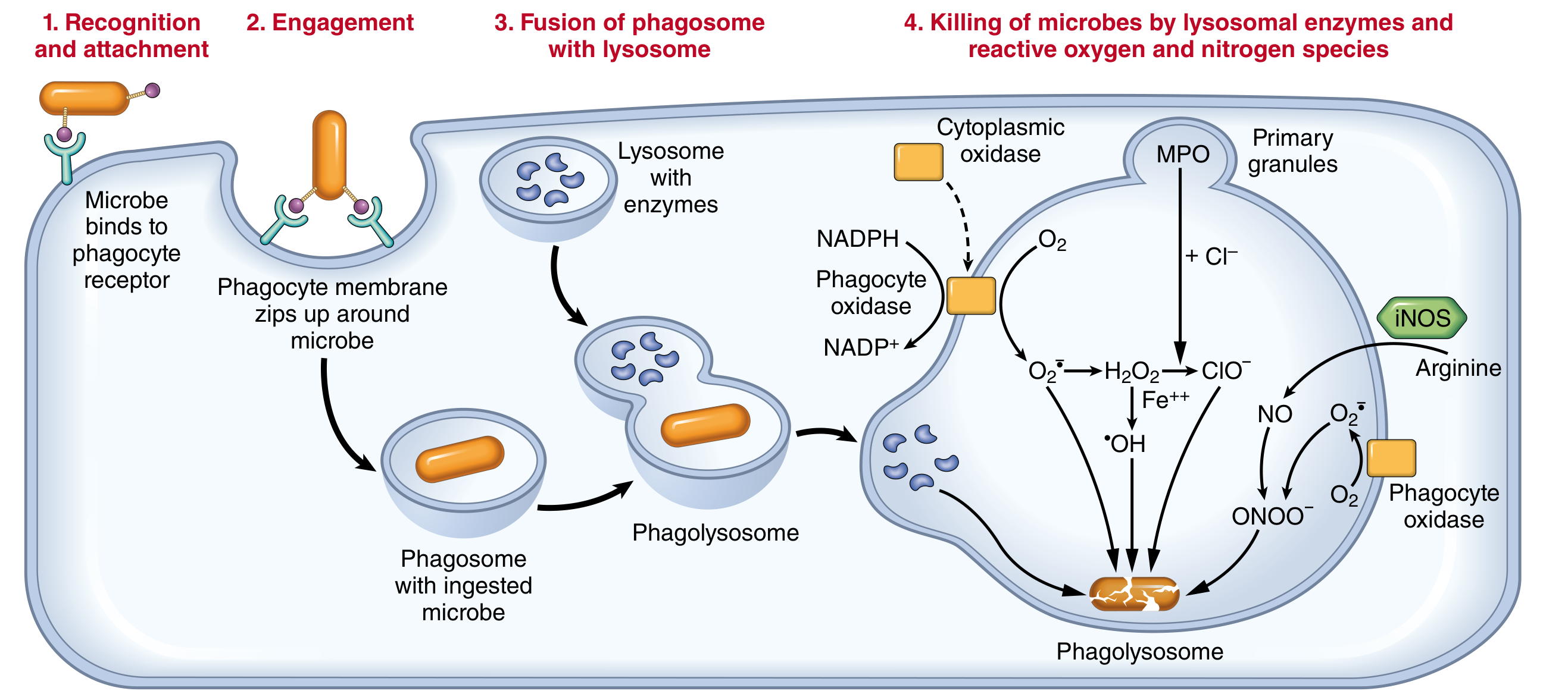
Phagocytosis
- Recognition and attachment - enhanced by opsonins (IgG, C3b) binding to Fc receptors and complement receptors on leukocytes
- Engulfment - cytoplasmic extensions flow around the particle forming a phagosome, which fuses with lysosomes to form a phagolysosome
- Killing and degradation
Intracellular Killing
- Reactive oxygen species (ROS): NADPH oxidase assembles in the phagosome membrane, oxidizing NADPH and generating superoxide (O2-) - the respiratory burst. Superoxide is converted to H2O2 and then to hypochlorous acid (HOCl) by myeloperoxidase (MPO), the most potent bactericidal mechanism in neutrophils.
- Reactive nitrogen species: Inducible nitric oxide synthase (iNOS) generates NO, which reacts with superoxide to form peroxynitrite (ONOO-)
- Lysosomal enzymes: elastase, cathepsins, defensins, lysozyme
Neutrophil Extracellular Traps (NETs)
5. Mediators of Inflammation
| Mediator | Source | Principal Action |
|---|---|---|
| Histamine | Mast cells, basophils | Vasodilation, increased permeability |
| Serotonin | Platelets | Vasodilation, increased permeability |
| Prostaglandins (PGE2, PGI2) | All cells (via COX) | Vasodilation, fever, pain |
| Leukotrienes (LTB4) | Leukocytes (via 5-LOX) | Chemotaxis |
| LTC4, LTD4, LTE4 | Mast cells, leukocytes | Increased permeability, bronchoconstriction |
| C3a, C5a | Complement cascade | Chemotaxis, opsonization, mast cell degranulation |
| TNF, IL-1 | Macrophages, mast cells | Endothelial activation, fever, acute-phase response |
| IL-8 (CXCL8) | Macrophages, endothelium | Neutrophil chemotaxis |
| Bradykinin | Kinin system (factor XII) | Vasodilation, pain, increased permeability |
| PAF | Leukocytes, endothelium | Platelet aggregation, leukocyte activation |
6. Morphologic Patterns of Acute Inflammation
| Pattern | Features | Example |
|---|---|---|
| Serous | Watery, protein-poor exudate; no significant neutrophils | Skin blister, pleural effusion in viral pleuritis |
| Fibrinous | Large protein exudate with fibrin deposition; "bread and butter" pericarditis | Acute pericarditis, lobar pneumonia |
| Purulent (Suppurative) | Abundant pus (neutrophils + dead cells + microbes); abscess formation | Bacterial pneumonia, brain abscess |
| Ulcer | Local defect of epithelial surface; necrosis of inflamed tissue | Peptic ulcer, pressure sore |
7. Outcomes of Acute Inflammation
- Complete resolution - the usual outcome when injury is limited and parenchyma can regenerate; involves macrophage clearance of debris and lymphatic resorption of edema
- Healing by scarring (fibrosis/organization) - when tissue destruction is extensive, or the tissue cannot regenerate (e.g., cardiac muscle); fibrin exudate is organized by connective tissue ingrowth
- Progression to chronic inflammation - when the injurious agent persists or normal healing is impaired; characterized by shift from neutrophils to mononuclear infiltrate (macrophages, lymphocytes)
Summary
- Vascular phase: vasodilation and increased permeability causing exudate formation
- Cellular phase: leukocyte margination, rolling (selectins), firm adhesion (integrins/ICAM), transmigration (PECAM-1), chemotaxis, and phagocytosis
- Mediator cascade: histamine, eicosanoids, cytokines, complement, and kinins amplify and direct the response
- Resolution or progression: complete healing if the agent is eliminated; scarring or chronic inflammation if not
Cellular events ah flow chart ah kudu
A detailed medical flowchart showing the cellular events of acute inflammation, with the following sequential steps and arrows connecting them: Title: "Cellular Events of Acute Inflammation" Flow: 1. INJURY / INFECTION (box at top) ↓ 2. STASIS OF BLOOD FLOW (slowing of blood in postcapillary venules) ↓ 3. MARGINATION (leukocytes move to periphery of vessel) ↓ 4. ROLLING (leukocytes loosely attach and roll along endothelium - mediated by P-selectin, E-selectin, L-selectin) ↓ 5. FIRM ADHESION (integrins LFA-1, MAC-1 bind ICAM-1, VCAM-1 on endothelium - induced by TNF, IL-1, chemokines) ↓ 6. TRANSMIGRATION / DIAPEDESIS (leukocytes squeeze through endothelial junctions - mediated by PECAM-1/CD31, then cross basement membrane using collagenases) ↓ 7. CHEMOTAXIS (leukocytes migrate along chemical gradient toward site of injury - agents: C5a, LTB4, IL-8, bacterial N-formyl peptides) ↓ 8. LEUKOCYTE ACTIVATION (by microbes, cytokines, immune complexes - neutrophils arrive first at 6-24h, then monocytes/macrophages at 24-48h) ↓ 9. PHAGOCYTOSIS Three sub-steps shown in a box: a) Recognition & Attachment (opsonins: IgG, C3b bind Fc receptors, complement receptors) b) Engulfment (phagosome formation → phagolysosome) c) Killing & Degradation ↓ 10. INTRACELLULAR KILLING (two branches): Branch A: ROS pathway → NADPH oxidase → Superoxide (O2-) → H2O2 → HOCl (via MPO) → BACTERIAL KILLING Branch B: RNS pathway → iNOS → Nitric Oxide (NO) → Peroxynitrite (ONOO-) → BACTERIAL KILLING Also: Lysosomal enzymes (elastase, defensins, lysozyme) ↓ 11. ELIMINATION OF OFFENDING AGENT ↓ 12. RESOLUTION / REPAIR Clean white background, professional medical textbook style, blue and orange color scheme for boxes and arrows, clear readable fonts, well-spaced layout
Cellular events in a flow chart
A clean detailed medical flowchart titled "Cellular Events of Acute Inflammation" on white background, professional medical textbook style. Blue header boxes, light blue process boxes, orange sub-step boxes, black arrows. Clear readable font. Sequential flow top to bottom: [INJURY / INFECTION / NECROSIS] - dark blue box at top Arrow down [STASIS OF BLOOD FLOW] - slowing of blood in postcapillary venules Arrow down [MARGINATION] - leukocytes move from central axial column to periphery of vessel wall Arrow down [ROLLING] - leukocytes loosely tumble along endothelium Note beside: Mediated by P-selectin, E-selectin, L-selectin Arrow down [FIRM ADHESION] - leukocytes stick tightly to endothelium Note beside: Integrins (LFA-1, MAC-1) bind ICAM-1, VCAM-1. Induced by TNF, IL-1 Arrow down [TRANSMIGRATION / DIAPEDESIS] - leukocytes squeeze between endothelial cells Note beside: Mediated by PECAM-1 (CD31). Cross basement membrane via collagenases Arrow down [CHEMOTAXIS] - migration along chemical gradient toward injury site Note beside: Chemotactic agents: C5a, LTB4, IL-8 (CXCL8), N-formyl peptides Arrow down [LEUKOCYTE ACTIVATION] - two timing notes: Neutrophils: 0-6 hours (first responders) Monocytes/Macrophages: 24-48 hours Arrow down [PHAGOCYTOSIS] - three numbered sub-steps in orange boxes side by side: 1. Recognition and Attachment - opsonins IgG and C3b bind Fc and complement receptors 2. Engulfment - phagosome forms then fuses with lysosome to form phagolysosome 3. Killing and Degradation Arrow down to two parallel branches: Left branch [ROS PATHWAY]: NADPH Oxidase → Superoxide O2- → H2O2 → HOCl via Myeloperoxidase (MPO) = RESPIRATORY BURST Right branch [RNS PATHWAY]: iNOS → Nitric Oxide NO → Peroxynitrite ONOO- Also below: Lysosomal enzymes - elastase, defensins, lysozyme, cathepsins Both branches merge to arrow down [ELIMINATION OF OFFENDING AGENT] Arrow down [RESOLUTION / REPAIR or CHRONIC INFLAMMATION] - green box
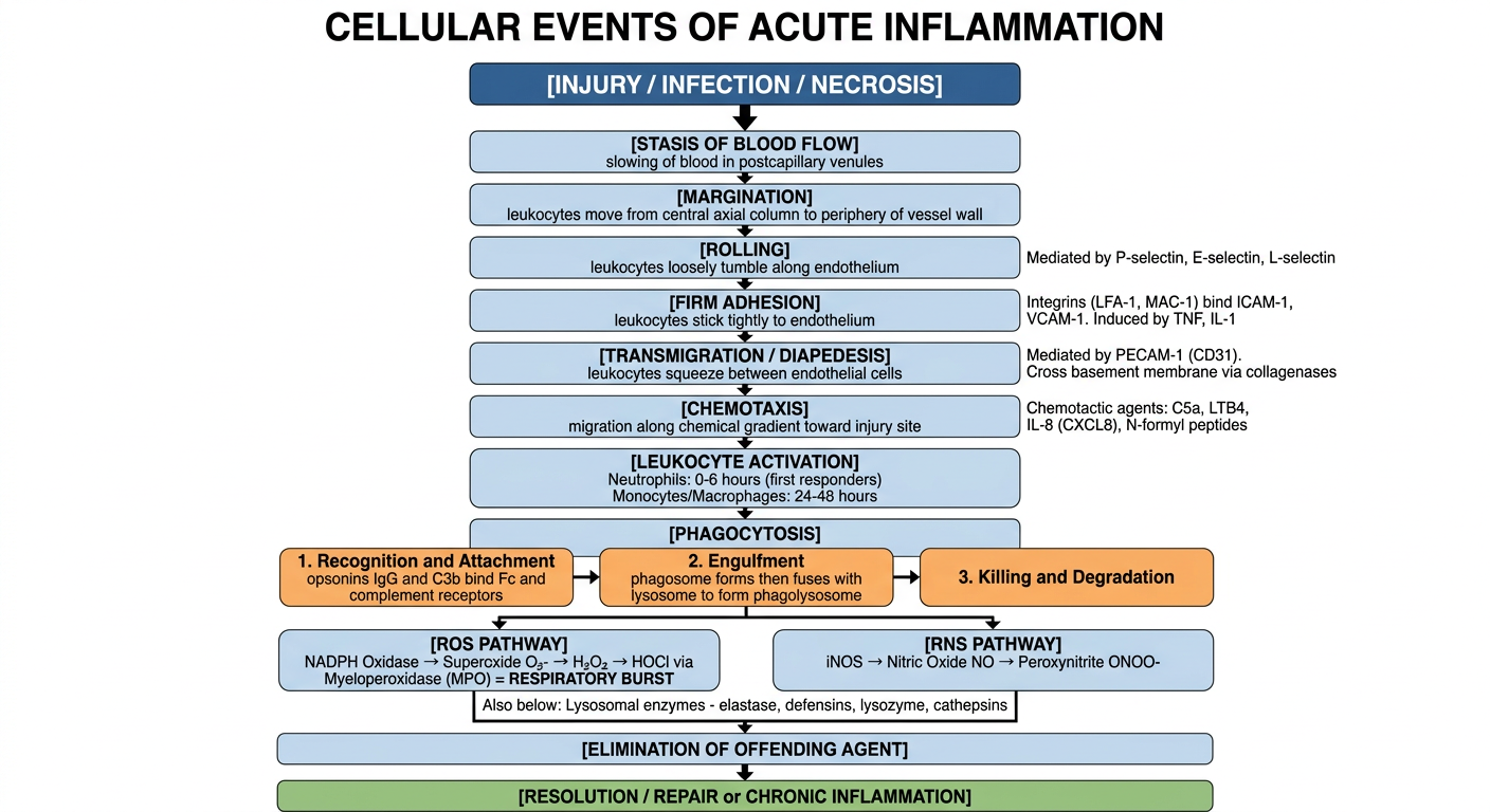
- Injury/Infection triggers slowing of blood flow
- Stasis causes leukocytes to shift to the vessel periphery - Margination
- Leukocytes begin Rolling along endothelium (selectins - P, E, L)
- Firm Adhesion occurs via integrins (LFA-1, MAC-1) binding ICAM-1/VCAM-1 (upregulated by TNF, IL-1)
- Transmigration/Diapedesis - leukocytes squeeze through junctions via PECAM-1 (CD31)
- Chemotaxis - migration toward injury guided by C5a, LTB4, IL-8, bacterial peptides
- Leukocyte Activation - neutrophils arrive first (0-6h), macrophages follow (24-48h)
- Phagocytosis - recognition → engulfment → phagolysosome formation
- Intracellular Killing via:
- ROS pathway (NADPH oxidase → O2- → H2O2 → HOCl via MPO) - the respiratory burst
- RNS pathway (iNOS → NO → peroxynitrite)
- Lysosomal enzymes
- Ends in Resolution or progression to Chronic Inflammation