Anatomy of the external nose and nasal cavity. The structure of the nasal mucosa. Nasal passages, their topography, which paranasal sinuses open in them.
Reading File
Finding Sources
Finding Sources
Finding Sources
Finding Sources
Finding Sources
Finding Sources
Finding Sources
Reading File
Finding Sources
Finding Sources
Finding Sources
Reading File
Reading File
Reading File
Reading File
Reading File
Reading File
Reading File
Reading File
Reading File
Reading File
Loading Image
Loading Image
Loading Image
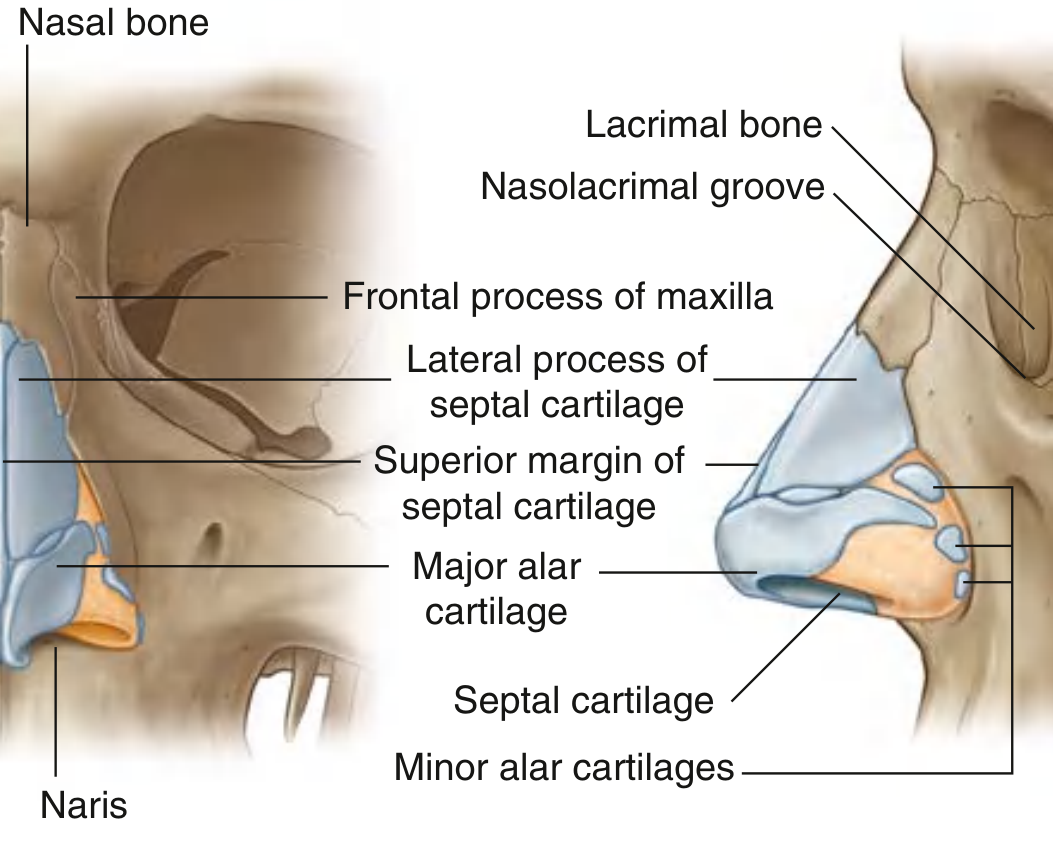
1) External nose: anatomy
The external nose is a pyramidal projection on the face with:
- Root (continuous with forehead)
- Dorsum
- Apex (tip)
- Alae
- Nares (nostrils) opening inferiorly
Its framework has:
- Bony part (superior): nasal bones, frontal bone, and maxilla
- Cartilaginous part (anterior/inferior): septal cartilage, lateral nasal cartilages, major and minor alar cartilages
This framework keeps the anterior nasal airway open.
Source: Gray’s Anatomy for Students, p. 1213-1214
Source: Gray’s Anatomy for Students, p. 1213-1214
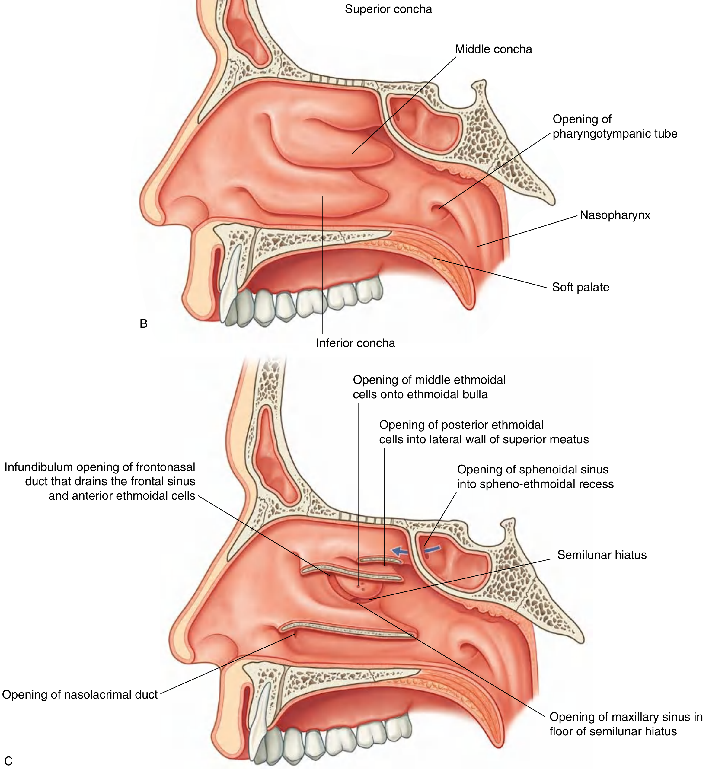
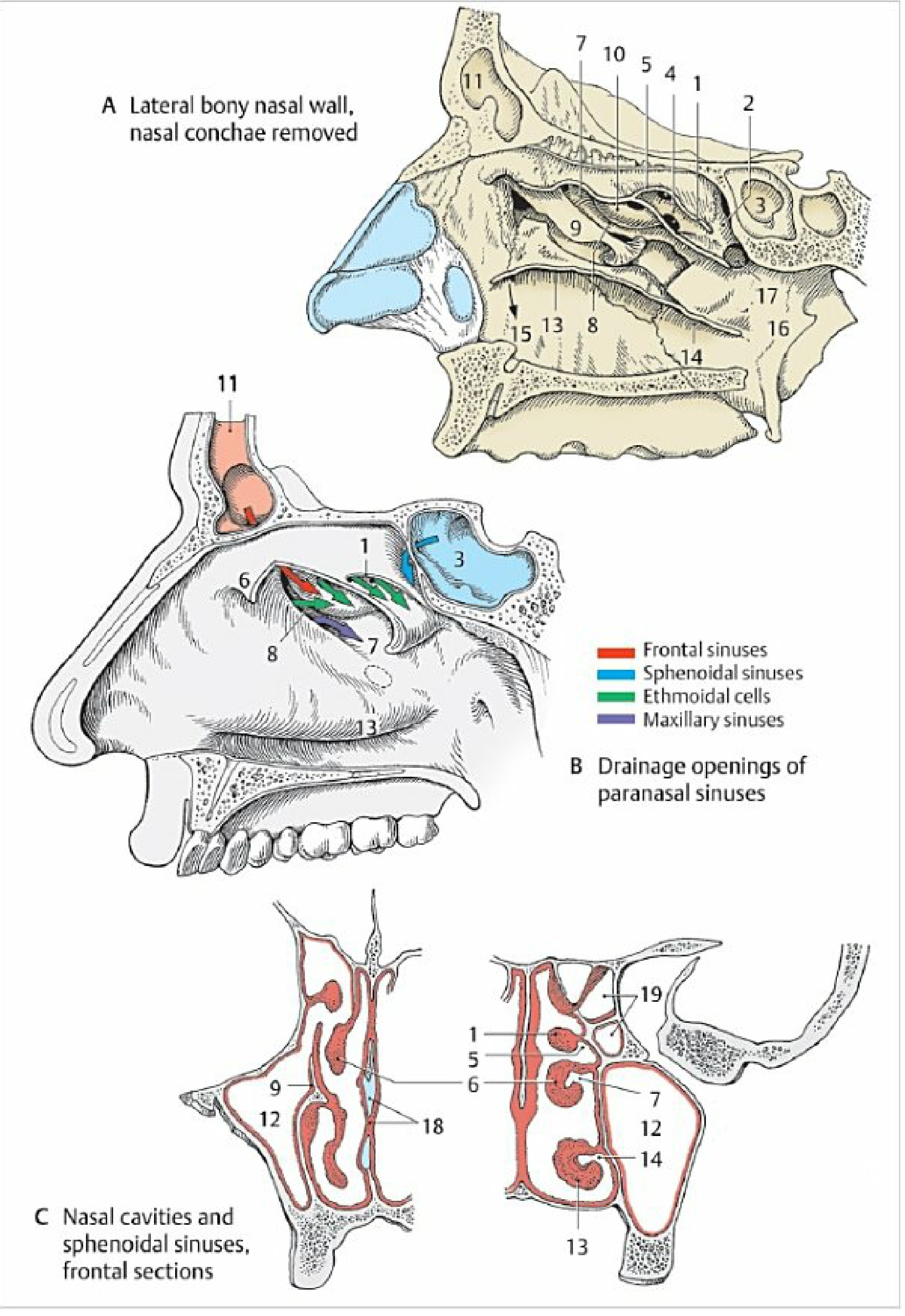
2) Nasal cavity and passages (topography)
Each nasal cavity extends from:
- Anterior nares to
- Posterior choanae (opening into nasopharynx)
Main walls:
- Medial wall: nasal septum
- Lateral wall: has conchae (superior, middle, inferior), creating passages:
- Superior meatus
- Middle meatus
- Inferior meatus
- Sphenoethmoidal recess (above superior concha)
Other key parts:
- Roof: highest at cribriform plate of ethmoid
- Floor: hard palate region
Source: Gray’s Anatomy for Students, p. 1217-1219; Color Atlas of Human Anatomy Vol. 2, p. 184-186
3) Structure of nasal mucosa
Nasal mucosa is divided into three regions:
- Nasal vestibule
- Lined by skin (stratified squamous keratinized epithelium)
- Respiratory region
- Pseudostratified ciliated columnar epithelium
- Cells: ciliated cells, goblet cells, brush cells, small granule cells, basal cells
- Lamina propria rich in venous plexuses and seromucous glands
- Function: warm, humidify, filter inspired air
- Olfactory region (roof, upper septum, upper lateral wall)
- Specialized pseudostratified olfactory epithelium
- Cells: olfactory receptor neurons (bipolar), supporting cells, basal cells, brush cells
- No goblet cells
- Lamina propria contains Bowman (olfactory) glands, vessels, olfactory nerve bundles
Sources:
- Histology: A Text and Atlas, p. 1763-1764, 1824, 1828
- Color Atlas of Human Anatomy Vol. 2, p. 175, 179
4) Which paranasal sinuses open where
Inferior meatus
- Nasolacrimal duct (not a sinus)
Middle meatus (major drainage pathway)
- Frontal sinus via frontonasal duct/infundibulum
- Maxillary sinus via ostium in semilunar hiatus
- Anterior ethmoidal cells (into infundibulum/frontonasal duct)
- Middle ethmoidal cells (onto/near ethmoidal bulla)
Superior meatus
- Posterior ethmoidal cells
Sphenoethmoidal recess
- Sphenoidal sinus
Sources: Gray’s Anatomy for Students, p. 1215, 1218-1219; Color Atlas of Human Anatomy Vol. 2, p. 184-186
Key diagrams
If you want, I can also provide this as a one-page exam table (columns: structure, epithelium, function, drainage site).
This is a shared conversation. Sign in to Orris to start your own chat.