
Reading File
This is a Pathology question paper from Siddaganga Medical College (II MBBS, 2024-25 batch). I'll now provide detailed answers to all questions using the medical library.
Searching sources...
Finding Sources
Finding Sources
Finding Sources
Reading File
Finding Sources
Finding Sources
Reading File
Finding Sources
Finding Sources
Loading Image
Finding Sources
I now have enough information from the textbooks to provide comprehensive answers. Let me compile the complete answer paper.Here are complete model answers for your Pathology question paper:
SIDDAGANGA MEDICAL COLLEGE - PATHOLOGY INTERNAL ASSESSMENT
Model Answers (II MBBS, 2024-25)
LONG ESSAY (1 × 10 = 10 Marks)
Q1. A 5-year-old boy with pallor, jaundice, failure to thrive; Hb 5 g%, PS - microcytic hypochromic RBCs + target cells, Reticulocyte count 7%
a) Most Probable Diagnosis (2 marks)
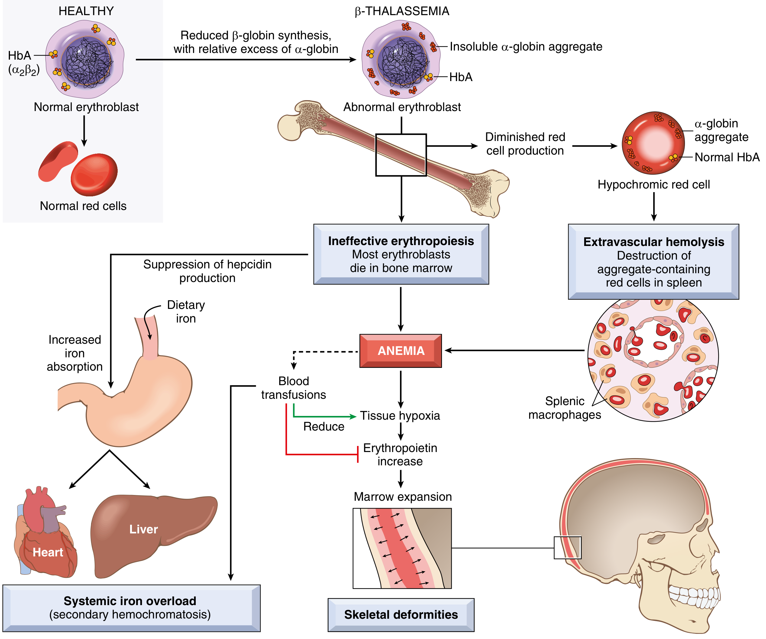
β-Thalassemia Major (Cooley's anemia)
Key clues:
- Age 5, failure to thrive - chronic illness from early childhood
- Hb 5 g% - severe anemia
- Microcytic hypochromic RBCs on peripheral smear - impaired HbA synthesis
- Target cells (codocytes) - characteristic of thalassemia
- Reticulocyte count 7% (elevated) - reflects hemolysis and compensatory erythropoiesis
- Jaundice - extravascular hemolysis
b) Etiopathogenesis (4 marks)
Genetics:
- Autosomal codominant disorder; mutations in β-globin gene (chromosome 11)
- Over 100 mutations described; most common: point mutations causing abnormal RNA splicing
- Two allele types: β⁰ (no β-globin) and β⁺ (reduced β-globin)
- β-Thalassemia major = homozygous state (β⁰/β⁰ or β⁰/β⁺)
Two-hit mechanism of anemia:
-
Inadequate HbA formation:
- Reduced/absent β-globin synthesis → insufficient HbA (α₂β₂) production
- Red cells are small (microcytic) and poorly hemoglobinized (hypochromic)
-
Toxic α-globin chain accumulation:
- Unpaired α-globin chains (now in excess) form insoluble precipitates
- These precipitates damage RBC membranes and erythroid precursors
- Massive apoptosis of erythroid precursors in bone marrow = Ineffective erythropoiesis
- The few mature RBCs produced are destroyed by splenic macrophages = Extravascular hemolysis → jaundice
Downstream consequences:
- Severe anemia → tissue hypoxia → ↑ erythropoietin → marrow expansion
- Marrow expansion → skeletal deformities (thalassemic facies: bossing of skull, maxillary prominence, "hair on end" X-ray)
- Ineffective erythropoiesis → low hepcidin → ↑ iron absorption → systemic iron overload (hemosiderosis)
- Repeated transfusions worsen iron overload → deposits in heart, liver, endocrine organs
c) Laboratory Investigations (4 marks)
| Investigation | Expected Finding |
|---|---|
| CBC | Hb severely low (5-7 g%), MCV low (<70 fL), MCH low, RDW high |
| Peripheral Smear | Microcytic hypochromic RBCs, target cells, nucleated RBCs (normoblasts), poikilocytosis, basophilic stippling, Cabot rings |
| Reticulocyte count | Elevated (5-10%) - compensatory |
| Serum bilirubin | Elevated (indirect/unconjugated) - hemolysis |
| Serum iron & ferritin | Elevated (iron overload) |
| TIBC | Decreased (iron saturation high) |
| Hemoglobin electrophoresis | ↑ HbF (>90%), ↑ HbA₂, absent/reduced HbA - DIAGNOSTIC |
| HPLC | Gold standard to quantify Hb fractions |
| Bone marrow biopsy | Erythroid hyperplasia, ineffective erythropoiesis |
| X-ray skull | "Hair-on-end" appearance due to marrow expansion |
| Molecular genetics | PCR for β-globin gene mutations (for definitive diagnosis/genetic counseling) |
REASONING QUESTIONS / SHORT ANSWERS (5 × 3 = 15 Marks)
Q2. 35-year-old HIV-positive with oral candidiasis, CD4+ T-cell count 120/μL
Reason: CD4+ T-lymphocytes are the primary coordinators of adaptive immunity, particularly cell-mediated immunity. HIV selectively infects and destroys CD4+ T-cells (via gp120 binding to CD4 receptor). When CD4 count drops below 200/μL, the patient enters AIDS. At CD4 <200/μL, protective Th1-mediated responses against opportunistic organisms like Candida albicans are lost. Normally, Th1 cytokines (IFN-γ, IL-2) activate macrophages and cytotoxic T-cells to clear fungal infections. Without this, Candida overgrows on mucosal surfaces. Oral candidiasis (thrush) is one of the earliest AIDS-defining illnesses and is almost universal when CD4 < 200/μL.
Q3. 45-year-old with low serum B12, anemia, and peripheral neuropathy
Reason: Vitamin B12 (cobalamin) is required for two critical reactions:
- Conversion of methylmalonyl-CoA → succinyl-CoA (via methylmalonyl-CoA mutase): Deficiency causes accumulation of methylmalonyl-CoA and its precursor propionic acid. Abnormal fatty acids are incorporated into neuronal myelin, leading to demyelination - specifically subacute combined degeneration (posterior columns + lateral corticospinal tracts), causing peripheral neuropathy and ataxia.
- Conversion of homocysteine → methionine (via methionine synthase): Deficiency impairs thymidylate synthesis → defective DNA synthesis → megaloblastic anemia (large, immature RBCs) and hypersegmented neutrophils.
Both processes are B12-dependent, explaining the combination of megaloblastic anemia AND peripheral neuropathy in B12 deficiency.
Q4. 60-year-old with homozygous α-1 antitrypsin deficiency; chest CT shows diffuse, uniform enlargement of air spaces affecting entire acini
Reason: α-1 Antitrypsin (AAT) is the principal inhibitor of neutrophil elastase in the lung. In AAT deficiency:
- Uninhibited neutrophil elastase and other proteases (MMP-9, MMP-12) destroy elastin and connective tissue in alveolar walls
- This leads to permanent, irreversible dilation of airspaces distal to the terminal bronchiole
- Homozygous ZZ genotype → severe AAT deficiency (<15% of normal)
- Affects entire acinus uniformly = Panacinar (panlobular) emphysema (vs. centriacinar in smokers, which affects respiratory bronchioles)
- The lower lobes are typically more affected in AAT-deficiency emphysema
- Progressive dyspnea results from loss of elastic recoil and air trapping
Q5. Echocardiography in 25-year-old with Rheumatic Heart Disease (RHD) shows thickening and fusion of mitral valve
Reason: In acute rheumatic fever, the pathogenesis is autoimmune - molecular mimicry between Group A Streptococcal M-protein antigens and cardiac valve proteins (particularly alpha-helical coiled-coil structures). Anti-streptococcal antibodies cross-react with valve endothelium, activating CD4+ T-cells and macrophages. This causes:
- Acute phase: Verrucous vegetations along valve leaflet lines of closure; Aschoff bodies (granulomatous inflammation with Anitschkow cells)
- Chronic/healed phase: Organization and fibrosis leads to leaflet thickening, commissural fusion, chordae tendineae shortening and thickening
- Result: Mitral stenosis - the classic sequela of RHD
- Mitral valve is most commonly affected (left-sided, higher pressure), followed by aortic valve
- The "fish-mouth" or "button-hole" deformity of the mitral valve results from commissural fusion and leaflet thickening seen on echo
Q6. Colorectal adenomas are considered premalignant
Reason: Colorectal adenomas are premalignant because of the well-established adenoma-carcinoma sequence:
- Adenomas harbor dysplastic epithelium by definition
- They follow a stepwise accumulation of mutations: APC gene loss (early) → KRAS mutation → SMAD4/DPC4 loss → TP53 mutation → carcinoma
- Risk factors for malignant transformation: size >2 cm, villous architecture (tubular < tubulovillous < villous in risk), high-grade dysplasia
- Villous adenomas have ~40% risk of malignant transformation
- They are detected by colonoscopy and must be removed - this is the basis of colorectal cancer screening programs
SHORT NOTES - APPLIED ASPECT (4 × 5 = 20 Marks)
Q7. 4-year-old boy: painful swelling of right knee after minor fall; PT-Normal, aPTT-Prolonged, Factor VIII assay 2% of normal
a) Diagnosis
Hemophilia A - X-linked recessive bleeding disorder due to Factor VIII deficiency
b) Genetic and Pathological Basis of Hemarthrosis
Genetic basis:
- Caused by mutations in F8 gene on X chromosome (Xq28)
- Males are affected (hemizygous); females are carriers
- ~45% due to intron 22 inversion; remainder from point mutations, deletions
- Severity: severe (<1% Factor VIII), moderate (1-5%), mild (5-30%)
- This case: 2% = severe hemophilia A
Pathological basis of hemarthrosis:
- Factor VIII is a critical cofactor for Factor IXa in the intrinsic coagulation pathway (tenase complex)
- Without Factor VIII, the intrinsic pathway is blocked → prolonged aPTT (intrinsic pathway) but normal PT (extrinsic pathway intact)
- Joints (knees, elbows, ankles) are susceptible because synovial vascularity is rich and minor trauma easily disrupts small vessels
- In the joint: repeated bleeding → hemosiderin deposition in synovium → synovial hypertrophy → pannus formation → cartilage and bone destruction = hemophilic arthropathy
- Acute: warm, swollen, painful joint (hemarthrosis)
- Chronic: iron-catalyzed free radical damage, inflammatory cytokines (IL-1, TNF-α) destroy cartilage
Lab summary: PT normal (extrinsic pathway intact), aPTT prolonged (intrinsic pathway deficient), Factor VIII assay confirms diagnosis.
Q8. 30-year-old male: weakness, pallor, bleeding gums; TLC 1,10,000 with many blasts positive for myeloperoxidase
a) Most Likely Diagnosis
Acute Myeloid Leukemia (AML)
b) Classification
WHO 2022 Classification of AML includes:
| Category | Key Features |
|---|---|
| AML with recurrent genetic abnormalities | t(8;21) - AML-M2; t(15;17) - APL (M3); inv(16) - M4Eo; NPM1, FLT3 mutations |
| AML with myelodysplasia-related changes | Prior MDS, MDS-related cytogenetics |
| Therapy-related AML | Post alkylating agent or topoisomerase II inhibitor |
| AML, not otherwise specified (NOS) | AML-M0 to M7 (FAB subtypes) |
This case fits AML-NOS: Blasts ≥20% of WBCs, myeloperoxidase (MPO) positivity confirms myeloid lineage.
FAB classification (still used in exams):
- M0: Undifferentiated; M1: Without maturation; M2: With maturation; M3: Acute promyelocytic (APL); M4: Myelomonocytic; M5: Monocytic; M6: Erythroleukemia; M7: Megakaryoblastic
Key pathological features:
- Replacement of normal marrow → anemia (pallor), thrombocytopenia (bleeding gums), neutropenia (infections)
- Auer rods - pathognomonic pink rod-like inclusions in myeloblasts (MPO-positive)
- MPO positive = myeloid origin (distinguishes from ALL, where MPO is negative)
Q9. 60-year-old man: weight loss, abdominal pain, anorexia; mass detected in stomach
a) Diagnosis
Carcinoma of the Stomach (Gastric Adenocarcinoma) - most likely
b) Morphology
Gross morphology:
- Intestinal type: Usually forms an ulcerated, exophytic mass with heaped-up margins (fungating/polypoid lesion), commonly in antrum/lesser curvature; associated with H. pylori, intestinal metaplasia
- Diffuse type: Infiltrating, produces a rigid, thickened "leather-bottle" stomach - linitis plastica; signet ring cells infiltrate all layers; poor prognosis
- Early gastric cancer: Confined to mucosa/submucosa regardless of node status
- Advanced gastric cancer: Invades muscularis propria and beyond
Microscopic morphology (intestinal type):
- Gland-forming adenocarcinoma
- Malignant columnar cells with nuclear pleomorphism, mitoses
- Desmoplastic stroma
Microscopic (diffuse type):
- Signet ring cells - large mucin vacuole displaces nucleus to periphery
- Poorly cohesive cells due to loss of E-cadherin (CDH1 mutation)
Routes of spread:
- Direct: through gastric wall
- Lymphatic: to regional lymph nodes; Virchow node (left supraclavicular)
- Hematogenous: liver most common
- Transcoelomic: peritoneal seeding; Krukenberg tumor (bilateral ovarian metastases from signet ring cells)
Q10. 42-year-old female: recurrent right upper quadrant pain; ultrasound shows multiple gallstones
a) Diagnosis
Cholelithiasis (Gallstone Disease) - most likely Cholesterol gallstones (most common type, especially in females)
"5 F" rule: Female, Fat, Fertile, Forty, Fair
b) Etiopathogenesis
Types of gallstones:
- Cholesterol stones (80%): Pure cholesterol or mixed
- Pigment stones (20%): Black (hemolysis) or brown (infection)
Pathogenesis of cholesterol gallstones:
Step 1 - Cholesterol supersaturation of bile:
- Liver secretes excess cholesterol relative to bile salts and lecithin
- Normal cholesterol solubility depends on the cholesterol saturation index (CSI)
- Obesity, estrogen (OCP, pregnancy), clofibrate → ↑ cholesterol secretion
- Ileal disease/resection → ↓ bile salt reabsorption → relative ↑ CSI
Step 2 - Nucleation:
- Supersaturated bile forms cholesterol monohydrate crystals
- Mucin glycoproteins act as pronucleating agents
- Gallbladder stasis (fasting, pregnancy, TPN) promotes crystal aggregation
Step 3 - Stone growth:
- Crystals aggregate and grow into macroscopic stones
- Reduced gallbladder motility (progesterone effect) → incomplete emptying → stone growth
Why females? Estrogen increases hepatic cholesterol secretion AND reduces bile acid synthesis; progesterone reduces gallbladder contractility - both promote stone formation.
Complications: Biliary colic, acute/chronic cholecystitis, choledocholithiasis, obstructive jaundice, ascending cholangitis, acute pancreatitis, gallbladder carcinoma (rare).
SHORT NOTES (7 × 5 = 35 Marks)
Q11. Carcinoid Tumor
- Definition: Well-differentiated neuroendocrine tumor (NET) arising from enterochromaffin (Kulchitsky) cells; can occur anywhere in GI tract, bronchus, or thymus
- Common sites: Appendix (most common, rarely malignant), ileum, rectum, bronchus
- Gross: Small, yellow-tan submucosal nodule; firm due to desmoplastic reaction
- Microscopy: Nests, ribbons, or rosettes of uniform small cells with round nuclei, "salt-and-pepper" chromatin, abundant pink granular cytoplasm; mitoses rare
- Histochemistry: Silver staining positive (argyrophilic/argentaffin); Chromogranin A and Synaptophysin positive (IHC markers)
- Products secreted: Serotonin (5-HT), histamine, bradykinin, substance P, VIP
- Carcinoid syndrome (occurs with liver metastases): Episodic flushing, diarrhea, bronchospasm, right-sided cardiac fibrosis (tricuspid/pulmonary valve)
- Malignant potential: Relates to size and site - appendiceal <2 cm rarely metastasize; ileal >2 cm frequently metastasize
- Marker: Serum Chromogranin A and 24-hr urine 5-HIAA (5-hydroxyindoleacetic acid) for diagnosis/follow-up
Q12. Morphology of Myocardial Infarction
Timeline of changes:
| Time | Gross | Microscopy |
|---|---|---|
| 0-4 hrs | No change | No change (EM: glycogen loss, relaxed myofibrils) |
| 4-12 hrs | Subtle pallor | Early coagulative necrosis; wavy fibers; edema |
| 12-24 hrs | Pale/mottled infarct | Coagulative necrosis; pyknotic nuclei; neutrophil infiltration begins |
| 1-3 days | Yellowed, soft | Coagulative necrosis complete; heavy neutrophil infiltrate (PMNs) |
| 3-7 days | Hyperemic border; central softening | Macrophage infiltration begins; removal of necrotic debris |
| 1-2 weeks | Yellow center, red-brown border | Granulation tissue (fibroblasts + capillaries); macrophages |
| 2-8 weeks | Progressive scarring | Collagen deposition, maturing scar |
| >2 months | White, firm fibrous scar | Dense acellular collagen scar |
Types:
- STEMI (transmural): Full thickness; associated with plaque rupture + occlusive thrombus
- NSTEMI (subendocardial): Inner 1/3 of wall; most vulnerable zone (farthest from epicardial supply)
Complications: Arrhythmias (most common, days 1-3), cardiac failure, cardiogenic shock, mural thrombus → embolism, ventricular aneurysm, papillary muscle rupture, Dressler syndrome (autoimmune pericarditis, 2-10 weeks later), cardiac rupture (3-7 days).
Q13. Morphology of Pleomorphic Adenoma
- Definition: Most common salivary gland tumor (~70%); also called "mixed tumor" - contains both epithelial and mesenchymal elements
- Site: Parotid gland (80%), then submandibular, minor salivary glands
- Gross: Encapsulated (but capsule incomplete/irregular), rubbery, lobulated, gray-white; cut surface shows myxoid/chondroid areas
- Microscopy (two components):
- Epithelial component: Ductal cells forming gland-like structures; surrounded by myoepithelial cells (spindle/stellate shaped)
- Stromal component: Myxoid, chondroid (cartilage-like), hyaline stroma - produced by myoepithelial cells (hence "mixed" appearance)
- Key feature: Myoepithelial cells are the key cell type - they can differentiate into both ductal epithelium AND stromal elements
- IHC: Cytokeratin (ductal), Vimentin, S-100, GFAP (myoepithelial)
- Behavior: Benign but risk of recurrence if incompletely excised (due to capsular pseudopods); 6-10% risk of malignant transformation to carcinoma ex pleomorphic adenoma over time
Q14. Classify Hodgkin Lymphoma (HL); Reed-Sternberg cells; IHC markers
Classification (WHO):
| Type | Frequency | Histology | RS cells | Prognosis |
|---|---|---|---|---|
| Nodular sclerosis (NSCHL) | 65-70% | Collagen bands, lacunar cells | Lacunar RS cells | Good |
| Mixed cellularity (MCCHL) | 20-25% | Mixed infiltrate (eos, plasma cells, lymphocytes) | Classic RS cells | Intermediate |
| Lymphocyte-rich (LRCHL) | 5% | Lymphocyte predominance | Rare RS cells | Good |
| Lymphocyte-depleted (LDCHL) | <1% | Sparse lymphocytes, fibrosis | Many RS cells | Poor |
| Nodular lymphocyte predominant (NLPHL) | 5% | Nodular, background lymphocytes | "Popcorn" (L&H) cells | Excellent |
Reed-Sternberg (RS) cells:
- Large, binucleated or bilobed cells
- Each nucleus has a large inclusion-like eosinophilic "owl-eye" nucleolus - pathognomonic
- Mononuclear variant: Hodgkin cell
- Lacunar cell (NSCHL): RS cell with retracted cytoplasm creating empty space (lacuna)
- "Popcorn" or LP cell (NLPHL): Multi-lobated nucleus
- RS cells are derived from germinal center B-cells
IHC markers for RS cells:
- CD15 (Leu-M1) - positive (also expressed on granulocytes)
- CD30 (Ki-1) - positive (marker of activated lymphoid cells)
- CD20 - negative (classic HL); positive in NLPHL
- CD45 (LCA) - negative (distinguishes from NHL)
- PAX5 - weakly positive (B-cell transcription factor)
- EBV (LMP-1) - positive in ~40% of classic HL (especially MCCHL)
Summary: CD15+, CD30+, CD45-, CD20- = classic HL RS cells
Q15. Etiopathogenesis of Atherosclerosis
Atherosclerosis = chronic intimal disease of large and medium arteries; characterized by atheromatous plaques.
Response-to-injury hypothesis (Ross):
Step 1 - Endothelial injury/dysfunction:
- Risk factors: Hyperlipidemia (LDL), hypertension, smoking, diabetes, homocysteine
- Injured endothelium → ↑ permeability, ↑ adhesion molecule expression (VCAM-1, ICAM-1, E-selectin)
Step 2 - Lipoprotein accumulation:
- LDL enters intima, gets oxidized (ox-LDL)
- Ox-LDL is chemotactic for monocytes and inhibits macrophage migration (traps them)
Step 3 - Monocyte recruitment:
- Monocytes adhere to endothelium (via VCAM-1/MCP-1 interaction), migrate into intima
- Differentiate into macrophages → engulf ox-LDL via scavenger receptors (SR-A, CD36)
- Become foam cells → accumulate to form fatty streak (earliest visible lesion)
Step 4 - Smooth muscle cell migration:
- Macrophages and platelets release PDGF, FGF → smooth muscle cells (SMC) migrate from media to intima
- SMC proliferate and secrete collagen, proteoglycans → fibrous cap
Step 5 - Plaque development:
- Mature plaque = fibrous cap (SMC + collagen) overlying lipid core (foam cells, cholesterol crystals, necrotic debris)
- Calcification occurs in advanced plaques
- Plaque rupture → thrombosis → MI/stroke
Risk factors (modifiable): Hyperlipidemia (most important), hypertension, smoking, diabetes, obesity, sedentary lifestyle
Non-modifiable: Age, male sex, genetic factors
Q16. Amebic Liver Abscess
- Causative organism: Entamoeba histolytica (protozoan)
- Route: Fecal-oral → trophozoites invade colonic mucosa → portal circulation → liver
- Site: Most commonly right lobe, posterosuperior aspect (portal blood from cecum/ascending colon)
- Gross morphology:
- Large solitary abscess (may reach 10-15 cm)
- Wall: shaggy, ragged inner lining (no true pyogenic wall)
- Content: "anchovy sauce" or "chocolate brown paste" - liquefied necrotic liver (NOT true pus; acellular)
- Composed of: necrotic hepatocytes, red blood cells, cellular debris
- Microscopy:
- Amebic trophozoites found at the periphery/wall of abscess (not in center)
- Trophozoites: 20-40 μm, with nucleus having central karyosome; engulf RBCs (erythrophagocytosis)
- Surrounding liver shows inflammation and fibrosis
- Complications: Rupture into peritoneal cavity, pleural space ("anchovie-like" pleuritis), pericardium; secondary bacterial infection
- Diagnosis: Serology (anti-amebic antibodies), stool examination, ultrasound (round/oval hypoechoic lesion), needle aspiration
- Treatment: Metronidazole (drug of choice) + diloxanide furoate (luminal agent)
Q17. Classify Cirrhosis; Mention Complications
Cirrhosis = diffuse hepatic fibrosis with nodule formation, disrupting normal hepatic architecture and vascular relationships
Classification:
A. Morphological:
| Type | Nodule size | Etiology |
|---|---|---|
| Micronodular | <3 mm, uniform | Alcoholic liver disease, biliary cirrhosis, hemochromatosis |
| Macronodular | >3 mm, variable | Viral hepatitis (B, C), Wilson's disease, post-necrotic |
| Mixed | Both types | Various |
B. Etiological:
- Alcoholic cirrhosis (most common in Western countries)
- Post-viral cirrhosis (HBV, HCV - most common in India/Asia)
- Biliary cirrhosis (primary or secondary)
- Metabolic: Wilson's disease, hemochromatosis, NAFLD/NASH
- Cardiac cirrhosis (chronic right heart failure)
- Cryptogenic (unknown cause, ~10%)
Complications (Child-Pugh classification determines prognosis):
- Portal hypertension → esophageal varices (bleeding), caput medusae, splenomegaly, hemorrhoids
- Ascites - due to portal hypertension + hypoalbuminemia + secondary hyperaldosteronism
- Spontaneous Bacterial Peritonitis (SBP) - infective complication of ascites
- Hepatic encephalopathy - ammonia accumulation, cerebral edema, asterixis
- Hepatorenal syndrome - functional renal failure
- Coagulopathy - reduced synthesis of clotting factors (I, II, V, VII, IX, X) → bleeding tendency
- Hypoalbuminemia → edema, ascites, poor wound healing
- Hepatocellular Carcinoma (HCC) - in ~3-5% of cirrhosis patients per year (especially HBV, HCV, hemochromatosis)
- Hepatopulmonary syndrome - intrapulmonary shunts → hypoxemia
- Hypersplenism - pancytopenia from splenic sequestration
Sources: Robbins & Kumar Basic Pathology (9780323790185); Fishman's Pulmonary Diseases; Fuster & Hurst's The Heart - all via Orris medical textbook library
This is a shared conversation. Sign in to Orris to start your own chat.