
Gait analysis Ortho in detail
Reading File
Now let me search the library for comprehensive gait analysis content in orthopaedics.
Finding Sources
Finding Sources
Reading File
Finding Sources
Finding Sources
Reading File
Reading File
Finding Sources
Reading File
Finding Sources
Finding Sources
Reading File
Reading File
Finding Sources
Reading File
Finding Sources
Reading File
Loading Image
Loading Image
I now have all the comprehensive information needed. Let me compile the full detailed answer.
Gait Analysis - Detailed Orthopaedic Review
1. Definitions and Terminology
Gait is the pattern of locomotion characterized by coordinated limb movements.
- Step: Distance between successive initial contact (IC) by the two lower limbs (one foot to the opposite foot)
- Stride: Distance between successive IC by the same lower limb (= two steps; one complete gait cycle)
- Cadence: Number of steps per unit of time (steps/min)
- Velocity: Ratio of distance to time (cadence x step length)
- Walking is defined by always having at least one foot in contact with the ground, plus a period of double-limb support
- Running differs from walking in that it eliminates double-limb support and introduces a float phase when neither foot touches the ground
2. The Gait Cycle
A complete gait cycle runs from foot strike to successive ipsilateral foot strike.
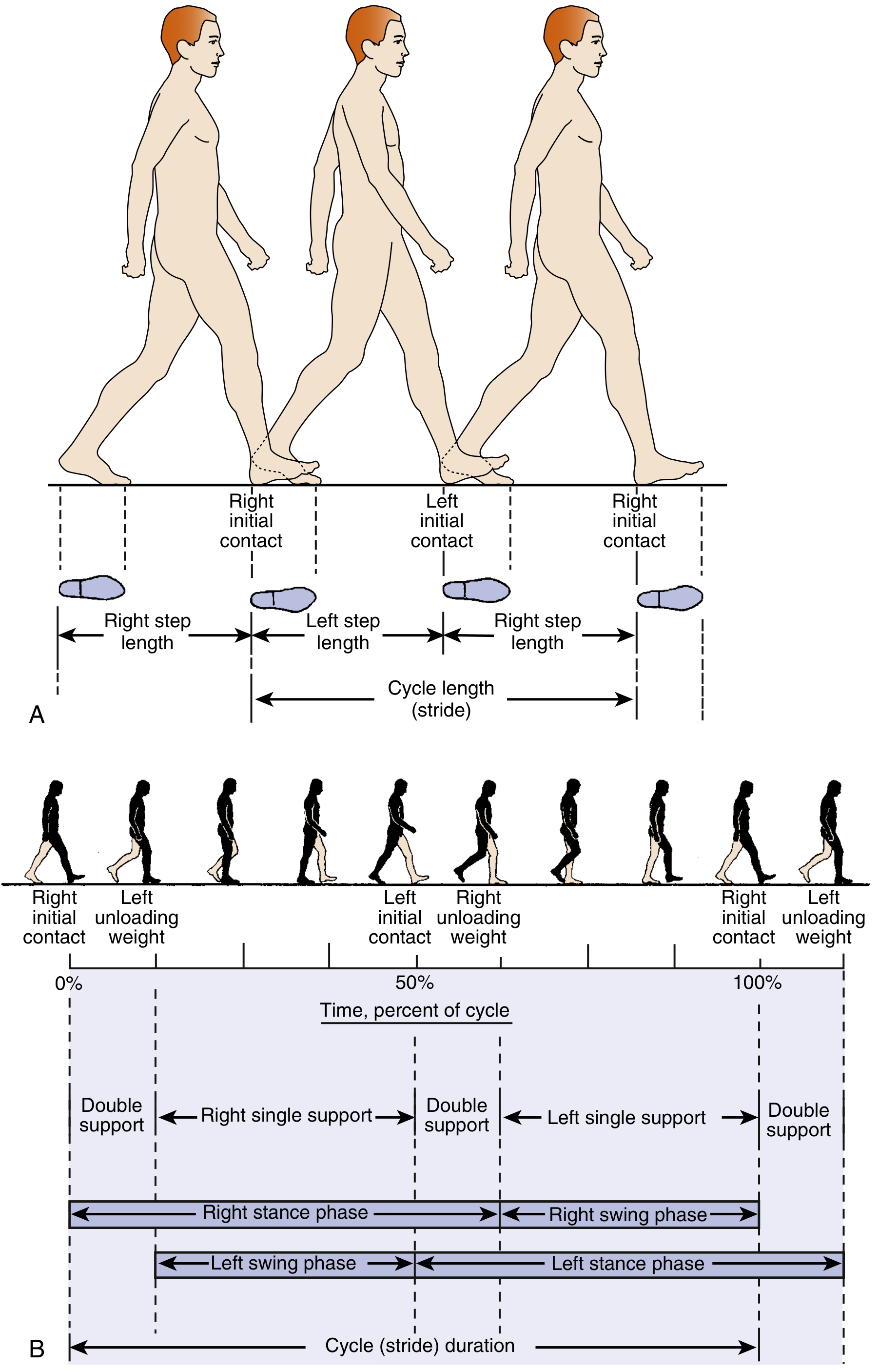
Fig. 10.1 - Dimensions of the walking cycle showing step length, stride (cycle length), stance, and swing phases with double-limb support intervals. (Miller's Review of Orthopaedics 9th Ed.)
The cycle is divided into two main phases:
| Phase | Duration | Description |
|---|---|---|
| Stance phase | 60% of cycle | Weight-bearing; from IC to toe-off |
| Swing phase | 40% of cycle | Non-weight-bearing; from toe-off to next IC |
There are two periods of double-limb support (during IC + loading response and during preswing), comprising 20-30% of the cycle; these periods are velocity-dependent.
3. Subdivisions of the Gait Cycle
Stance Phase (60%)
| Sub-phase | Event | Key Feature |
|---|---|---|
| Initial Contact (IC) / Heel Strike | Reference foot contacts ground | Foot dorsiflexed; tibialis anterior contracts eccentrically |
| Loading Response (LR) | IC of reference foot → initial swing of contralateral foot | Knee flexes ~15° to dampen load; subtalar joint everted (unlocks transverse tarsal joint for shock absorption) |
| Midstance (MSt) | Initial swing of advancing foot → body's CoG directly over supporting forefoot | Single-leg stance; CoG passes over weight-bearing leg |
| Terminal Stance (TSt) | Heel rise → IC of contralateral foot | Gastrocnemius and plantar flexors contract; subtalar joint inverts, rigidifying midfoot for propulsion |
| Preswing (PSw) | IC of contralateral limb → stance foot lifts off ground | Maximal subtalar inversion; maximum midfoot rigidity at toe-off |
Swing Phase (40%)
| Sub-phase | Event | Key Feature |
|---|---|---|
| Initial Swing (ISw) | Foot leaves ground → swinging foot opposite stance foot | Hip flexors propel leg forward |
| Midswing (MSw) | Swinging limb forward → tibia perpendicular to ground | Tibialis anterior and dorsiflexors contract to clear foot |
| Terminal Swing (TSw) | Tibia perpendicular → foot makes IC | Hamstrings contract eccentrically to decelerate limb before heel strike |
4. Kinetics and Kinematics
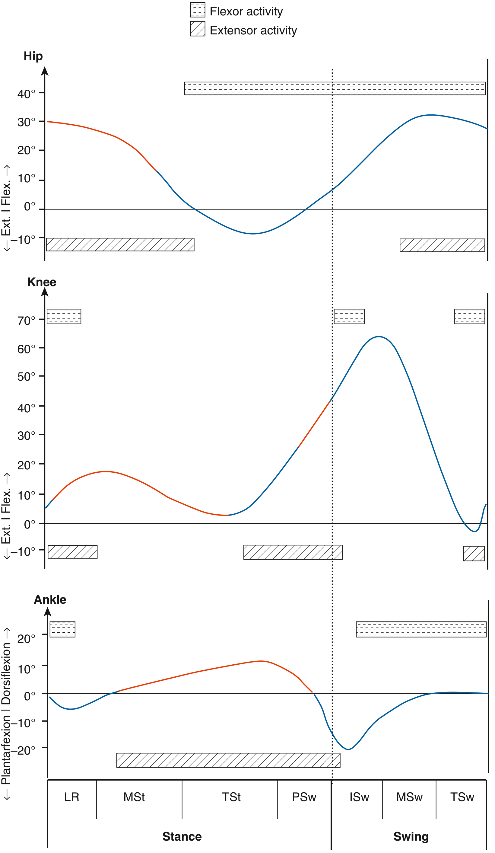
Fig. 10.2 - Joint angles (hip, knee, ankle) through each phase of the gait cycle with corresponding muscle activity. Red portions = ground reaction force anterior to hip, posterior to knee, and anterior to ankle during stance. (Miller's Review of Orthopaedics 9th Ed.)
Key joint motion values:
- Hip: ~30° flexion at IC → extends to ~10° extension at TSt → 30°+ flexion during swing
- Knee: ~5° flexion at IC → 15-18° flexion at LR (shock absorption) → near-full extension at MSt → ~65° flexion in swing
- Ankle: ~5° plantar flexion at IC → 0° at neutral through MSt → ~10° dorsiflexion at TSt → ~20° plantar flexion at PSw/toe-off → returns to neutral during swing
Ground Reaction Force (GRF)
- The mean load-bearing vector that changes in magnitude and direction throughout the cycle
- It determines the moment/torque exerted on each joint
- During stance: GRF is anterior to the hip, posterior to the knee, and anterior to the ankle
Joint Loading
- Knee forces: 4-7x body weight during stance; 70% of load passes through the medial compartment
- During water walking: significant decrease in joint moments due to buoyancy
5. Muscle Actions During Gait
Most muscle activity during gait is eccentric (muscle active while lengthening):
| Muscle | Type of Contraction | Function |
|---|---|---|
| Gluteus medius | Eccentric | Controls pelvic tilt during midstance |
| Gluteus maximus | Concentric | Powers hip extension |
| Iliopsoas | Concentric | Powers hip flexion (swing phase) |
| Hip adductors | Eccentric | Control lateral sway (late stance) |
| Quadriceps | Eccentric | Stabilize knee at initial contact and preswing |
| Hamstrings | Eccentric | Control rate of knee extension at swing (deceleration at TSw) |
| Tibialis anterior | Eccentric (IC) / Concentric (swing) | Slows plantar flexion rate at IC; dorsiflexes ankle in swing |
| Gastrocnemius-soleus | Eccentric | Slows dorsiflexion rate during stance; powers push-off |
6. Determinants of Gait (6 Classic Determinants - Saunders/Inman)
Six processes minimize vertical and lateral displacement of the center of mass, maximizing energy efficiency:
- Pelvic rotation - Pelvis externally rotates from IC to preswing, internally during preswing and swing; reduces total vertical displacement needed for limb advancement
- Pelvic list (tilt) - Non-weight-bearing contralateral side drops 5°, reducing superior deviation of CoM
- Knee flexion at loading - ~15° stance-phase knee flexion dampens impact at initial loading
- Foot and ankle motion - Subtalar damping at LR; stability at midstance; propulsion efficiency at push-off
- Knee motion - Works with foot/ankle to decrease unnecessary limb motion; flexes at IC, extends at MSt
- Control of pelvic lateral displacement - ~5 cm motion over weight-bearing limb; narrows base of support, increases stance stability
Center of mass displacement:
- Vertical: sinusoidal curve
- Lateral: sinusoidal curve with amplitude of ~6 cm
- Body's CoM is located 2 cm anterior to S2
- Trunk's CoG is just anterior to T10 (~33 cm above hip joints in 184 cm individual)
7. Pathologic Gait Patterns
Factors causing abnormal gait: muscle weakness, neurological conditions, pain, limb deformity, joint disease.
Antalgic Gait
- Cause: Pain (hip/knee arthritis, stress fracture, infection)
- Mechanism: Patient shortens the stance phase on the painful limb to reduce load time; contralateral swing phase is more rapid
- Pattern: Dot-dash cadence (short stance, rapid swing); head dips on affected side
Trendelenburg Gait
- Cause: Weakness of hip abductors (gluteus medius); positive Trendelenburg sign = pelvis sags >2 cm during single-leg stance
- Mechanism: Failure to stabilize pelvis during single-limb support; pelvis drops on the unsupported (contralateral) side
- Pattern: Trunk lurches toward the affected side (compensated Trendelenburg = "waddling" if bilateral)
- Clinical associations: Superior gluteal nerve injury, polio, OA hip, total hip arthroplasty complications
High-Stepping / Steppage Gait (Foot Drop)
- Cause: Weakness of ankle dorsiflexors (tibialis anterior); common peroneal nerve palsy, L4/L5 root lesion
- Mechanism: Inability to clear foot during swing; exaggerated knee and hip flexion to clear foot from ground
- Pattern: Foot slaps ground at IC; hyperextension moment through knee in stance
Scissor Gait
- Cause: Overactive/spastic hip adductors (cerebral palsy, spastic diplegia)
- Mechanism: Adductor overactivity causes knees to cross midline; legs cross like scissors
- Pattern: Narrow base, knees crossing, typical in spastic CP; often combined with equinus
Crouch Gait
- Cause: Hamstring contracture, plantar flexor weakness, or cerebral palsy
- Mechanism: Excessive knee flexion throughout stance; increased dorsiflexion
- Pattern: "Crouching" posture; increased joint loading
Circumduction Gait
- Cause: Long limb, stiff knee, abductor shortening/overuse
- Mechanism: Swinging limb cannot flex normally, so patient circumducts it in an arc laterally to clear the floor
Hip Hiking Gait
- Cause: Long limb, stiff knee, hamstring weakness
- Mechanism: Elevation of pelvis on swing side to clear a stiff or long limb
Stiff / Ankylosed Hip Gait
- Cause: Hip arthrodesis or fusion
- Pattern: Head rocks to and fro; compensatory lumbosacral motion
Ataxic Gait
- Cause: Cerebellar pathology
- Pattern: Wide base, irregular cadence, lurching, incoordinated
| Gait Type | Primary Cause | Key Feature |
|---|---|---|
| Antalgic | Pain | Short stance phase; dot-dash cadence |
| Trendelenburg | Abductor weakness | Trunk lurches to affected side |
| Steppage/High-stepping | Foot drop (peroneal nerve) | Exaggerated hip/knee flexion; foot slap |
| Scissor | Adductor spasticity (CP) | Knees cross midline |
| Crouch | Hamstring contracture / CP | Excessive knee flexion throughout |
| Circumduction | Stiff knee / long limb | Lateral arc of swinging limb |
| Hip hiking | Stiff knee / long limb | Pelvis elevates on swing side |
| Ataxic | Cerebellar disease | Wide base, irregular, lurching |
8. Gait Analysis Methods
Clinical Observation (Most Common)
- Watch patient walk from front, sides, and back
- Study one joint at a time: pelvis, hip, knee, ankle, foot
- Note stride length, cadence, rotational alignment, trunk position, and side-to-side differences
Quantitative Gait Analysis
Modern labs use:
- High-speed motion capture cameras from multiple angles
- Retroreflective markers aligned with palpable skeletal landmarks
- Force platforms (measure GRF)
- Kinematic data presented as 3D waveforms for each joint
- Electromyography (EMG) - documents muscle activation timing; identifies out-of-phase firing
- Pedobarography - plantar pressure/foot pressure mapping
- Oxygen consumption measurement (energy expenditure)
Quantitative gait analysis in preoperative planning for CP: when given to experienced observers after clinical recommendations were made, the surgical recommendation changed 52% of the time - Campbell's Operative Orthopaedics 15th Ed 2026.
Key Clinical Uses
- Preoperative planning in cerebral palsy and lower extremity surgery
- Postoperative outcome assessment
- Making further recommendations for bracing and physiotherapy
- Identifying gait deviations and muscle out-of-phase firing patterns
9. Gait Abnormalities by Phase (Summary Table)
| Phase | Deviation | Possible Cause |
|---|---|---|
| IC-MSt | Foot slap | Tibialis anterior weakness |
| IC-MSt | Genu recurvatum | Quadriceps spasticity/shortness; plantar flexor spasticity |
| IC-MSt | Excessive trunk extension | Gluteus maximus weakness; hip extensor weakness |
| IC-PSw | Excessive trunk flexion | Hip flexion contracture; quadriceps weakness |
| IC-PSw | Crouch (excessive knee flexion) | Hamstring contracture; plantar flexor weakness |
| IC-PSw | Medial femoral rotation | Medial hamstring tightness |
| LR-PSw | Wide base of support | Abductor contracture |
| MSt-PSw | Excessive foot pronation | Valgus forefoot; pes planus; tibialis posterior weakness |
| MSt-PSw | Bouncing/insufficient push-off | Achilles contracture; plantar flexor weakness |
| Swing | Steppage gait | Dorsiflexion weakness; equinus deformity |
| Swing | Circumduction | Abductor shortening; knee stiffness; long limb |
| Swing | Hip hiking | Hamstring weakness; long limb |
10. Metabolic Cost of Gait
- Metabolic cost of walking increases with proximal-level amputations and is inversely proportional to the length of the residual limb and number of functional joints preserved
- Body weight is carried 70% by head, neck, trunk, and arms
- Trunk's CoG is ~33 cm above the hip joints
- Whole-body CoM is 2 cm anterior to S2
Sources:
- Miller's Review of Orthopaedics 9th Ed, Chapter 10 (Gait Biomechanics)
- Campbell's Operative Orthopaedics 15th Ed 2026, Gait Analysis Section
- Bailey and Love's Short Practice of Surgery 28th Ed, Table 35.1 (Types of limp)
- Imaging Anatomy: Bones, Joints, Vessels and Nerves (Thieme Atlas), Gait section
This is a shared conversation. Sign in to Orris to start your own chat.