
Aplastic crisis
"aplastic crisis" AND parvovirus
parvovirus B19 aplastic crisis giant pronormoblasts bone marrow
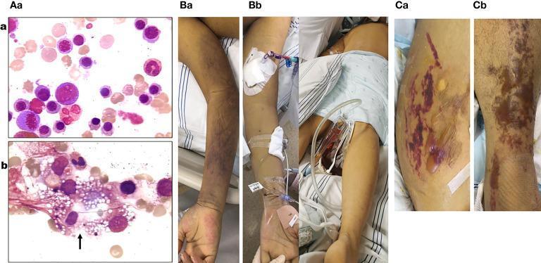
This composite medical image includes bone marrow smears and clinical photographs documenting the progression of a systemic illness, likely Parvovirus B19 infection. Diagnostic Images (Aa, Ab): Bone marrow smears (400x magnification) demonstrate a transition from hyperplastic marrow with active erythropoiesis (Aa) to a significant decrease in erythroblast density and maturation inhibition (Ab). Image Ab prominently displays a phagocytic cell containing numerous vacuoles and cellular debris (black arrow), indicative of hemophagocytic syndrome. Clinical Photographs (Ba-Cb): Serial photographs show evolving cutaneous manifestations. Initial findings (Ba, Bb) demonstrate blue-purple ecchymosis and diffuse mottling on the limbs and trunk. Later stages (Ca, Cb) reveal progressive purple-red ecchymotic lesions associated with the development of large, strained bullae filled with serous fluid on an erythematous base. The images illustrate the clinical and pathological manifestations of acute aplastic crisis and hemophagocytic lymphohistiocytosis (HLH) within the context of hematologic and infectious disease.
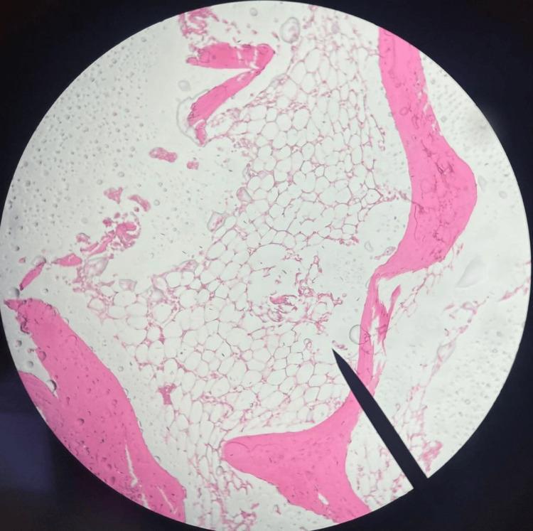
This diagnostic image is a low-magnification light microscopy photomicrograph of a bone marrow trephine biopsy, likely stained with hematoxylin and eosin (H&E). The specimen exhibits marked hypocellularity, a hallmark of severe aplastic anemia. The hematopoietic space is predominantly occupied by clear, vacuolated adipocytes (fatty marrow), with a notable absence of normal myeloid, erythroid, and megakaryocytic cell lines. Eosinophilic (pink) trabecular bone spicules are visible at the periphery and interspersed within the marrow cavity, providing structural context. The clinical significance of this finding is the demonstration of bone marrow failure, where the functional marrow has been replaced by adipose tissue. The absence of infiltrative abnormal cells or significant fibrosis is a key negative finding for differentiating this from myelodysplastic syndromes or myelofibrosis.
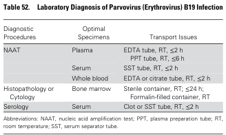
Table 52. Laboratory Diagnosis of Parvovirus (Erythrovirus) B19 Infection <table><thead><tr><th>Diagnostic Procedures</th><th>Optimal Specimens</th><th>Transport Issues</th></tr></thead><tbody><tr><td rowspan="3">NAAT</td><td>Plasma</td><td>EDTA tube, RT, ≤2 h<br>PPT tube, RT, ≤6 h</td></tr><tr><td>Serum</td><td>SST tube, RT, ≤2 h</td></tr><tr><td>Whole blood</td><td>EDTA or citrate tube, RT, ≤2 h</td></tr><tr><td>Histopathology or Cytology</td><td>Bone marrow</td><td>Sterile container, RT; ≤24 h;<br>Formalin-filled container, RT</td></tr><tr><td>Serology</td><td>Serum</td><td>Clot or SST tube, RT, ≤2 h</td></tr></tbody></table> Abbreviations: NAAT, nucleic acid amplification test; PPT, plasma preparation tube; RT, room temperature; SST, serum separator tube.
Aplastic Crisis
Definition
Pathophysiology
Etiology and At-Risk Groups
| Condition | Reason at Risk |
|---|---|
| Sickle cell disease | Shortened RBC lifespan (~20 days) |
| Thalassemia (major/intermedia) | Ineffective erythropoiesis + hemolysis |
| Hereditary spherocytosis | Rapid hemolysis |
| G6PD deficiency | Episodes of acute hemolysis |
| Autoimmune hemolytic anemia | High RBC turnover |
| Post-bone marrow transplant | Suppressed baseline erythropoiesis |
| Hematologic malignancies | Impaired marrow reserve |
"The anemic crises associated with low or absent reticulocytes in hereditary spherocytosis and sickle cell disease are virtually always secondary to B19 parvovirus infection." - Goldman-Cecil Medicine
Clinical Features
- Prodrome (viremic phase): Fever, malaise, myalgia, headache - 7-10 days before the hematologic crisis
- Crisis phase: Rapid-onset fatigue, pallor, dyspnea, worsening anemia
- Rash (erythema infectiosum/"slapped cheek") is rarely seen during aplastic crisis - it appears in the post-viremic immune phase, by which time the aplastic crisis has usually resolved
- Hemoglobin may fall below 4 g/dL
- No reticulocytosis - this is the key distinguishing feature (reticulocyte count is very low or zero)
- WBC and platelet counts may also fall, especially in patients with a functioning spleen
- Rarely: agranulocytosis, thrombocytopenia, pancytopenia, hemophagocytic syndrome (HLH)
Laboratory Findings
| Parameter | Finding |
|---|---|
| Hemoglobin | Severely low (can be <4 g/dL) |
| Reticulocyte count | Very low or absent (reticulocytopenia) |
| WBC | May decrease |
| Platelets | May decrease |
| Bilirubin | Markedly elevated (from hemolysis + hyperbilirubinemia) |
| LDH | Elevated |
- Hypoplasia or aplasia of the erythroid series
- Absence of maturing erythroid precursors
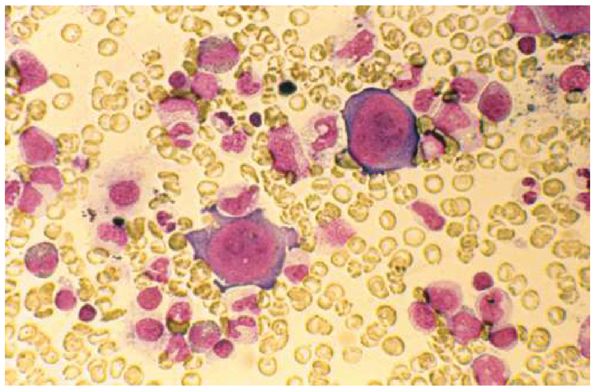
- Pathognomonic finding: "giant pronormoblasts" - large erythroid precursors with pale nuclei and cytoplasmic vacuoles, representing the cytopathic effect of B19
Diagnosis
- Serology (IgM/IgG): Unreliable in aplastic crisis because antibodies have not yet developed (still viremic). IgM appears 2-3 months post infection.
- PCR for B19 DNA (preferred): Most sensitive - viral loads can reach ~10¹¹ genome copies/mL during viremia. Detects B19 from serum, blood cells, or tissue.
- Bone marrow biopsy: Shows giant pronormoblasts and erythroid aplasia (if done)
Management
- First-line for symptomatic severe anemia (Hb <6 g/dL or symptomatic at higher levels)
- Often life-saving; may be urgently required
- The crisis is self-limited, so transfusion is a bridge to allow immune recovery
- Fluids, oxygen supplementation, rest
- Monitor for complications (heart failure, CVA from severe anemia)
- Not indicated for standard aplastic crisis (self-limited with intact immune system)
- Reserve for immunocompromised patients with persistent/chronic B19 infection causing pure red cell aplasia (e.g., HIV, transplant recipients, congenital immunodeficiency). Commercial IVIG contains neutralizing anti-B19 antibodies.
- Patients with aplastic crisis are viremic and infectious - they should be isolated from immunocompromised persons and pregnant women (risk of hydrops fetalis)
Natural History and Prognosis
- Recovery of erythropoiesis typically occurs within 1-2 weeks after onset, once the immune system produces anti-B19 antibodies and clears the viremia
- Reticulocyte count rises first, followed by hemoglobin recovery
- Generally a once-in-a-lifetime event - long-lasting protective immunity develops after infection
- Can be fatal if untreated, particularly from high-output cardiac failure or stroke precipitated by severe anemia
- In Egyptian patients with hemoglobinopathies, a large series documented reversible bone marrow failure, acute kidney injury, and extreme hyperbilirubinemia with B19 seroconversion
Distinction from Related Conditions
| Feature | Aplastic Crisis (B19) | Vaso-occlusive Crisis (SCD) | Sequestration Crisis | Hemolytic Crisis |
|---|---|---|---|---|
| Cause | Parvovirus B19 | Hypoxia, dehydration, cold | Blood pooling in spleen | Infection/G6PD |
| Reticulocytes | Very low/absent | Elevated | Elevated | Elevated |
| Hemoglobin fall | Gradual (days) | Stable | Rapid | Gradual |
| Bone marrow | Erythroid aplasia | Normal | Normal | Hyperplastic |
| Pain | Absent | Prominent | Mild-moderate | Absent |
| Treatment | Transfusion | Analgesia, hydration | Urgent transfusion | Supportive |
Special Populations
- Immunocompromised (HIV, transplant, congenital immunodeficiency): Cannot clear B19 - leads to chronic pure red cell aplasia with transfusion dependence. IVIG is effective.
- Pregnancy: B19 during pregnancy can cross the placenta - fetal anemia, hydrops fetalis, fetal death. Risk of fetal loss <10% of primary maternal infections, highest before 20 weeks.
- Healthy individuals: Erythropoiesis arrests but anemia is subclinical and unnoticed.