
Cerebral palsy treatment
"cerebral palsy"[MeSH Terms] AND treatment
cerebral palsy classification motor function treatment
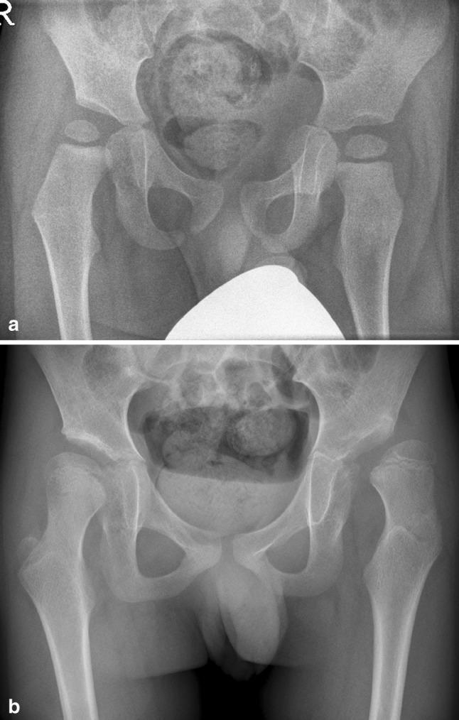
Two anteroposterior (AP) pelvic radiographs demonstrating the longitudinal progression of hip displacement in a pediatric patient with Gross Motor Function Classification System (GMFCS) level V cerebral palsy. Figure (a) shows the initial state at 22 months, where the right femoral head exhibits early subluxation with 29% migration, while the left remains seated. Figure (b) displays the same patient at 95 months (nearly 8 years), illustrating significant disease progression. Both hips show increased coxa valga, with the right hip progressing to 34% migration and the left hip reaching 76% migration, indicating severe lateral displacement and loss of acetabular coverage. The image serves as a clinical example of how high head-shaft angles (HSA) and neuromuscular imbalances contribute to progressive hip luxation over time in non-ambulatory patients. Key diagnostic features visible include the increasing lateral migration of the femoral epiphyses relative to Perkins' line and the persistent vertical orientation of the femoral necks.
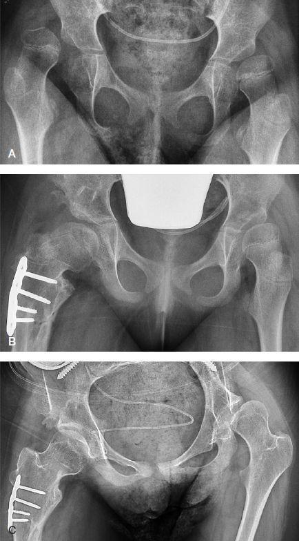
A series of three anteroposterior pelvic radiographs (A, B, C) documenting the longitudinal management of hip dysplasia in a pediatric patient with Gross Motor Function Classification System (GMFCS) level V cerebral palsy. Radiograph A (baseline) shows complete lateral and superior dislocation of the right hip with a high migration percentage (MP 100%) and subluxation of the left hip (MP 51%). Radiograph B, taken 14 months post-surgical intervention, displays the results of a right femoral varus osteotomy and pelvic Dega osteotomy. Visible hardware includes a lateral femoral compression plate secured with five screws. Both hips show improved containment with mild residual subluxation. Radiograph C, at 7.3 years follow-up, demonstrates a well-seated right hip joint with stable surgical hardware and remodeled femoral neck-shaft angle. In contrast, the untreated left hip shows significant progression of subluxation (MP 64%) with flattening of the acetabular roof and lateralization of the femoral head, illustrating the natural history of hip displacement in non-ambulatory patients without prophylactic bony surgery.
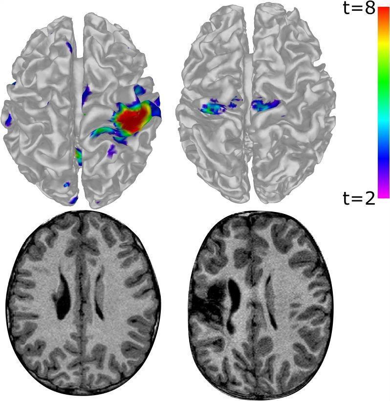
This composite diagnostic image features functional magnetic resonance imaging (fMRI) surface maps (top row) and corresponding T1-weighted axial structural scans (bottom row) of two pediatric patients with unilateral cerebral palsy. The left column illustrates a child with higher motor function performing a hand-tapping task with the less-impaired hand; fMRI demonstrates robust, focal, unilateral activation in the contralateral sensorimotor cortex, specifically at the 'hand knob' landmark. The right column illustrates a child with significant brain damage performing the task with the more-impaired hand. The top-right fMRI shows atypical, bilateral, and more medial/anterior activation concentrated in the supplementary motor areas (SMA), suggesting functional reorganization. The bottom-right T1 image reveals a clear periventricular lesion with altered signal intensity and tissue loss compared to the relatively preserved cortical morphology in the bottom-left image. A color scale indicates t-values from 2 to 8, representing the statistical significance of hemodynamic response. This comparison highlights the neuroplastic adaptation of motor control pathways following early-life brain injury.
Cerebral Palsy: Treatment
Four Core Principles (Box 35.2, Campbell's Operative Orthopaedics 2026)
- CNS injury is nonprogressive, but secondary deformities from abnormal muscle forces are progressive.
- Available treatments correct secondary deformities only - not the underlying brain injury.
- Deformities worsen during rapid growth, so timing interventions around growth spurts matters.
- Treatments should minimize negative effects on the child's socialization and education.
Note: Children with CP have skeletal ages approximately 2 years advanced over chronological age - factor this into surgical planning.
1. Rehabilitative/Non-Pharmacological Therapies
Physical Therapy
- Ambulatory patients (GMFCS I-III): muscle strengthening, contracture prevention, gait and balance training
- Non-ambulatory patients (GMFCS IV-V): improving sitting balance, hygiene, and ease of caregiver tasks
Occupational and Speech Therapy
Orthotics and Casting
- Ankle-foot orthoses (AFOs) - the most frequently used; control equinus and crouch gait
- Hip abduction braces - for hip surveillance and subluxation prevention
- Hand and wrist splints
- Spinal braces - for scoliosis
2. Pharmacological Management
Oral Antispasmodics (first-line for diffuse spasticity)
| Drug | Mechanism | Notes |
|---|---|---|
| Baclofen (oral) | GABA-B agonist at spinal cord | Poor BBB penetration, short half-life (3-4 h); requires high systemic doses; may cause sedation |
| Tizanidine | Alpha-2 agonist | Alternative to baclofen |
| Dantrolene | Inhibits muscle SR calcium release | Acts peripherally; useful for diffuse spasticity |
| Benzodiazepines (diazepam) | GABA-A potentiation | Useful short-term; sedating |
| Clonidine / Gabapentin | Second-line | Dual benefit for spasticity plus sleep disruption, dysautonomia, and pain |
Chemodenervation - Botulinum Toxin A (BoNT-A)
- First-choice for focal or segmental spasticity
- Injected directly into the muscle at the motor endplate; blocks acetylcholine release
- Effect begins ~24 hours after injection; lasts up to 4-6 months
- Safer than phenol/alcohol because it binds selectively to the neuromuscular junction
- Repeat injections every 3-6 months (no sooner than 3 months to prevent resistance)
- Combined with rehabilitative therapies, BoNT-A can delay or avoid the need for orthopedic surgery
- Large systemic doses can cause respiratory depression - careful dosing is mandatory
Intrathecal Baclofen (ITB)
- Programmable pump implanted subcutaneously in the abdominal wall; requires refilling every 2-3 months
- A meta-analysis of 14 studies confirmed reduction of lower-limb spasticity, improved function, and manageable complications
- Reversible - an advantage over SDR
- Also used for dystonia in dyskinetic CP (although evidence is mixed - may occasionally worsen dystonia)
- Complications: catheter disconnection (~9%), catheter dislodgement (~8%), infection (~9.3%), CSF leak (~4.9%), overdose (decreased trunk tone, sedation, respiratory depression)
- 10-20% of patients require further surgery or pump removal
Associated Comorbidities Requiring Pharmacotherapy
- Seizures (~30% of CP patients): antiepileptics - note that many have CNS side effects affecting learning and ambulation
- Osteopenia (~80% with Z-score <-2): bisphosphonates and growth hormone shown safe and effective in small studies for increasing bone mineral density
- Sleep disruption / pain (gabapentin, clonidine as dual-purpose agents)
3. Neurosurgical Treatment
Selective Dorsal Rhizotomy (SDR)
- Age 4-10 years (when relearning gait is easier)
- GMFCS levels I-III with good selective motor control and minimal weakness
- Underlying etiology: periventricular leukomalacia (PVL) in the setting of prematurity
- Basal ganglia and thalamus spared on imaging (to avoid worsening dystonia)
- Adequate cognition to participate in post-op rehabilitation
4. Orthopedic Surgery
Soft-Tissue Procedures
- Muscle-tendon lengthening: for flexible/dynamic deformities; corrects relative shortening caused by spasticity during growth (skeleton outgrows the musculotendinous unit)
- Techniques: aponeurotic recession, Z-plasty, complete tenotomy
- Recessions preferred - avoid overlengthening and subsequent weakness
- Tendon transfers: to remove a deforming muscle force or create a passive tendon sling. Note: spastic muscles typically remain spastic after transfer, so goals are limited
Bony Procedures
- Osteotomies: for severe rigid deformities or joint subluxation/dislocation; commonly combined with soft-tissue releases
- Hip: femoral varus osteotomy + pelvic osteotomy (Dega/Salter) for hip displacement (common in GMFCS IV-V; hip surveillance programs are standard of care)
- Femoral derotation osteotomy for internal rotation gait
- Tibial/foot osteotomies for severe foot deformity
- Arthrodesis: for severe joint destruction (e.g., foot)
- Proximal femoral resection arthroplasty: for painful dislocated hips in non-ambulatory patients
Event-Based vs. "Birthday Surgery"
5. Recent Evidence (2024-2025)
Treatment by GMFCS Level (Summary)
| GMFCS Level | Ambulatory Status | Key Priorities |
|---|---|---|
| I | Walks without limitation | Tone management, AFOs, gait optimization |
| II | Walks with limitations | PT, AFOs, BoNT-A, consider SDR |
| III | Walks with assistive device | Intensive PT/OT, BoNT-A/ITB, hip surveillance, consider SDR or orthopedic surgery |
| IV | Limited self-mobility | ITB, hip and spine surveillance, seating/positioning, caregiver support |
| V | Transported | ITB, pain management, hip prevention surgery, nutrition, anti-epileptics |
Multidisciplinary Team
- Campbell's Operative Orthopaedics 15th Ed 2026 (primary orthopaedic reference)
- Bradley and Daroff's Neurology in Clinical Practice (neurological management)
- Katzung's Basic and Clinical Pharmacology 16th Ed (pharmacological management)
- Recent PubMed: PMID 40494559 (nonsurgical therapies NMA, 2025) | PMID 39300490 (hippotherapy SR, 2024)
Osnid d
Could you clarify what you mean by 'Osnid d'?
Osnid D drug medication composition uses
Osnate-D (Osnid D) - Drug Profile
Composition (per tablet)
| Component | Amount |
|---|---|
| Ossein Mineral Complex | 830 mg |
| - Calcium (elemental) | ~177.6 mg |
| - Phosphorus | ~82.2 mg |
| - Collagen | ~224 mg |
| - Other proteins | ~66.4 mg |
| - Residual mineral salts (Mg, F, Fe, Zn, Cu, Ni) | 24.9 mg |
| - Hydroxyapatite equivalent | ~440 mg |
| Vitamin D | 400 IU |
Mechanism of Action
- Calcium + Phosphorus: Primary mineral constituents of bone hydroxyapatite; essential for mineralization of osteoid and maintaining bone density.
- Collagen and bone proteins: Provide the organic scaffold of bone; may influence osteoblast activity.
- Vitamin D (400 IU): Promotes intestinal calcium and phosphorus absorption; regulates parathyroid hormone (PTH); essential for bone mineralization. Without adequate Vitamin D, calcium cannot be properly absorbed regardless of dietary intake.
Indications (Uses)
- Osteoporosis - treatment and prevention
- Osteomalacia (soft bones due to vitamin D / calcium deficiency in adults)
- Rickets (in children)
- Pregnancy and lactation - increased mineral requirements
- Menopause - prevention of bone loss due to estrogen decline
- Growth periods in children and adolescents
- Corticosteroid-induced osteoporosis (long-term steroid use depletes calcium)
- Bone pain or fragility fractures
Dosage
| Patient Group | Dose |
|---|---|
| Adults | 1-2 tablets daily (before meals), or as directed |
| Children | As determined by physician |
| Pregnant/lactating | As directed; usually 1-2 tablets/day |
Side Effects
- Constipation
- Nausea, bloating, upset stomach
- Gas
- Vomiting, loss of appetite
- Unusual fatigue or weakness
- Mood changes (confusion)
- Increased thirst and urination
- Kidney problems / kidney stones
Contraindications
- Hypercalcemia (high blood calcium)
- Hypercalciuria (excess calcium in urine)
- Nephrolithiasis (kidney stones) - use with caution
- Severe renal impairment
- Sarcoidosis or other granulomatous diseases (risk of vitamin D toxicity)
- Hypersensitivity to any component
Important Drug Interactions
| Interacting Drug | Effect |
|---|---|
| Digoxin | Hypercalcemia from excess vitamin D can precipitate cardiac arrhythmias |
| Thiazide diuretics | May cause hypercalcemia in hypoparathyroid patients |
| Corticosteroids | Steroids reduce calcium absorption and deplete bone; Osnate-D may help counteract this |
| Antiseizure drugs | Induce hepatic enzymes that convert vitamin D to inactive metabolites - reduced vitamin D effect |
| Rifampin | Increases vitamin D metabolism, reduces blood levels |
| Cholestyramine / Colestipol | Impair vitamin D absorption - give Osnate-D as far apart in time as possible |
| Orlistat (weight-loss drug) | Reduces fat-soluble vitamin D absorption |
| Magnesium-containing antacids | Risk of hypermagnesemia, especially in renal failure |
Key Precautions
- Monitor blood/urine calcium levels during long-term use or in patients with renal impairment.
- Patients with a history of kidney stones should use only under medical supervision.
- Ensure adequate fluid intake to reduce risk of nephrolithiasis.
- The 400 IU of Vitamin D per tablet is a moderate dose; higher supplementation may be needed for severe deficiency (to be prescribed separately by a physician).