
Colles fracture every thing with pictures
Colles fracture wrist X-ray radiograph
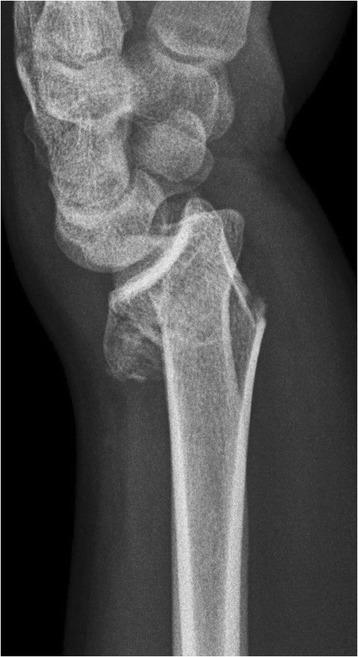
A lateral view diagnostic X-ray of the wrist demonstrating an unstable distal radius fracture, characteristic of a Colles-type injury. The radiograph reveals an oblique fracture line through the distal radial metaphysis with significant dorsal displacement and dorsal angulation of the distal fragment. Visible metaphyseal impaction and comminution are present on the dorsal aspect, creating a bone void or compression defect. This displacement results in secondary incongruity of the radiocarpal joint, as the carpal bones (including the scaphoid and lunate) remain aligned with the dorsally tilted distal radius fragment rather than the radial shaft. The image serves as a clinical example of metaphyseal instability requiring surgical intervention, such as volar locking plate fixation, to restore anatomical alignment and articular surface congruity.
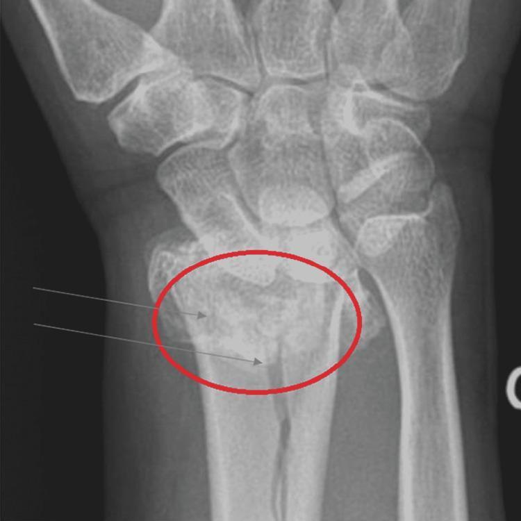
This medical diagnostic image is an anteroposterior (AP) x-ray radiograph of the distal forearm and wrist, demonstrating a Colles fracture. The radiograph depicts the distal radius, distal ulna, carpal bones, and proximal metacarpals. A red circle and grey arrows highlight a transverse fracture involving the distal radial metaphysis. Key visual findings include bony discontinuity with classic dorsal (posterior) displacement and impaction of the distal radial fragment, characteristic of a fall on an outstretched hand (FOOSH) injury. The distal ulna appears intact in this view, though some radial shortening may be present due to the fracture's impaction. The surrounding soft tissues show generalized swelling. This image serves as a clinical teaching tool for identifying common orthopaedic fractures, particularly in the context of underlying metabolic bone disease such as osteoporosis, as indicated by the cortical thinning visible in the long bones.
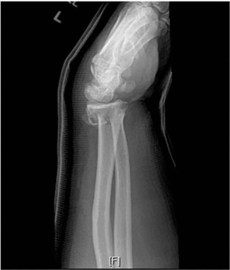
A diagnostic lateral X-ray of the left wrist demonstrating an acute, comminuted fracture of the distal radius. The distal radial fragment exhibits significant dorsal angulation and dorsal displacement, characteristic of a Colles-type injury pattern. There is associated cortical disruption and multi-fragmentary bone involvement at the metaphyseal-diaphyseal junction. Additionally, a displaced fracture of the ulnar styloid process is visible. The radiograph is taken through a dorsal splint or cast material, which is visible as a radiopaque shadow bordering the posterior aspect of the forearm and hand. This image is clinically significant for illustrating common orthopedic trauma following a fall on an outstretched hand (FOOSH), highlighting the loss of normal volar tilt and the resulting dorsal deformity. It serves as a classic educational example of unstable distal radius fractures requiring surgical consideration due to dorsal angulation and comminution.
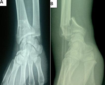
This diagnostic image consists of two preoperative X-ray views of the wrist, illustrating an unstable intra-articular distal radius fracture. View A is an anteroposterior (AP) radiograph showing comminution and a clear sagittal fracture line extending into the radiocarpal joint space. View B is a lateral radiograph demonstrating a Goyrand-Smith fracture, characterized by significant volar (palmar) displacement and angulation of the distal fracture fragment relative to the radial shaft. The alignment between the distal radius, carpal bones, and distal ulna is severely disrupted, with the carpus following the volar displacement of the distal radial fragment. This case highlights key orthopedic findings for surgical planning, specifically the involvement of the articular surface and the direction of fragment displacement, which distinguishes the Smith fracture from the more common dorsal displacement seen in Colles fractures.
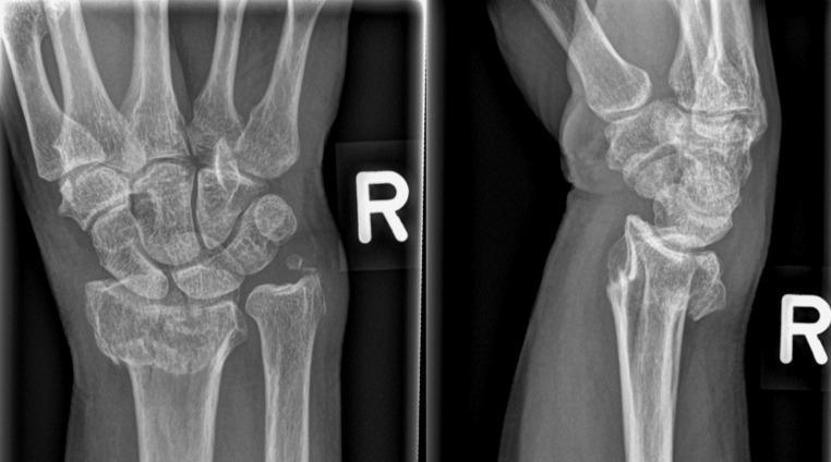
This diagnostic image consists of a two-view conventional radiograph (X-ray) of the right wrist, presenting both anteroposterior (AP) and lateral views. The image demonstrates a distal radius fracture with significant displacement. In the AP view (left), a transverse fracture line is visible across the distal metaphysis of the radius, accompanied by an associated fracture of the ulnar styloid process. The lateral view (right) clearly illustrates dorsal (posterior) displacement and dorsal angulation of the distal radial fragment, characteristic of a Colles-type fracture pattern. There is a disruption of the normal radiocarpal alignment, with the carpal bones following the dorsally displaced distal radius segment. Key anatomical structures visible include the distal radius and ulna, the proximal and distal carpal rows, and the bases of the metacarpals. This visual material is primarily used in orthopedic and emergency medicine education to teach fracture identification, terminology for displacement and angulation, and the assessment of wrist joint congruity.
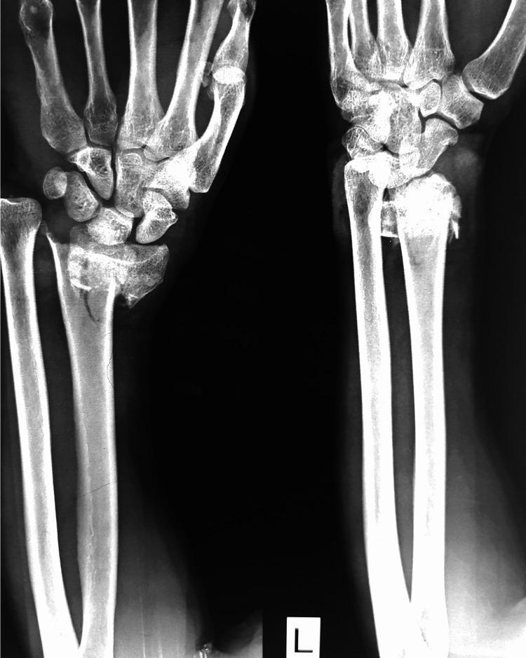
This diagnostic image is a plain radiograph (X-ray) of the left wrist in two views (oblique and lateral) demonstrating a complex distal radius fracture. The visual evidence shows a comminuted fracture of the distal radial metaphysis with intra-articular extension. Key pathological features include significant radial shortening, dorsal displacement, and dorsal angulation of the distal fracture fragments (characteristic of a Colles-type injury pattern). The alignment of the radiocarpal joint is disrupted, with the carpal bones shifted dorsally in relation to the main radial shaft. There is visible fragmentation and irregularity at the fracture site, indicating instability. The distal ulna appears intact, though the distal radioulnar joint (DRUJ) relationship is altered due to the radial shortening. This image illustrates a common orthopedic trauma scenario requiring surgical evaluation for anatomic reduction and stabilization, often via external fixation or internal plate osteosynthesis.
Colles fracture dinner fork deformity clinical appearance wrist
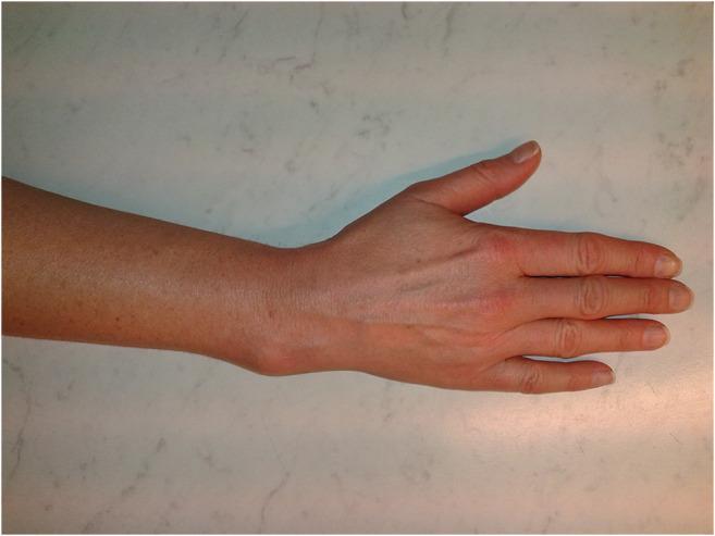
Clinical photograph showing a dorsal view of the right forearm, wrist, and hand of a 48-year-old female. The image demonstrates a significant structural abnormality at the distal forearm characterized by a prominent dorsal-radial convexity, consistent with a malunited distal radius fracture (Colles-type deformity). The wrist exhibits a 'dinner fork' like contour deviation where the distal fragment has healed with dorsal angulation. The skin over the deformity is intact without acute inflammatory signs, though superficial veins are visible on the dorsum of the hand. The fingers are held in a neutral, extended position with no apparent secondary digital deformities. This visual serves as an educational example of the long-term sequelae of non-surgically treated or inadequately reduced distal radius fractures, highlighting the functional and aesthetic impact of bone malunion in orthopedics.
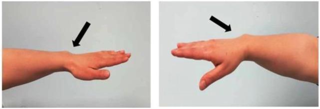
This clinical photograph consists of two side-profile views of a human forearm and wrist, illustrating the classic morphological features of a Madelung deformity. The images demonstrate a characteristic 'dinner fork' appearance caused by the dorsal subluxation of the distal ulna and the volar displacement of the hand relative to the forearm. In both panels, black arrows highlight a prominent bony protrusion on the dorsal aspect of the wrist. This skeletal anomaly is associated with bowing of the radius and an increased palmar and ulnar tilt of the distal radial articular surface. The first view shows the arm in a neutral extension, while the second view emphasizes the deformity with the hand in a more pronounced position. Such findings are hallmark clinical signs of Léri-Weill dyschondrosteosis (LWD), often linked to SHOX gene mutations. The image is intended for educational purposes in orthopedic surgery, genetics, and pediatric endocrinology to help identify dysharmonic short stature and associated skeletal dysplasias.
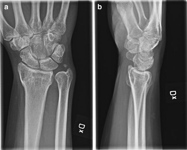
Two radiographic views of a left wrist demonstrate a malunited distal radius fracture in a 48-year-old female. Image (a) is an anterior-posterior (AP) view showing radial shortening and a loss of normal radial inclination. The distal radioulnar joint (DRUJ) appears widened. Image (b) is a sagittal (lateral) view clearly depicting significant dorsal angulation of the distal radial articular fragment, characteristic of a 'dinner-fork' deformity common in malunited Colles-type fractures. This malalignment results in dorsal tilt of the radiocarpal joint surface, affecting the articulation with the carpal bones (scaphoid and lunate). The lunate shows compensatory dorsal subluxation relative to the radial shaft. This imaging provides the clinical basis for a corrective osteotomy to restore anatomical alignment, improve wrist range of motion, and reduce functional limitations.
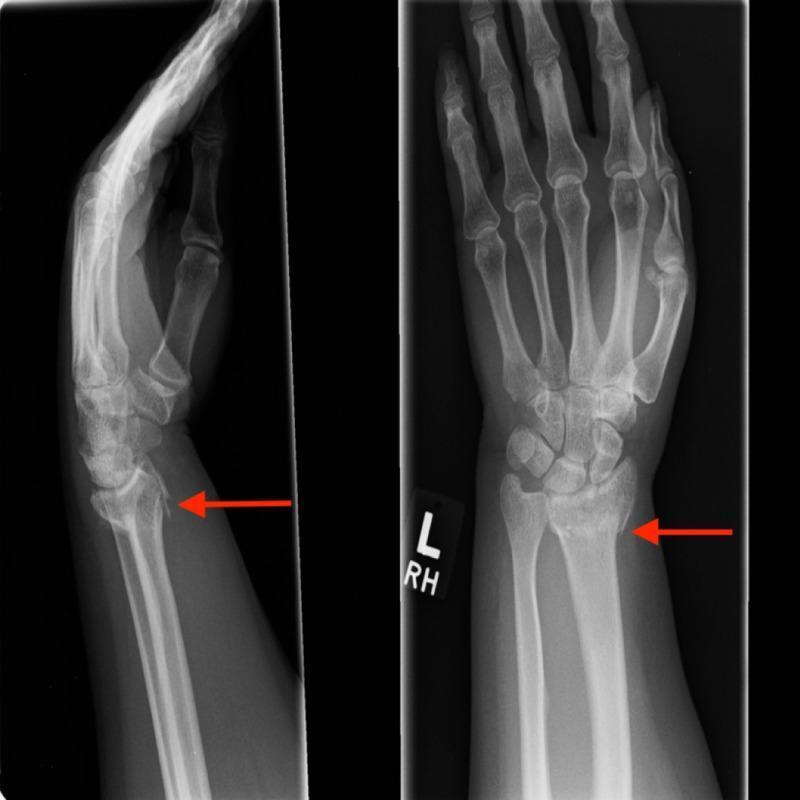
Two-view radiographic study (Lateral and Posteroanterior) of the left wrist and hand demonstrating an extra-articular distal radius fracture. The lateral view on the left shows a Colles-type injury pattern with prominent dorsal angulation and dorsal comminution of the fracture fragments. The red arrow highlights the displacement and cortical disruption at the dorsal aspect of the distal radius. The PA view on the right identifies the fracture line extending across the metaphysis of the distal radius (red arrow), showing radial shortening and mild radial deviation. The carpal bones, including the scaphoid and lunate, appear intact without evidence of intra-articular extension. The distal radioulnar joint alignment is altered secondary to the radial displacement, though the ulna appears visually intact. This imaging is diagnostic for a common fracture resulting from a fall on an outstretched hand (FOOSH), illustrating typical deformities such as the 'dinner fork' deformity seen in clinical practice.
distal radius fracture reduction splinting sugar tong cast management
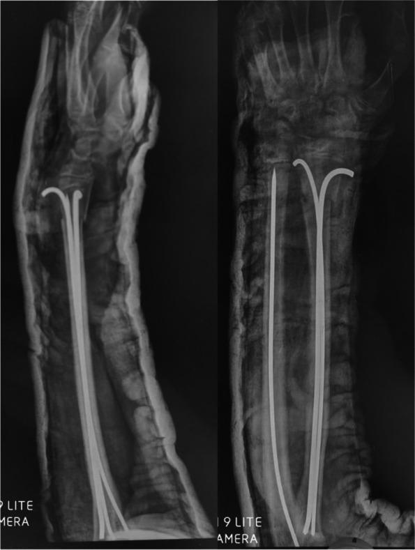
Anteroposterior (AP) and lateral X-ray views of a human forearm showing immediate postoperative results of a pediatric distal forearm fracture reduction. The diagnostic imaging demonstrates internal fixation using multiple intramedullary Kirschner wires (K-wires). In the radius, two pre-bent K-wires are visible, extending through the medullary canal from the distal to the proximal region. A single straight K-wire is positioned within the medullary canal of the ulna. The alignment of both the radius and ulna shows successful surgical reduction with restored anatomical continuity. An external sugar-tong splint or plaster cast is visible as a radio-opaque layer surrounding the soft tissue of the forearm. The clinical focus of this image is to illustrate the surgical technique for stabilizing pediatric long bone fractures using a minimally invasive intramedullary pinning approach, ensuring maintenance of reduction while allowing for secondary bone healing.
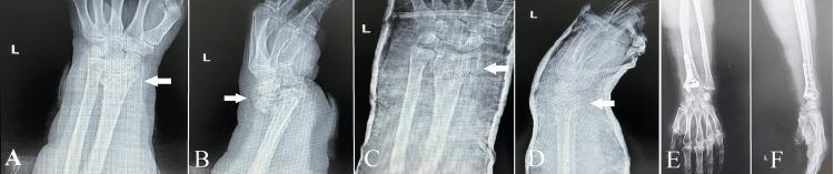
This composite of diagnostic radiographs illustrates the management sequence of a complex distal radius fracture in an elderly patient. Panels A and B (Anteroposterior and Lateral views) demonstrate a severely comminuted, intra-articular fracture of the distal radial epiphysis with significant dorsal displacement and multiple bone fragments. White arrows highlight the fracture lines and articular involvement. Panels C and D show the post-reduction state, where the wrist is immobilized in a radiopaque cast; alignment is improved, but the comminution remains visible. Panels E and F display the post-osteosynthesis outcome, showing definitive internal fixation with a volar locking plate and screws. The final images demonstrate anatomic reconstruction of the articular surface and restoration of radial length and inclination. This educational series highlights the transition from initial emergency presentation through closed reduction to surgical stabilization in orthopedic traumatology, specifically focusing on geriatric fracture management and the use of internal fixation hardware to achieve stability in poor-quality bone.
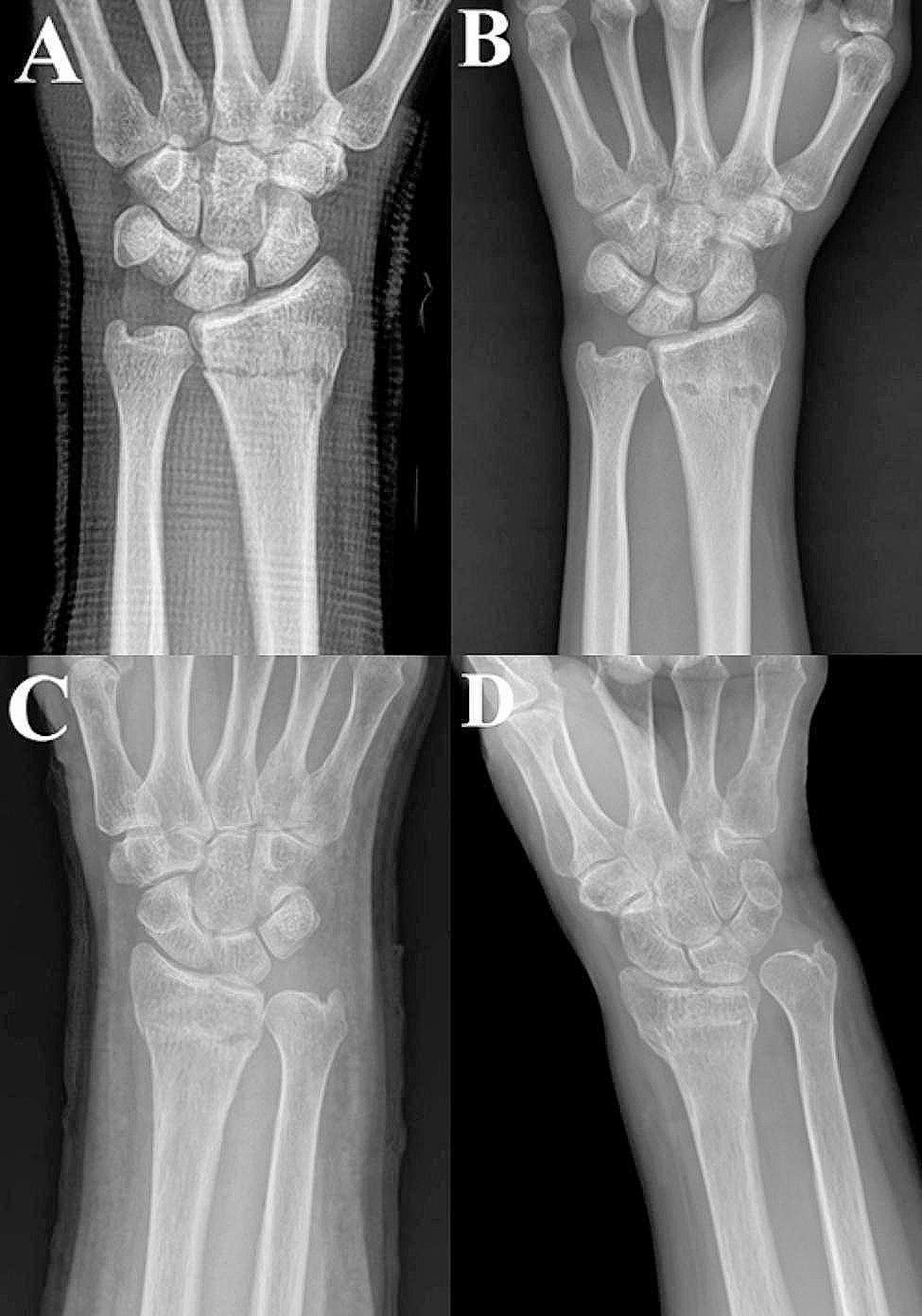
This diagnostic image consists of four anteroposterior (AP) x-ray radiographs of the wrist joint, categorized as comparison charts for orthopedic evaluation. Panels A and B demonstrate a successful radiological outcome. Panel A shows the wrist immediately following a closed reduction of a distal radius fracture, immobilized in a radiopaque Muenster splint; the distal radius and ulna exhibit anatomical alignment and neutral ulnar variance. Panel B shows the final follow-up of the same patient, revealing stable maintenance of the reduction and healed cortical surfaces. In contrast, panels C and D illustrate a poor radiological result. Panel C shows the immediate post-reduction state with acceptable initial alignment. However, panel D, the final follow-up, displays significant malunion characterized by radial shortening, increased positive ulnar variance, and dorsal/radial angulation of the distal radius fragment. This comparison highlights the potential for secondary displacement in distal radius fractures during conservative management with splinting. The series is intended for medical education on fracture management, radiological assessment of reduction quality (radial height and inclination), and monitoring for malunion.
Colles' Fracture - Complete Guide
Definition
- Dorsal displacement and angulation of the distal fragment
- Radial deviation of the wrist
- Proximal displacement (radial shortening)
- Frequent associated fracture of the ulnar styloid
Epidemiology & Mechanism
- Most common in postmenopausal women (osteoporotic bone - often considered a pathological fracture in this group)
- Mechanism: Fall on an outstretched hand (FOOSH) with the wrist in extension
- Compression forces on the dorsal side produce dorsal comminution
- May also affect younger patients with high-energy trauma
Classic "Dinner Fork" Deformity
- Wrist deviated radially (prominent lower end of ulna on ulnar side)
- Swelling and tenderness over distal radius
- Palmar paresthesias from pressure on the median nerve
- Pain with any wrist movement
Radiographic Appearance
X-ray - AP and Lateral Views (Classic Colles')
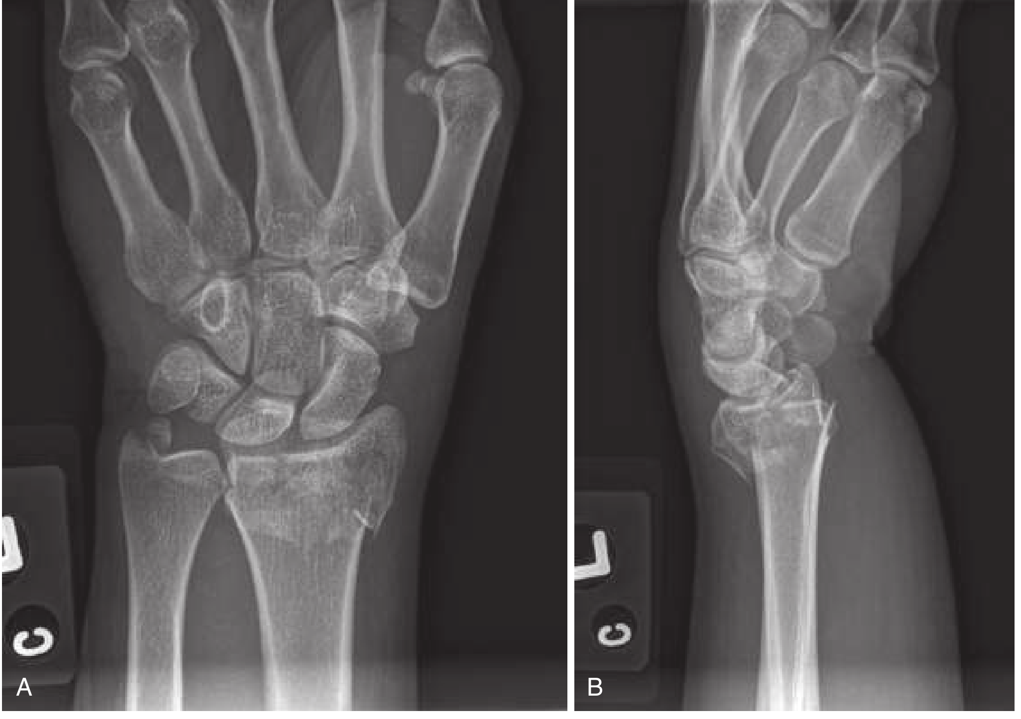
Grainger & Allison Radiology Textbook Image (Fig 45.39)
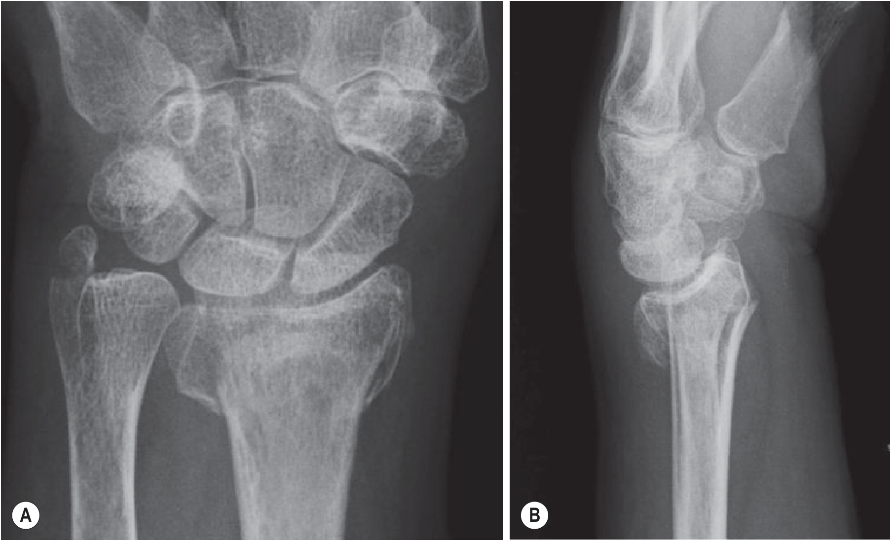
Radiographic Key Findings (Summary Table)
| View | Findings |
|---|---|
| PA / AP | Distal radial metaphysis fracture; radial shortening; loss of radial inclination; intra-articular extension; ulnar styloid fracture |
| Lateral | Dorsal angulation of the distal radial articular surface (loss of normal volar tilt); dorsal displacement of fragment; dorsal comminution |
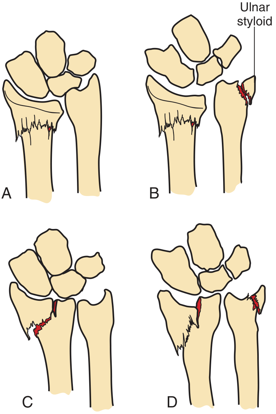
Diagram of Distal Radius Fracture Types
Instability Criteria
- Dorsal angulation >20 degrees
- Intra-articular involvement
- Marked comminution
- >1 cm (5 mm) of radial shortening (ulnar-positive variance)
- Distal radioulnar joint instability
Associated Injuries
- Ulnar styloid fracture - in ~60% of distal radius fractures; may indicate triangular fibrocartilage complex (TFCC) injury
- Median nerve injury - most common nerve complication (17%); may cause acute carpal tunnel syndrome
- TFCC injury - from avulsion of the ulnar styloid
- Carpal instability - especially scapholunate dissociation
- Radial and ulnar nerve injuries (less common)
Management
Step 1 - Assess the Need for Reduction
- Lateral film: A line through anterior and posterior lips of the radial articular surface - if tilted dorsally beyond neutral, reduction is indicated
- AP film: If radius is shorter than ulna (positive ulnar variance), reduction is indicated
- Extra-articular, non-displaced fractures may be treated without reduction
Step 2 - Anesthesia for Reduction
- Hematoma block: 10 mL of 1% lidocaine injected into the fracture hematoma via the dorsum of the distal radius with a 22-gauge needle - easy, effective, reduces ED stay
- Bier block (IV regional anesthesia)
- Regional nerve blocks: Median, radial, ulnar, or brachial plexus
- Procedural sedation
Step 3 - Closed Reduction Technique
- Apply finger traps to index and middle fingers (relaxes deforming muscular forces, may reduce fracture passively)
- Apply traction while the fracture fragment is pushed distally and palmarly
- Forearm held firmly proximally as counterforce
- Goal: Restore volar tilt, radial inclination, and proper radial length
- Minimum acceptable: neutral (0 degrees) angulation; volar tilt ideally restored
Step 4 - Immobilization
- Double sugar-tong splint (elbow to wrist + elbow to axilla) - preferred after reduction
- Allows finger movement while immobilizing the wrist
- No circumferential cast for at least 24 hours - edema may cause neurovascular compromise
- If a short arm cast is applied, it must be bivalved
- Immobilization for 4-6 weeks
- Follow-up X-rays at 1, 2, and 6 weeks to monitor displacement
Step 5 - Indications for Surgery
- Failed closed reduction / unstable fractures
- Severely comminuted or intra-articular fractures
- Open fractures
- Neurovascular compromise
- High-demand patients (young, active, dominant hand)
- Fractures not reducible to acceptable alignment
Comparison with Related Distal Radius Fractures
| Fracture | Distal Fragment Direction | Mechanism |
|---|---|---|
| Colles' | Dorsal displacement + dorsal angulation | FOOSH (extended wrist) |
| Smith's ("Reverse Colles'") | Volar displacement + volar angulation | Fall on flexed wrist |
| Barton's | Rim fracture + radiocarpal subluxation | Shearing injury |
| Chauffeur's / Hutchinson | Radial styloid (intra-articular) | Axial compression via scaphoid |
Complications
| Complication | Notes |
|---|---|
| Malunion | Most common; leads to stiff, weak, painful wrist |
| Carpal tunnel syndrome | Most common nerve injury; median nerve compression (17%) |
| DRUJ instability | From ulnar styloid avulsion/TFCC injury |
| Radiocarpal instability | Especially with intra-articular fractures |
| Post-traumatic arthritis | From intra-articular involvement |
| CRPS (Sudeck's atrophy) | Chronic regional pain syndrome |
| Tendon rupture | Extensor pollicis longus most vulnerable |
| Iatrogenic median nerve injury | From extreme wrist flexion during splinting |
- Colles' fracture = within 2 cm of radial articular surface + dorsal displacement + radial deviation
- Dinner fork deformity on exam; median nerve at risk
- Unstable = >20° angulation, intra-articular, comminuted, >1 cm shortening
- Reduce under hematoma block + finger traps; splint in double sugar-tong; no circumferential cast early
- Follow up in 2-3 days with orthopedics after ED reduction