
from Guyton and Hall textbook of medical physiology, 4th South Asia edition write a 5 mark short note on Heart Block-enlist types ,explain AV Nodal Block
Reading File
Finding Sources
Finding Sources
Finding Sources
Finding Sources
Quote the exact sentences from Guyton and Hall textbook of medical physiology, 4th South Asia edition and write a 5 mark short note on Heart Block-enlist types ,explain AV Nodal Block
Reading File
Finding Sources
Finding Sources
Reading File
Reading File
Loading Image
Loading Image
Loading Image
I now have all the exact text and ECG images from Guyton and Hall. Here is the complete short note with direct quotes:
Heart Block
(Guyton and Hall Textbook of Medical Physiology - Exact Quotes)
Definition / Introduction
"The only means whereby impulses ordinarily can pass from the atria into the ventricles is through the AV bundle, also known as the bundle of His. Conditions that can either decrease the rate of impulse conduction in this bundle or block the impulse entirely are as follows..."
- Guyton & Hall, Chapter 13
Types of Heart Block
Heart block is classified into two broad categories:
A. Sinoatrial (SA) Block
B. Atrioventricular (AV) Block
- First-degree AV block (Incomplete)
- Second-degree AV block (Incomplete) - Mobitz Type I (Wenckebach) & Mobitz Type II
- Third-degree AV block (Complete)
Causes of AV Block
The textbook lists the following exact causes:
"1. Ischemia of the AV node or AV bundle fibers often delays or blocks conduction from the atria to the ventricles. Coronary insufficiency can cause ischemia of the AV node and bundle in the same way that it can cause ischemia of the myocardium.2. Compression of the AV bundle by scar tissue or by calcified portions of the heart can depress or block conduction from the atria to the ventricles.3. Inflammation of the AV node or AV bundle can depress conduction from the atria to the ventricles. Inflammation results frequently from different types of endocarditis or myocarditis..."
AV Nodal Block - Degrees Explained
First-Degree Block (Incomplete AV Block)
"This so-called P-R interval usually decreases in length with a faster heart rate."
The P-R interval is prolonged beyond the normal 0.20 seconds. Every atrial impulse still reaches the ventricles, but with a delayed conduction time.
ECG - First-degree AV Block (Prolonged P-R interval, Lead II):
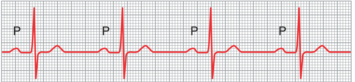
Figure 13.5: Prolonged P-R interval caused by first-degree atrioventricular heart block (lead II)
Second-Degree Block (Incomplete AV Block)
"This condition is called second-degree heart block. There are two types of second-degree AV block - Mobitz type I (also known as Wenckebach periodicity) and Mobitz type II. Type I block is characterized by progressive prolongation of the P-R interval until a ventricular beat is dropped..."
"Fig. 13.6 shows progressive P-R interval prolongation typical of type I (Wenckebach) block. Note prolongation of the P-R interval preceding the dropped beat, followed by a shortened P-R interval after the dropped beat."
- Mobitz Type I (Wenckebach): Progressive P-R prolongation until one QRS is dropped, then the cycle resets.
- Mobitz Type II: Sudden dropping of a QRS complex without preceding P-R prolongation; more serious and may progress to complete block.
ECG - Type I Second-Degree AV Block (Wenckebach - dropped beat):
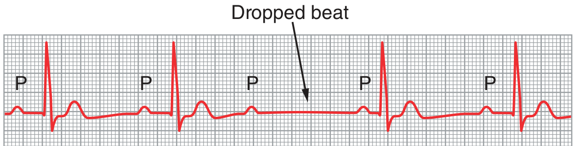
Figure 13.6: Type I second-degree atrioventricular block showing progressive P-R prolongation prior to the dropped beat.
Third-Degree Block (Complete AV Block)
"When the condition causing poor conduction in the AV node or AV bundle becomes severe, complete block of the impulse from the atria into the ventricles occurs. In this case, the ventricles spontaneously establish their own signal, usually originating in the AV node or AV bundle distal to the block. Therefore, the P waves become dissociated from the QRS and T complexes... Note that the rate of rhythm of the atria in this ECG is about 100 beats/min, whereas the rate of ventricular beat is less than 40 beats/min. Furthermore, there is no relationship between the rhythm of the P waves and that of the QRS-T complexes because the ventricles have 'escaped' from control by the atria and are beating at their own natural rate, controlled most often by rhythmical signals generated distal to the AV node or AV bundle where the block occurs."
ECG - Complete AV Block (P waves dissociated from QRS-T, Lead II):
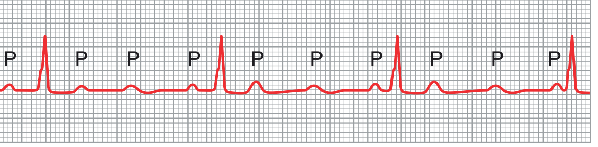
Figure 13.7: Complete atrioventricular block (lead II)
Stokes-Adams Syndrome (Ventricular Escape)
"In some patients with AV block, the total block comes and goes; that is, impulses are conducted from the atria into the ventricles for a period of time and then, suddenly, impulses are not conducted... Each time AV conduction ceases, the ventricles often do not start their own beating until after a delay of 5 to 30 seconds. This delay results from the phenomenon called overdrive suppression... However, after a few seconds, some part of the Purkinje system beyond the block... begins discharging rhythmically at a rate of 15 to 40 times/min, acting as the pacemaker of the ventricles. This phenomenon is called ventricular escape."
"Because the brain cannot remain active for more than 4 to 7 seconds without blood supply, most people faint a few seconds after complete block occurs because the heart does not pump any blood for 5 to 30 seconds, until the ventricles 'escape.' After escape, however, the slowly beating ventricles (typically beating less than 40 beats/min) usually pump enough blood to allow rapid recovery from the faint and then to sustain the person. These periodic fainting spells are known as the Stokes-Adams syndrome."
Treatment
"Therefore, if there is a severe AV nodal or Purkinje system block leading to dizziness, fainting, or other signs of cardiac compromise such as heart failure, a small electronic pacemaker can be implanted to maintain a sufficient heart rate..."
Source: Guyton and Hall Textbook of Medical Physiology (ISBN: 9780443111013), Chapter 13 - Cardiac Arrhythmias and Their Electrocardiographic Interpretation. All quoted text is reproduced directly from the textbook.
from Guyton and Hall textbook of medical physiology, 4th South Asia edition write a 5 mark short note on Heart Block-enlist types ,explain AV Nodal Block
I already have all the exact text and ECG images from the Guyton and Hall textbook retrieved in this session. Here is the complete 5-mark short note with direct quotes:
Heart Block
Source: Guyton and Hall Textbook of Medical Physiology (ISBN: 9780443111013), Chapter 13
Definition
"The only means whereby impulses ordinarily can pass from the atria into the ventricles is through the AV bundle, also known as the bundle of His. Conditions that can either decrease the rate of impulse conduction in this bundle or block the impulse entirely..."
Heart block refers to a delay or complete failure of impulse conduction anywhere along the intracardiac conduction pathway.
Types of Heart Block
I. Sinoatrial (SA) Block
II. Atrioventricular (AV) Block
- First-Degree AV Block (Incomplete)
- Second-Degree AV Block (Incomplete)
- Mobitz Type I (Wenckebach periodicity)
- Mobitz Type II
- Third-Degree AV Block (Complete)
AV Nodal Block - Explained
Causes
"1. Ischemia of the AV node or AV bundle fibers often delays or blocks conduction from the atria to the ventricles. Coronary insufficiency can cause ischemia of the AV node and bundle in the same way that it can cause ischemia of the myocardium.
First-Degree AV Block
The P-R interval is prolonged (> 0.20 sec) but every P wave is still followed by a QRS. Conduction is slowed but not interrupted.
"This so-called P-R interval usually decreases in length with a faster heart rate."
Fig. 13.5 - Prolonged P-R interval caused by first-degree atrioventricular heart block (lead II)
Second-Degree AV Block
"This condition is called second-degree heart block. There are two types of second-degree AV block - Mobitz type I (also known as Wenckebach periodicity) and Mobitz type II. Type I block is characterized by progressive prolongation of the P-R interval until a ventricular beat is dropped..."
"Fig. 13.6 shows progressive P-R interval prolongation typical of type I (Wenckebach) block. Note prolongation of the P-R interval preceding the dropped beat, followed by a shortened P-R interval after the dropped beat."
- Mobitz Type I (Wenckebach): P-R interval progressively lengthens until a QRS is dropped, then the cycle resets.
- Mobitz Type II: QRS drops suddenly without any preceding P-R prolongation - more dangerous, may progress to complete block.
Fig. 13.6 - Type I second-degree AV block showing progressive P-R prolongation prior to the dropped beat
Third-Degree (Complete) AV Block
"When the condition causing poor conduction in the AV node or AV bundle becomes severe, complete block of the impulse from the atria into the ventricles occurs. In this case, the ventricles spontaneously establish their own signal, usually originating in the AV node or AV bundle distal to the block. Therefore, the P waves become dissociated from the QRS and T complexes... Note that the rate of rhythm of the atria in this ECG is about 100 beats/min, whereas the rate of ventricular beat is less than 40 beats/min. Furthermore, there is no relationship between the rhythm of the P waves and that of the QRS-T complexes because the ventricles have 'escaped' from control by the atria and are beating at their own natural rate, controlled most often by rhythmical signals generated distal to the AV node or AV bundle where the block occurs."
Fig. 13.7 - Complete atrioventricular block (lead II)
Stokes-Adams Syndrome and Ventricular Escape
"In some patients with AV block, the total block comes and goes; that is, impulses are conducted from the atria into the ventricles for a period of time and then, suddenly, impulses are not conducted... Each time AV conduction ceases, the ventricles often do not start their own beating until after a delay of 5 to 30 seconds. This delay results from the phenomenon called overdrive suppression... However, after a few seconds, some part of the Purkinje system beyond the block... begins discharging rhythmically at a rate of 15 to 40 times/min, acting as the pacemaker of the ventricles. This phenomenon is called ventricular escape."
"Because the brain cannot remain active for more than 4 to 7 seconds without blood supply, most people faint a few seconds after complete block occurs because the heart does not pump any blood for 5 to 30 seconds, until the ventricles 'escape.' After escape, however, the slowly beating ventricles (typically beating less than 40 beats/min) usually pump enough blood to allow rapid recovery from the faint and then to sustain the person. These periodic fainting spells are known as the Stokes-Adams syndrome."
Treatment
"Therefore, if there is a severe AV nodal or Purkinje system block leading to dizziness, fainting, or other signs of cardiac compromise such as heart failure, a small electronic pacemaker can be implanted to maintain a sufficient heart rate..."
Source: All quoted text reproduced directly from Guyton and Hall Textbook of Medical Physiology, Chapter 13 - Cardiac Arrhythmias and Their Electrocardiographic Interpretation. ECG figures are from the same chapter (Figs. 13.5, 13.6, 13.7).
This is a shared conversation. Sign in to Orris to start your own chat.