
Pulmonary tb detailed
"pulmonary tuberculosis"[MeSH Terms] AND "diagnosis"[MeSH Terms]
pulmonary tuberculosis chest X-ray cavitation
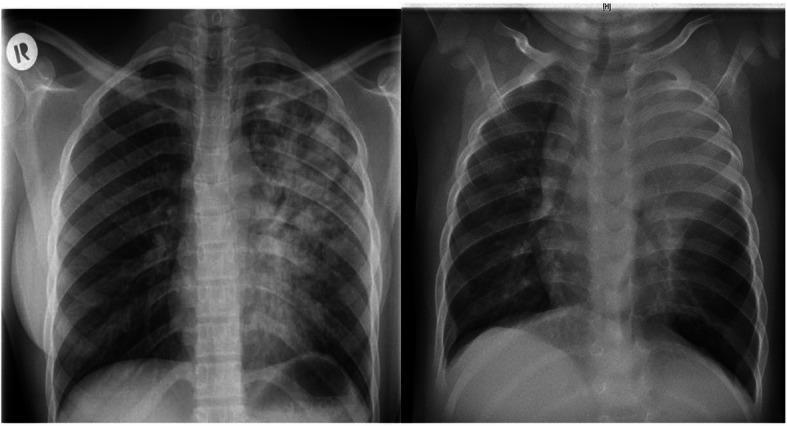
This side-by-side comparison displays two posteroanterior (PA) chest X-rays illustrating the diverse radiographic presentations of pulmonary tuberculosis (TB) in adult versus pediatric patients. The adult image (left) demonstrates extensive, heterogeneous, patchy infiltrates and areas of consolidation predominantly involving the left lung field, typical of post-primary or reactivation TB. Visible radiolucencies suggest cavitation within the upper lobe. In contrast, the pediatric image (right) shows a different pattern characterized by diffuse, relatively homogeneous opacification throughout both lung fields. Notable features in the pediatric X-ray include significant widening of the superior mediastinum, which is highly suggestive of bilateral mediastinal lymphadenopathy, a hallmark of primary TB in children. Both cases exhibit reduced lung clarity, but the pediatric case shows more pronounced bronchial involvement and hilar prominence without the distinct cavitary lesions seen in the adult. These images serve as a comparative educational resource for identifying age-specific diagnostic markers of Mycobacterium tuberculosis infection.
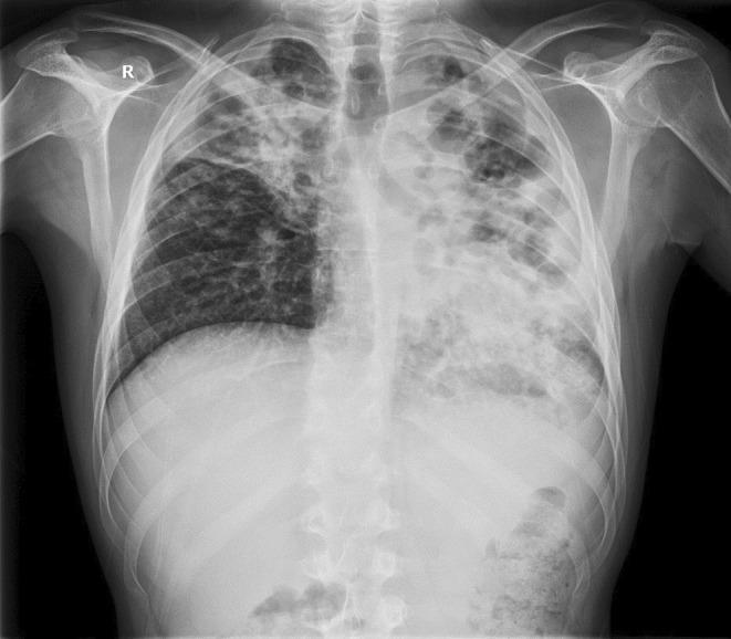
This posteroanterior (PA) chest X-ray demonstrates extensive bilateral pulmonary pathology, highly characteristic of advanced post-primary tuberculosis with superinfection. The left lung field shows a significantly distorted architecture with large, irregular, and oval-shaped lucencies in the upper and middle zones, consistent with pulmonary cavitation. The left lower zone exhibits dense alveolar opacification with visible air bronchograms and spotty shadowing, indicating consolidation. The right lung displays inhomogeneous opacities in the upper and middle lobes, accompanied by volume loss evidenced by the retraction of the right hilum and horizontal fissure. The mediastinal structures appear shifted, and there is visible distortion of the normal hilar anatomy. This diagnostic image illustrates severe parenchymal destruction, cavitary disease, and bilateral infiltrates, which are key clinical markers for evaluating disease severity and treatment response in infectious respiratory medicine.
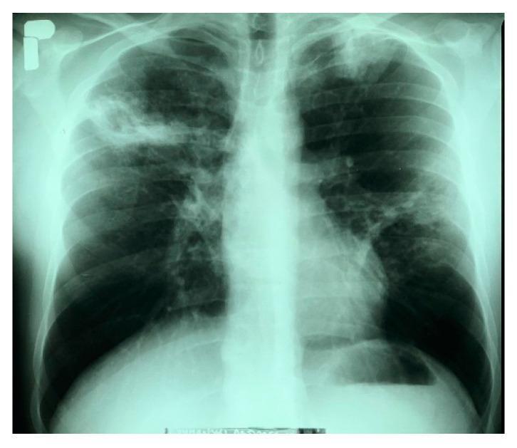
This diagnostic image is a posteroanterior (PA) chest X-ray demonstrating bilateral pulmonary pathology consistent with active tuberculosis. In the right lung, there is a large, heterogeneous infiltrative lesion in the upper field containing a distinct cavitary lesion measuring approximately 35 x 30 mm with irregular borders. The left lung shows an infiltrative opacity at the apex and multiple nodular, fibrous lesions distributed throughout the middle lung field, suggesting a chronic or granulomatous process. The cardiac silhouette appears within normal limits, and the left costophrenic angle is sharp and clear. This radiograph serves as a classic educational example of secondary pulmonary tuberculosis, highlighting hallmark features such as apical predilection, parenchymal infiltration, and cavitation. These findings are clinically significant for the assessment of disease severity and the risk of hemoptysis in patients with Mycobacterium tuberculosis complex infections.
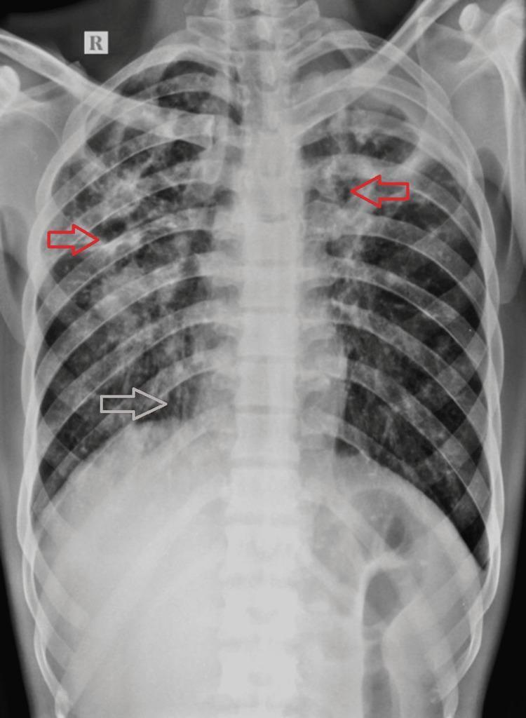
Posteroanterior (PA) chest X-ray demonstrating extensive bilateral pulmonary pathology, highly characteristic of chronic or post-primary pulmonary tuberculosis. The imaging shows widespread, asymmetrical air-space opacities scattered throughout both lung fields, with a greater density and involvement on the right side. Multiple well-defined cavitary lesions are visible, predominantly located in the bilateral upper zones and the right mid-zone (highlighted by red arrows), appearing as radiolucent areas surrounded by thick, irregular walls. In the right lower and upper zones, there is evidence of tractional bronchiectasis (highlighted by white arrows), indicated by tubular lucencies and architectural distortion. The trachea shows a mild shift toward the right, suggesting volume loss or fibrotic changes in the right lung parenchyma. These findings illustrate the chronic sequelae of mycobacterial infection, including lung destruction, cavitation, and secondary airway remodeling.
tuberculosis granuloma histology pathology
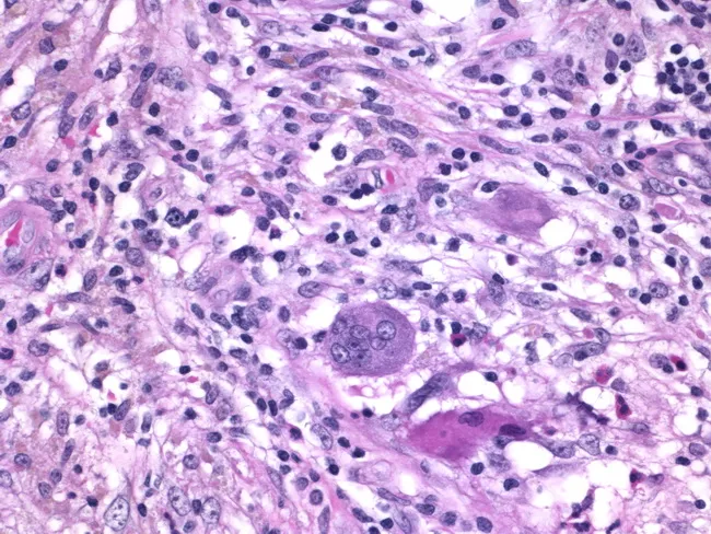
Histology of granulomatous mastitis on bright-field light microscopy. A dense inflammatory infiltrate forms granulomas centered on Langhans-type giant cells surrounded by epithelioid histiocytes, scattered lymphocytes, and occasional eosinophils. The image depicts a high-power view of the center of a granuloma within breast tissue biopsy; multinucleated giant cells with horseshoe-shaped nuclei are evident. The granulomatous architecture includes a peripheral rim of lymphocytes and histiocytes with minimal necrosis described. Staining by Hematoxylin and Eosin highlights basophilic nuclei and eosinophilic cytoplasm; pink collagenous stroma and adipose breast tissue may be visible in the background. Clinically, granulomatous mastitis can mimic infectious etiologies or neoplasia; the presence of Langhans giant cells strongly suggests a granulomatous process and warrants infectious workup for tuberculosis and fungal pathogens, as well as consideration of sarcoidosis or granulomatous reaction to foreign material. This image is diagnostic for granulomatous inflammation in breast tissue when correlated with clinical data and microbiologic studies; useful for education, differential diagnosis, and research on breast granulomatous disease. Additional educational value lies in distinguishing nonnecrotizing granulomas from caseating TB patterns, correlating histology with mammography and ultrasound, and guiding microbiologic testing and patient management in suspected granulomatous breast disease. The image supports teaching of granuloma biology and pathology.
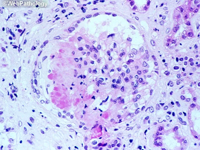
This is a light microscopy image of a formalin-fixed, paraffin-embedded tissue section stained with Hematoxylin and Eosin (H&E). The sample reveals a well-formed granulomatous lesion characterized by a surrounding cuff of lymphocytes and numerous epithelioid histiocytes with elongated, crescent-shaped nuclei. Within the granuloma, multinucleated giant cells (Langhans-type) are visible, and a central amorphous pink necrotic area (caseous necrosis) is evident. The tissue architecture demonstrates organized granulomatous inflammation with a necrotic core, consistent with infectious granulomas. The differential includes mycobacterial disease (tuberculosis) and certain fungal infections (histoplasmosis, coccioidomycosis), though non-necrotizing granulomas can occur in sarcoidosis. The staining highlights chromatin-rich nuclei and cytoplasmic eosinophilia; occasional fibroblasts and mild edema extend into the perigranulomatous region. Clinically, granulomatous tissue with caseation is highly suggestive of TB in the appropriate clinical context and warrants further diagnostic workup (acid-fast bacilli staining, fungal stains, culture, PCR). This image serves as teaching material for histopathology education, differential diagnosis of granulomatous inflammation, and correlation with radiologic and microbiologic findings in suspected infectious etiologies. Additional keywords for indexing: epithelioid macrophages, granuloma, necrosis, caseation, tuberculosis, mycobacteria, fungal infection, lymphohistiocytic rim, tissue biopsy, pathology slide, H&E, histology teaching. Clinical correlation with imaging and sputum analysis improves diagnostic yield.
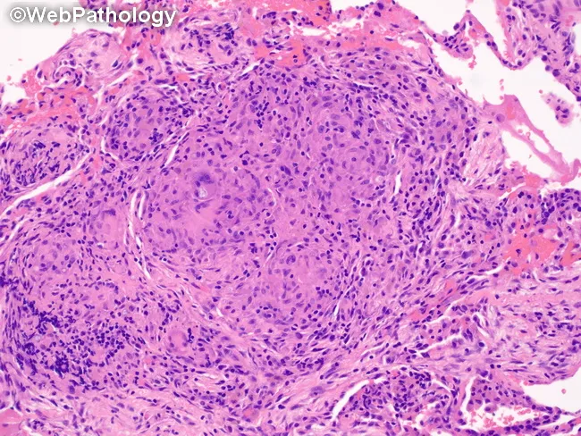
Pulmonary tissue from a wedge resection stained with Hematoxylin and Eosin, analyzed by brightfield light microscopy. The specimen shows robust granulomatous inflammation within the lung parenchyma consistent with a granulomatous infectious process. Nodular aggregates of epithelioid histiocytes are surrounded by a rim of lymphocytes and scattered plasma cells; multinucleated Langhans-type giant cells are present, producing a palisading appearance. The granulomas appear well circumscribed within the interstitium and may encroach upon adjacent alveolar spaces. Central necrosis is variably present and not conspicuously predominant in this field, a feature compatible with coccidioidal granulomatous reaction (coccidioidomycosis) that may display BCG-like granulomas in tissue specimens. The background alveolar architecture is preserved in portions, with mild interstitial inflammation and occasional eosinophils. Although fungal organisms are not definitively visualized in this image, the histology is compatible with fungal infection of the lung in the clinical context of pulmonary coccidioidomycosis, potentially associated with chronic granulomatous response. This image emphasizes characteristic granuloma morphology useful for differential diagnosis from mycobacterial tuberculosis or sarcoidosis, and provides histopathologic confirmation to guide clinical management, including antifungal therapy decisions and serology correlation. It is suitable for education on pulmonary granulomatous disease and wedge resection pathology.
Pulmonary Tuberculosis - Detailed Overview
1. Etiology and Microbiology
- Acid-fast bacillus (AFB): The cell wall contains mycolic acids and a lipid-rich layer (arabinogalactan-mycolate complex), making it resistant to decolorization by acid after Ziehl-Neelsen staining
- Growth: Grows on Lowenstein-Jensen or Middlebrook media; colonies take 3-8 weeks to appear at 37°C
- Generation time: ~24 hours (slow compared to most bacteria)
- Transmission: Airborne, via inhalation of droplet nuclei (1-5 micrometers) expelled by an infectious person coughing, sneezing, or speaking
- Other members of the M. tuberculosis complex include M. bovis (transmitted via unpasteurized dairy), M. africanum, and M. microti
2. Epidemiology
- The WHO estimates 1.7 billion people (a quarter of the world's population) are latently infected
- In 2018: ~10 million new active cases and 1.2 million deaths per year worldwide
- TB was the leading infectious disease killer prior to being overtaken by SARS-CoV-2
- 35% of cases worldwide occur in women; prevalence peaks in young adults
- High-burden countries: Southeast Asia has an incidence of 220/100,000/year; most high-income countries have <10/100,000/year
- HIV co-infection: 10% of TB cases globally occur in people with HIV; TB is the leading cause of death in persons living with HIV
- US incidence: 8,920 active cases in 2019 (2.7/100,000); ~70% of US cases occur in immigrants from high-incidence countries
- Drug resistance: ~3% of new cases and 18% of previously treated cases have drug-resistant TB; MDR-TB (resistant to both isoniazid and rifampin) is most prevalent in India (27%), China (14%), and Russia (8%)
3. Risk Factors
| Risk Factor | Mechanism |
|---|---|
| HIV infection | Profound T-cell immunosuppression |
| Household contact with active TB | High bacillary exposure |
| TNF-α inhibitors (infliximab, etanercept) | Inhibit granuloma integrity |
| Corticosteroids (≥15 mg/day prednisone for ≥1 month) | Broad immunosuppression |
| Diabetes mellitus | Impaired cell-mediated immunity |
| Malnutrition / underweight | Reduced immune competence |
| Silicosis | Direct lung parenchymal damage |
| Residence in homeless shelter, correctional facility | Overcrowding and exposure |
| End-stage renal disease, organ transplant | Immunosuppression |
| Birth/residence in high-incidence country | High prevalence exposure |
4. Pathogenesis
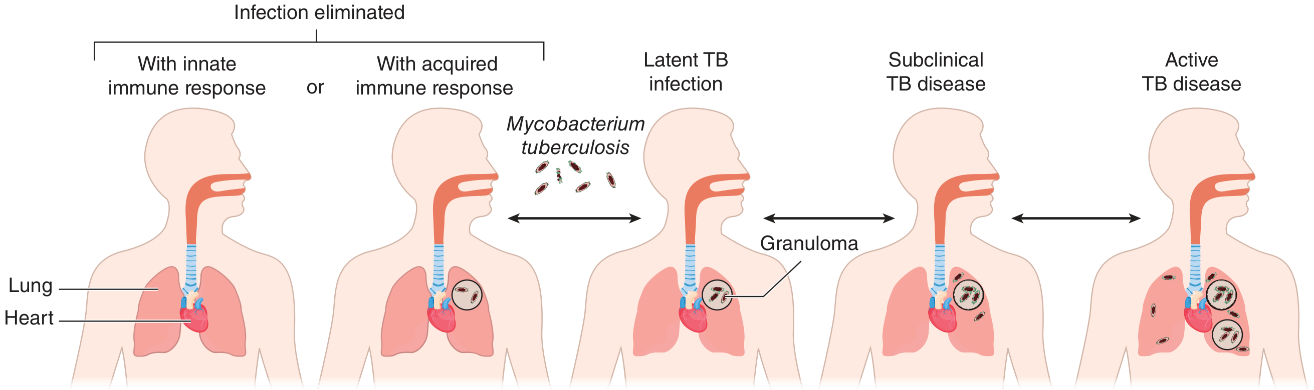
- Inhalation: Droplet nuclei containing M. tuberculosis are inhaled and reach the alveoli of the lower/middle lung zones (optimal for deposition)
- Macrophage uptake: Alveolar macrophages phagocytose the bacilli; M. tuberculosis survives by inhibiting phagosome-lysosome fusion and resisting oxidative killing
- Innate immune response: Infected macrophages release cytokines (TNF-α, IL-1, IL-6, IL-12), recruiting additional macrophages and monocytes
- Granuloma formation (2-8 weeks): Antigen-presenting cells activate CD4+ T-helper 1 (Th1) cells, which produce IFN-γ, activating macrophages to kill bacilli. A granuloma forms - a structured collection of epithelioid macrophages, Langhans giant cells (multinucleated), surrounded by a cuff of lymphocytes
- Caseous necrosis: Central necrosis within the granuloma produces "cheese-like" (caseous) material - an avascular, hypoxic, acidic environment that limits but does not eliminate M. tuberculosis
- Ghon focus: The initial parenchymal lesion in the mid-lung field. Combined with ipsilateral hilar lymphadenopathy = Ghon complex. Ghon complex + calcified hilar nodes = Ranke complex
- Containment (latency): In ~90% of immunocompetent individuals, the immune response controls infection. The bacilli persist in a dormant/metabolically inactive state within granulomas
- Reactivation: Impairment of cell-mediated immunity (any cause) can allow dormant bacilli to resume replication, leading to active disease
5. Histopathology
- Epithelioid macrophages (abundant eosinophilic cytoplasm, elongated nuclei)
- Langhans giant cells (multinucleated with peripheral/horseshoe-arranged nuclei - different from foreign body giant cells which have randomly arranged nuclei)
- Central caseous necrosis (amorphous, acellular, granular, eosinophilic)
- Peripheral lymphocytic cuff
- Fibrosis over time - healing leads to calcification
6. Clinical Presentation
Primary TB (first infection):
- Usually asymptomatic or mild, self-limited illness
- Ghon complex forms; may show subtle radiographic changes
- Progressive primary infection (especially in children, elderly, immunocompromised) can result in dense lower/middle lobe consolidation, pleural effusion, or miliary spread
Reactivation (Post-Primary) TB:
- Most common presentation in adults
- Symptoms develop insidiously over weeks to months:
| Symptom | Frequency/Notes |
|---|---|
| Persistent cough | Most common; may be productive; up to 25% of culture-confirmed TB have no cough |
| Fever | Often low-grade, usually afternoon/evening |
| Night sweats | Classic, though nonspecific |
| Weight loss / anorexia | Reflects chronicity and systemic inflammation |
| Fatigue, malaise | Common |
| Hemoptysis | Occurs with advanced cavitary disease or erosion into blood vessels |
| Chest pain / dyspnea | Less common; more common with pleural involvement |
- Abnormal breath sounds (post-tussive rales, especially over upper zones)
- Amphoric breath sounds (hollow, cave-like resonance) suggest cavitation
- Dullness to percussion over pleural effusion
- Lymphadenopathy is uncommon in immunocompetent adults
7. Radiology
Chest X-Ray Findings
- Dense consolidation in middle/lower lobes
- Hilar lymphadenopathy (particularly prominent in children)
- Pleural effusion
- Miliary pattern (1-3 mm diffuse nodules) in hematogenous spread
- Apical and posterior segment upper lobe involvement (right > left)
- Cavitation - thin or thick-walled cavities; size reflects disease severity
- Fibrocavitary changes, fibrotic scars, loss of lung volume
- Tree-in-bud opacities - endobronchial spread to lower lobes (active disease)
- Calcification and pleural thickening - healed/latent disease
- Atypical distribution; may lack cavitation
- More likely to show lymphadenopathy and diffuse infiltrates
- CT scan required if chest X-ray is normal but smear is positive
8. Diagnosis
A. Sputum Examination
- At least 2-3 sputum specimens for AFB smear microscopy and culture should be collected (ideally on separate days, including one early-morning specimen)
- AFB smear (Ziehl-Neelsen or auramine-rhodamine): Rapid, inexpensive; sensitivity 40-60% for pulmonary TB; specificity is limited (cannot distinguish M. tuberculosis from NTM)
- Mycobacterial culture: Gold standard; Lowenstein-Jensen or MGIT liquid culture; sensitivity ~80-85%; takes 2-8 weeks; required for drug susceptibility testing (DST)
B. Nucleic Acid Amplification Tests (NAAT)
- Xpert MTB/RIF (GeneXpert): Most widely used; detects M. tuberculosis DNA and rifampin resistance mutations simultaneously in 90 minutes
- Smear-positive: NAAT confirms TB diagnosis rapidly
- Smear-negative, intermediate-to-high probability: positive NAAT = presumptive TB
- Smear-negative, low probability: positive NAAT needs caution (false positives possible)
- Negative NAAT does NOT exclude TB - insufficient sensitivity to replace culture
- WHO recommendation: Xpert MTB/RIF as the preferred initial test in HIV-infected patients, those with MDR-TB risk factors, and seriously ill patients
C. Tuberculin Skin Test (TST / Mantoux)
- Intradermal injection of 5 tuberculin units (TU) of PPD at the volar forearm
- Read at 48-72 hours (up to 96 hours); measure induration (not erythema)
- Interpretation thresholds:
| Induration | Positive in |
|---|---|
| ≥5 mm | HIV-infected, recent contacts of active TB, immunosuppressed (TNF antagonists, transplant), abnormal CXR with fibrotic changes |
| ≥10 mm | Recent immigrants from high-incidence countries, IV drug users, prisoners, healthcare workers, silicosis, diabetes, malnutrition |
| ≥15 mm | Low-risk individuals (no known risk factors) |
- Limitations: False positive with prior BCG vaccination or NTM exposure; false negative in anergy (advanced immunosuppression, miliary TB, sarcoidosis, malnutrition)
- Booster effect: TST may turn positive after first test "primes" memory T-cells; relevant in serial testing programs
D. Interferon-Gamma Release Assays (IGRA)
- QuantiFERON-TB Gold Plus and T-SPOT.TB: In vitro whole-blood tests measuring IFN-γ released by sensitized T-cells in response to ESAT-6 and CFP-10 antigens (absent in BCG strains and most NTM)
- Advantages over TST: Not affected by BCG vaccination; fewer false positives; single visit; more specific
- Preferred in persons with prior BCG vaccination or those unlikely to return for TST reading
- Cannot distinguish latent from active TB
E. Bronchoscopy
- Indicated when sputum cannot be collected or is non-diagnostic
- Bronchoalveolar lavage (BAL) and bronchial washings for smear, culture, and NAAT
- Transbronchial biopsy for histology
F. Laboratory Tests
- CBC: Leukocytosis and normochromic normocytic anemia common
- ESR and CRP: Elevated, nonspecific
- Liver function tests: Baseline before starting hepatotoxic drugs
- No specific blood test diagnoses active pulmonary TB
9. Treatment
Latent TB Infection (LTBI) Regimens
| Regimen | Duration | Route | Notes |
|---|---|---|---|
| Isoniazid (INH) + Rifapentine (3HP) | 3 months, once weekly × 12 doses | DOT preferred | First-line; includes HIV+ patients; risk of hypotension/syncope (1/1000) |
| Rifampin (4R) | 4 months, daily | Self-administered | For HIV-negative adults; higher completion, less hepatotoxicity; significant drug interactions |
| Isoniazid + Rifampin (3HR) | 3 months, daily | Self-administered | Comparable to 6-month INH |
| Isoniazid (6H) | 6 months, daily | Self-administered | Alternative; effective for HIV-negative; conditionally recommended for HIV-positive |
| Isoniazid (9H) | 9 months, daily | Self-administered | Alternative for HIV-positive or negative; add pyridoxine 25-50 mg daily |
Active Pulmonary TB Treatment
- R - Rifampin (RIF): 10 mg/kg/day (max 600 mg)
- I - Isoniazid (INH): 5 mg/kg/day (max 300 mg)
- P - Pyrazinamide (PZA): 25-30 mg/kg/day (max 2g)
- E - Ethambutol (EMB): 15-20 mg/kg/day (max 1.6g)
- Rifampin + Isoniazid
- Total: 6 months for drug-susceptible TB without cavitation or positive cultures at 2 months
- Cavitary disease on CXR AND culture-positive at 2 months
- Pyrazinamide cannot be used during intensive phase
Drug Side Effects - Key Monitoring Points
| Drug | Main Adverse Effects | Monitoring |
|---|---|---|
| Isoniazid | Hepatitis, peripheral neuropathy (B6 deficiency), drug-induced lupus | LFTs, add pyridoxine 25-50 mg/day |
| Rifampin | Hepatitis, orange discoloration of secretions, drug interactions (CYP450 inducer), thrombocytopenia | LFTs, drug-drug interactions |
| Pyrazinamide | Hepatitis, hyperuricemia, gout, arthralgias | LFTs, uric acid |
| Ethambutol | Optic neuritis (dose-dependent, reversible if stopped early) | Monthly visual acuity + color vision |
Multidrug-Resistant (MDR-TB) and Extensively Drug-Resistant (XDR-TB)
- MDR-TB: Resistant to at least isoniazid + rifampin
- XDR-TB: Resistant to isoniazid, rifampin, fluoroquinolones, AND at least one second-line injectable (amikacin, streptomycin)
- Pre-XDR-TB: MDR + resistance to either a fluoroquinolone OR a second-line injectable (but not both)
- Intensive phase (5-7 months): 5 active drugs: moxifloxacin or levofloxacin + bedaquiline + linezolid + clofazimine + cycloserine
- Continuation phase: 4 active drugs
- Total duration: 15-21 months after culture conversion
- Bedaquiline + Pretomanid + Linezolid (BPaL) for 26 weeks
- Showed promising outcomes in patients with MDR and XDR-TB
10. Special Situations
TB-HIV Co-infection
- Start antiretroviral therapy (ART) within 2-8 weeks of starting TB treatment (except TB meningitis, where ART is delayed 8 weeks due to IRIS risk)
- TB-IRIS (Immune Reconstitution Inflammatory Syndrome): Occurs in 5-50% of patients when ART is started; characterized by paradoxical worsening (fever, lymphadenitis, respiratory symptoms) due to restored immune response against M. tuberculosis antigens
- Rifampin interaction: Induces CYP3A4, significantly reducing PI (protease inhibitor) levels; preferred ART backbone with rifampin is efavirenz-based
TB in Pregnancy
- INH, rifampin, ethambutol are safe in pregnancy
- Pyrazinamide: Used by WHO; some US guidelines previously avoided it (concern for teratogenicity) but now generally acceptable
- Streptomycin is contraindicated (ototoxicity to fetus)
- Pyridoxine supplementation is particularly important
Pediatric TB
- Children more likely to have primary progressive disease
- Classic presentation: hilar/mediastinal lymphadenopathy, lower/middle lobe involvement
- Gastric aspirates used for bacteriological confirmation in young children who cannot produce sputum
11. Prevention
-
BCG vaccine:
- Live attenuated M. bovis vaccine; given at birth in high-burden countries
- Highly effective against severe pediatric forms (miliary TB, TB meningitis): 70-80% efficacy
- Modest protection against pulmonary TB in adults: ~50%
- Does NOT prevent latent infection; main benefit is preventing severe disseminated disease in children
-
Infection control:
- Negative-pressure isolation rooms with HEPA filtration for infectious patients
- N95 respirators (fit-tested) for healthcare workers
- Surgical masks worn by infectious patients
- Natural ventilation in resource-limited settings (6 air changes/hour minimum)
-
Contact tracing: All household contacts of active pulmonary TB should be evaluated for LTBI and active TB; treatment of LTBI contacts reduces transmission
-
Active case finding: Especially in high-risk populations (correctional facilities, homeless shelters, immunocompromised persons)
12. Complications
| Complication | Mechanism |
|---|---|
| Hemoptysis | Erosion into blood vessels; Rasmussen aneurysm (pseudoaneurysm of pulmonary artery in cavity wall) |
| Pneumothorax | Rupture of subpleural cavity |
| Empyema | Cavitary rupture into pleural space or contiguous spread |
| Miliary TB | Hematogenous dissemination; millet-seed nodules on CXR |
| TB meningitis | CNS dissemination; high mortality |
| Bronchiectasis | Parenchymal destruction and airway remodeling |
| Amyloidosis (AA) | Chronic inflammation leading to serum amyloid A deposition |
| Aspergilloma | Aspergillus colonization of old cavities |
| Cor pulmonale | Chronic hypoxia from extensive lung destruction |
Summary
- Goldman-Cecil Medicine International Edition (2-Volume Set), Chapter 299 - Tuberculosis, pp. 3249-3265
- Murray & Nadel's Textbook of Respiratory Medicine (2-Volume), Chapter on Diagnosis of Pulmonary Tuberculosis, pp. 1186-1195