
can u give be a list of oral diseases in which different wbcs increase and decrease
Reading File
Now let me search the medical library for relevant content on WBC changes in oral diseases.
Finding Sources
Finding Sources
Finding Sources
Finding Sources
Finding Sources
Finding Sources
Finding Sources
Finding Sources
Searching the Web
oral diseases WBC white blood cell changes increase decrease neutrophil lymphocyte eosinophil table
Now I have enough information to compile a comprehensive answer from medical knowledge. Let me provide a thorough, well-organized response.
Here is a comprehensive list of oral diseases and conditions with the associated changes in white blood cell (WBC) types:
WBC Changes in Oral Diseases
Quick Reference Guide
| WBC Type | Normal Range | Increased (↑) | Decreased (↓) |
|---|---|---|---|
| Neutrophils | 40-70% | Acute bacterial infections | Cyclic/agranulocytosis |
| Lymphocytes | 20-40% | Viral stomatitis, TB | HIV/AIDS oral lesions |
| Eosinophils | 1-4% | Allergic, parasitic, autoimmune | - |
| Monocytes | 2-8% | Chronic oral infections | - |
| Basophils | 0-1% | Allergic reactions | - |
1. NEUTROPHIL CHANGES
Neutrophilia (↑ Neutrophils)
| Oral Disease/Condition | Reason |
|---|---|
| Acute dentoalveolar abscess | Acute bacterial infection triggers bone marrow to release more neutrophils as first-line defense |
| Acute necrotizing ulcerative gingivitis (ANUG) | Rapid bacterial invasion (Fusobacterium, Treponema) causes acute neutrophilic response |
| Pericoronitis | Bacterial accumulation around wisdom tooth flap causes acute inflammation |
| Osteomyelitis of jaw | Bone infection from dental source causes marked neutrophilia |
| Ludwig's angina | Severe spreading cellulitis of floor of mouth causes systemic neutrophilia |
| Cellulitis of face/neck | Spreading odontogenic infection causes prominent neutrophilia |
| Acute parotitis | Bacterial parotid gland infection |
Neutropenia (↓ Neutrophils)
| Oral Disease/Condition | Reason |
|---|---|
| Cyclic neutropenia | Characteristic oral manifestation - periodic recurrent ulcers coincide with neutrophil count troughs every ~21 days |
| Agranulocytosis (oral manifestation) | Severe neutropenia causes painful necrotic oral ulcers without an inflammatory border (no pus) |
| Chronic benign neutropenia | Recurrent aphthous-like ulcers, gingivitis |
| Chediak-Higashi syndrome (oral) | Functional neutropenia leads to severe periodontitis and recurrent oral infections |
| Lazy leukocyte syndrome | Neutrophil dysfunction leads to recurrent gingivitis/stomatitis |
| Kostmann syndrome | Congenital agranulocytosis presents with mouth sores early in life |
2. LYMPHOCYTE CHANGES
Lymphocytosis (↑ Lymphocytes)
| Oral Disease/Condition | Reason |
|---|---|
| Primary herpetic gingivostomatitis (HSV-1) | Viral infection triggers lymphocytic (T-cell) response |
| Infectious mononucleosis (EBV) | Atypical lymphocytes (Downey cells) are classic; tonsillitis and oral ulcers are prominent features |
| Herpangina (Coxsackievirus) | Viral infection with lymphocytosis; vesicles on soft palate/pharynx |
| Hand, foot, and mouth disease | Coxsackievirus causes oral vesicles + lymphocytosis |
| Oral tuberculosis | Chronic granulomatous infection with lymphocytic infiltration |
| Recurrent aphthous ulcers | Immunologically mediated; local T-lymphocyte infiltration (though systemic count may be normal) |
| Oral lichen planus | T-lymphocyte-mediated autoimmune reaction - dense subepithelial lymphocytic band is the histological hallmark |
Lymphocytopenia (↓ Lymphocytes)
| Oral Disease/Condition | Reason |
|---|---|
| HIV/AIDS - oral manifestations | Destruction of CD4+ T-lymphocytes leads to oral candidiasis, hairy leukoplakia, Kaposi sarcoma in the mouth |
| Oral candidiasis (in immunosuppressed) | Secondary to lymphopenia from chemotherapy, AIDS, corticosteroids |
3. EOSINOPHIL CHANGES
Eosinophilia (↑ Eosinophils)
| Oral Disease/Condition | Reason |
|---|---|
| Oral eosinophilic ulcer | Idiopathic - histology shows dense eosinophilic infiltrate (formerly called traumatic eosinophilic granuloma) |
| Oral pemphigus / Pemphigoid | Autoimmune blistering diseases affecting oral mucosa; eosinophilia in pemphigoid is well-recognized |
| Eosinophilic granuloma (Langerhans cell histiocytosis - oral) | Destructive jaw lesion with eosinophilic infiltrate; causes "floating teeth" appearance |
| Oral Crohn's disease | Leucocytosis and eosinophilia are characteristic blood findings; oral cobblestoning, lip fissuring |
| Allergic contact stomatitis | Reaction to dental materials (nickel, acrylics) causes eosinophilia |
| Parasitic infections with oral involvement | Helminth-related; eosinophilia is a key marker |
| Kimura's disease (oral) | Rare chronic inflammatory disease with systemic eosinophilia; affects parotid/perioral region |
| Eosinophilic granulomatosis with polyangiitis - EGPA (oral) | Vasculitis with marked eosinophilia; oral mucosal lesions occur |
4. MONOCYTE CHANGES
Monocytosis (↑ Monocytes)
| Oral Disease/Condition | Reason |
|---|---|
| Oral tuberculosis | Chronic granulomatous infection - monocytes differentiate into granuloma-forming macrophages |
| Chronic periodontitis (severe) | Chronic bacterial challenge drives monocyte/macrophage response |
| Infectious mononucleosis | EBV infects B-lymphocytes but monocytosis also occurs along with atypical lymphocytes |
| Oral histoplasmosis | Fungal infection - monocyte-macrophage system heavily involved |
5. BASOPHIL CHANGES
Basophilia (↑ Basophils)
| Oral Disease/Condition | Reason |
|---|---|
| Severe allergic reactions (oral allergy syndrome) | IgE-mediated hypersensitivity to certain foods triggers basophil/mast cell degranulation |
| Angioedema of lip/tongue | Basophil and mast cell activation drives histamine release causing swelling |
6. BLAST CELLS / IMMATURE WBCs
| Oral Disease/Condition | WBC Change | Notes |
|---|---|---|
| Acute myeloid leukemia (oral) | Blasts ↑ markedly | Oral manifestations = gingival hyperplasia (leukemic infiltration), petechiae, spontaneous gingival bleeding - a classic presentation |
| Acute lymphoblastic leukemia (oral) | Lymphoblasts ↑ | Gingival swelling, pallor, bleeding; common in children |
| Chronic myeloid leukemia (CML) | All WBCs markedly ↑ (leukocytosis) | Gingival hyperplasia, oral infections due to dysfunctional WBCs |
Summary: Key Points to Remember
- Bacterial oral infections (abscess, ANUG, osteomyelitis) → Neutrophilia
- Viral oral infections (HSV, EBV, Coxsackievirus) → Lymphocytosis
- Cyclic/congenital neutropenia → Neutropenia → recurrent oral ulcers
- AIDS/HIV oral disease → Lymphocytopenia (CD4 count falls)
- Eosinophilic ulcer, pemphigoid, Crohn's → Eosinophilia
- Leukemia (oral manifestations) → Leukocytosis with blasts → gingival hyperplasia + spontaneous bleeding
- Agranulocytosis → Neutropenia → necrotic oral ulcers without pus
Note: In many oral diseases, the WBC change is a systemic reflection of the underlying disease process. Always correlate with a full CBC differential and clinical findings. The local tissue infiltrate (histology) may differ from peripheral blood counts.
different tupes of blood group and a short note on bombay blood group
The ABO and Rh systems are the most clinically important blood group systems. There are actually 43 recognized blood group systems by the ISBT. The Bombay blood group (hh) is extremely rare and lacks the H antigen, meaning these individuals cannot receive blood from any ABO group.
Searching sources...
Finding Sources
Finding Sources
Reading File
Reading File
Finding Sources
Finding Sources
Finding Sources
Reading File
Finding Sources
Reading File
Finding Sources
I now have comprehensive textbook material to give a detailed, well-cited answer.
Types of Blood Groups
Blood groups are defined by the presence or absence of specific antigens on the surface of red blood cells (RBCs). As noted in [Harper's Illustrated Biochemistry](Henry's Clinical Diagnosis), approximately 30+ recognized blood group systems exist, but only a few are clinically important.
The Major Blood Group Systems
1. ABO Blood Group System (Most Important)
The ABO system divides humans into 4 main blood groups based on the presence of A and B agglutinogens (antigens) on RBCs and naturally occurring antibodies (agglutinins) in the serum.
| Blood Group | Antigen on RBC | Antibody in Serum | Can Donate To | Can Receive From |
|---|---|---|---|---|
| A | A antigen | Anti-B | A, AB | A, O |
| B | B antigen | Anti-A | B, AB | B, O |
| AB | A and B antigens | None | AB only | A, B, AB, O (Universal Recipient) |
| O | None (H substance) | Anti-A and Anti-B | A, B, AB, O (Universal Donor) | O only |
Subgroups: Group A has two important subgroups - A1 and A2, which also appear in AB, giving A1B and A2B. Weaker rare subgroups A3, A4, and A5 also exist.
Secretor Status: About 80% of the population are "secretors" - they secrete ABH substances in saliva, tears, sweat, and other body fluids. This has forensic significance.
- Parikh's Textbook of Medical Jurisprudence, p. 439
2. Rh Blood Group System (Second Most Important)
The Rh system involves three sets of closely linked antigens: C/c, D/d, and E/e. The D antigen is the most strongly immunogenic.
- Rh positive (Rh+): Person has the D antigen (~85% of population)
- Rh negative (Rh-): Person lacks the D antigen (~15% of population)
Clinical importance:
- An Rh- mother carrying an Rh+ fetus can develop anti-D antibodies, causing erythroblastosis fetalis (hemolytic disease of the newborn) in subsequent pregnancies
- Prevented by administering anti-D immunoglobulin (Rho-GAM) to the mother
- Guyton & Hall Medical Physiology; Langman's Medical Embryology
3. MNS Blood Group System (ISBT No. 002)
- Discovered in 1927, the second system identified after ABO
- Contains 49 antigens; the four commonly encountered clinically are M, N, S, and s
- M and N antigens are fairly evenly distributed in the population
- Antibodies (anti-M, anti-N) are usually cold-reacting and not clinically significant, but anti-S and anti-s can cause hemolytic transfusion reactions
- Henry's Clinical Diagnosis and Management by Laboratory Methods
4. Kell Blood Group System (ISBT No. 006)
- The K (Kell) antigen is highly immunogenic - second only to D in clinical importance
- About 9% of Caucasians are K-positive
- Anti-K can cause severe hemolytic transfusion reactions and hemolytic disease of the newborn
- The McLeod phenotype (absence of Kx antigen) is associated with acanthocytosis and a neuromuscular disorder
5. Duffy Blood Group System (ISBT No. 008)
- Antigens: Fya and Fyb
- The Duffy antigens serve as receptors for Plasmodium vivax (malarial parasite)
- Most West Africans are Duffy null (Fy a-b-) and are therefore naturally resistant to P. vivax malaria - a classic example of natural selection in blood group genetics
- Anti-Fya can cause hemolytic transfusion reactions
6. Kidd Blood Group System (ISBT No. 009)
- Antigens: Jka and Jkb
- Anti-Jka is notorious for causing delayed hemolytic transfusion reactions - the antibody titer drops quickly and may be undetectable before transfusion
- The Kidd null phenotype (Jk null) is associated with an inability to concentrate urine maximally (the Kidd glycoprotein is a urea transporter)
7. Lewis Blood Group System (ISBT No. 007)
- Antigens: Lea and Leb
- Unlike most systems, Lewis antigens are not synthesized by RBCs directly - they are produced in the GI tract and adsorbed onto the RBC surface from plasma
- Lewis antigens are closely related to ABH secretor status
- Lea is more common in non-secretors; Leb is found in secretors
8. P1PK Blood Group System (ISBT No. 003)
- Includes P1 antigen
- The Pk (p) null phenotype individuals make anti-PP1Pk (formerly Tja), which is associated with recurrent early abortions
- P antigen is a receptor for parvovirus B19 and uropathogenic E. coli
9. Lutheran Blood Group System
- Antigens: Lua and Lub
- The null phenotype (In(Lu)) is associated with acanthocytosis
10. Diego Blood Group System
- Related to the Band 3 protein (the main anion exchanger of RBCs)
- Dia antigen is more common in Mongoloid/East Asian populations - has anthropological/forensic significance
11. HLA System (Leukocyte Antigens)
- Located on leukocytes and platelets (not primarily RBCs)
- Four established systems: HLA-A, HLA-B, HLA-C, and HLA-D
- Critical for organ transplantation compatibility
- P.C. Dikshit Textbook of Forensic Medicine
Short Note on the Bombay Blood Group (Oh Phenotype)
Discovery
The Bombay blood group was first discovered in Bombay (Mumbai), India in 1952 by Dr. Y.M. Bhende. It is also called the Oh phenotype (the "O" superscripted h stands for Bombay in honor of the place of discovery) or the hh phenotype.
Genetics and Biochemistry
The ABO system works in a two-step process:
- The H gene (FUT1) produces an enzyme that adds fucose to glycolipids/glycoproteins on RBCs, creating the H antigen (the precursor substance)
- The A and B genes (ABO) then modify the H antigen to create A or B antigens
- In group O individuals, the H antigen remains unmodified (they are hh in terms of A/B genes, but HH or Hh for the H gene)
In Bombay blood group individuals:
- They carry two non-functional copies of the FUT1 gene (hh genotype)
- The enzyme fucosyltransferase is absent or non-functional
- No H antigen is produced
- Without H antigen as a precursor, no A or B antigens can be formed either
- Therefore, Bombay individuals appear to be group O on routine ABO typing, but are fundamentally different
Serology (What Tests Show)
| Feature | Group O (normal) | Bombay (Oh) |
|---|---|---|
| Antigen on RBC | H antigen (no A or B) | No H, no A, no B |
| Antibodies in serum | Anti-A + Anti-B | Anti-A + Anti-B + Anti-H |
| Reaction with Anti-H serum | Agglutination (positive) | No agglutination (negative) |
| Reaction with Group O blood | Compatible | Incompatible |
Anti-H Antibody - The Dangerous Part
Bombay individuals produce alloanti-H - a clinically significant antibody that:
- Is a warm-reacting IgG antibody (unlike the harmless cold anti-H seen in some A1 individuals)
- Reacts strongly against ALL normal human RBCs (since every normal person has H antigen)
- Can cause severe, life-threatening hemolytic transfusion reactions if given any normal blood - including group O
This means Bombay individuals cannot receive blood from any normal donor regardless of ABO type. They can only receive blood from other Bombay phenotype donors.
- Henry's Clinical Diagnosis and Management by Laboratory Methods
- The Essentials of Forensic Medicine and Toxicology, 36th ed.
Prevalence
- Extremely rare worldwide: 1 in 1,000,000 in most populations
- Relatively less rare in South Asia (India, especially Maharashtra): approximately 1 in 10,000
- The higher frequency in India is likely due to founder effects and consanguinity in certain communities
Clinical Importance
- Transfusion: Bombay patients can ONLY receive blood from other Oh donors - this is almost impossible in an emergency, making autologous blood banking (pre-operative self-donation) extremely important
- Pregnancy: Bombay mothers can develop anti-H, causing hemolytic disease of the fetus and newborn
- Misidentification: These individuals type as group O on routine testing - the error is only caught if anti-H testing is done, which is NOT standard practice. This can have fatal consequences in transfusion
- Forensic significance: Can cause confusion in paternity testing and forensic blood grouping if the Bombay phenotype is not recognized
Para-Bombay Phenotype
A related but distinct condition where H antigen is absent from RBCs but present in secretions (saliva, etc.). These individuals have a non-functional FUT1 but a functional secretory FUT2 gene.
Summary Table of Key Blood Group Systems:
| System | Key Antigens | Clinical Significance |
|---|---|---|
| ABO | A, B, H | Most important; universal donor/recipient concept |
| Rh | D, C, c, E, e | Hemolytic disease of newborn; transfusion reactions |
| MNS | M, N, S, s | Occasional transfusion reactions |
| Kell | K, k | Severe hemolytic reactions; HDN |
| Duffy | Fya, Fyb | P. vivax malaria resistance (Fy null) |
| Kidd | Jka, Jkb | Delayed hemolytic reactions |
| Lewis | Lea, Leb | Related to secretor status |
| Bombay (H) | H antigen absent | Universal incompatibility; extremely rare |
normal and abnormal hemoglobins short summry
Normal hemoglobin is a tetramer of globin chains plus heme groups. Adults have mainly HbA (α2β2), with small amounts of HbA2 and HbF. Abnormal hemoglobins arise from mutations in globin genes - sickle cell (HbS), thalassemias, HbC, HbE, and methemoglobin are the most important clinically.
Searching sources...
Finding Sources
Finding Sources
Finding Sources
Finding Sources
Reading File
Reading File
Finding Sources
Finding Sources
Reading File
Reading File
Excellent content gathered. Let me fetch the key hemoglobin structure diagram to include visually.
Loading Image
Loading Image
Here is a comprehensive but concise summary of normal and abnormal hemoglobins, fully sourced from the medical library.
Hemoglobin - Normal and Abnormal: A Summary
Structure of Hemoglobin
Hemoglobin is a globular tetrameric protein made of four subunits. Each subunit contains:
- A globin polypeptide chain (folded into 7-8 helical regions)
- A heme prosthetic group - an iron-containing porphyrin ring that carries one O₂ molecule
Each hemoglobin molecule can therefore carry 4 molecules of oxygen.
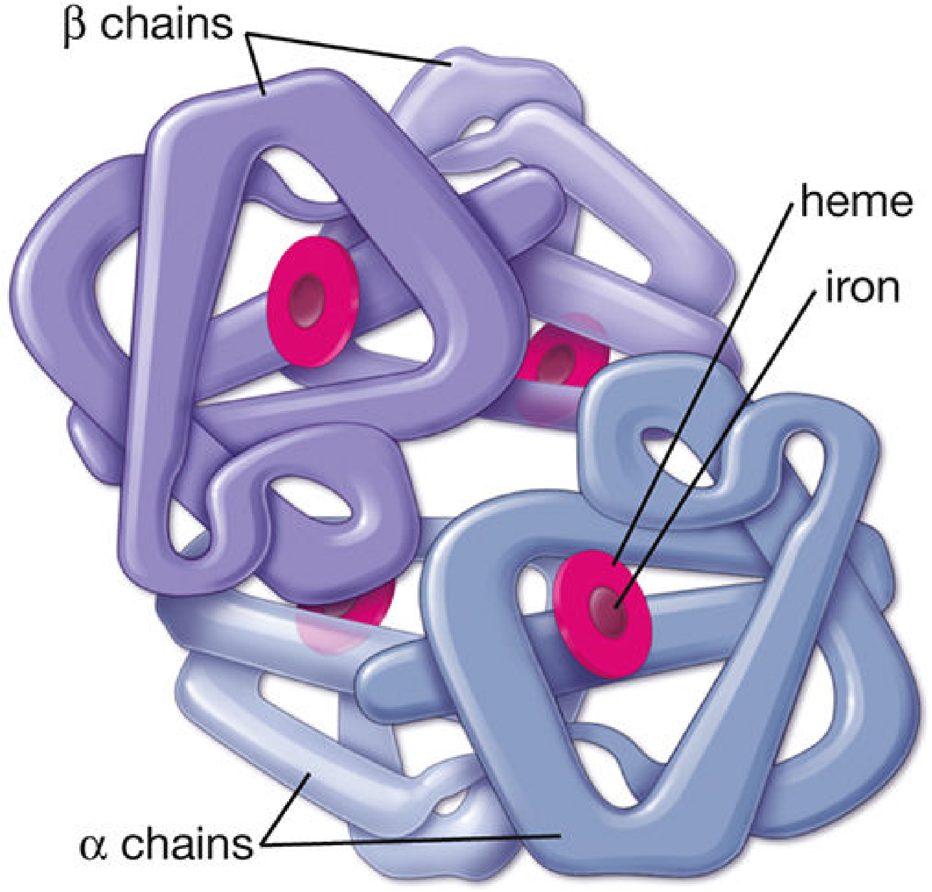
Structural diagram of hemoglobin A (HbA): two α-chains + two β-chains, each centered around a heme-iron moiety - Histology: A Text and Atlas
PART 1: NORMAL HEMOGLOBINS
The type of hemoglobin present changes throughout development based on which globin genes are expressed - a process called globin switching.
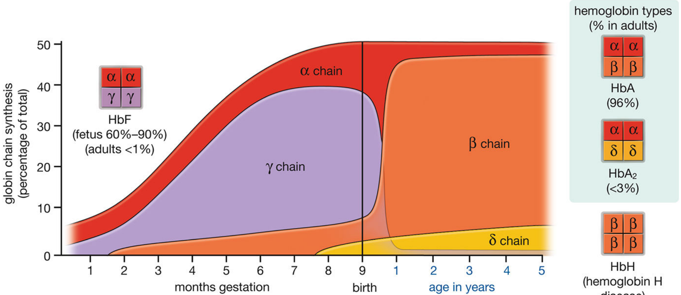
Globin chain synthesis across development - Thompson & Thompson Genetics
Normal Hemoglobins at a Glance
| Hemoglobin | Chains | When Present | Normal % in Adults |
|---|---|---|---|
| HbA | α₂β₂ | Predominant adult form | ~96% |
| HbA2 | α₂δ₂ | Minor adult form | 1.5 - 3% |
| HbF | α₂γ₂ | Fetal + traces in adults | <1% in adults |
| Hb Gower 1 | ζ₂ε₂ | Embryonic (yolk sac, 3-8 wks) | 0 after birth |
| Hb Gower 2 | α₂ε₂ | Embryonic | 0 after birth |
| Hb Portland | ζ₂γ₂ | Embryonic | 0 after birth |
Globin gene locations:
- α-like genes (α, ζ): Chromosome 16
- β-like genes (β, δ, γ, ε): Chromosome 11
Key Points on Normal Hb
-
HbA (α₂β₂): β-chain synthesis begins near birth; by 3 months of age almost all hemoglobin is HbA. It carries O₂ cooperatively (sigmoidal O₂ dissociation curve) due to the T-state (deoxy) ↔ R-state (oxy) conformational shift.
-
HbA2 (α₂δ₂): β and δ chains differ in only 10 of 146 amino acids. HbA2 is elevated in β-thalassemia trait (diagnostic marker).
-
HbF (α₂γ₂): Has higher O₂ affinity than HbA (because γ-chain binds 2,3-BPG less avidly), allowing the fetus to extract O₂ from maternal blood. Constitutes ~70% of total Hb at birth. Drops to <1% in adults but is elevated in β-thalassemia and sickle cell disease, where it has a protective/ameliorating role.
-
Histology: A Text and Atlas, Thompson & Thompson Genetics and Genomics in Medicine
PART 2: ABNORMAL HEMOGLOBINS
Abnormal hemoglobins fall into two broad categories:
- Structural variants - mutations that change the amino acid sequence of globin chains (qualitative defects)
- Thalassemias - mutations that reduce the quantity of globin chain synthesis (quantitative defects)
A. Structural Hemoglobin Variants
1. Hemoglobin S (HbS) - Sickle Cell Hemoglobin
| Feature | Detail |
|---|---|
| Mutation | Single point mutation: Adenine → Thymine at codon 6 of β-globin gene |
| Amino acid change | Glutamic acid (hydrophilic) → Valine (hydrophobic) at position 6 of β-chain |
| Chain composition | α₂β₂ˢ |
| Gene location | Chromosome 11 |
| Pathophysiology | Deoxygenated HbS polymerizes into long rigid fibers → RBC distorts into sickle shape → vaso-occlusion, hemolysis |
| Genotypes | HbSS (sickle cell anemia - most severe), HbSC disease, HbS/β-thalassemia |
| Features | Vaso-occlusive crises, dactylitis, splenic sequestration, acute chest syndrome, stroke, chronic hemolytic anemia |
| Protective factor | HbF reduces sickling - basis for hydroxyurea therapy |
- Robbins Pathologic Basis of Disease, Harrison's Principles of Internal Medicine 22E
2. Hemoglobin C (HbC)
| Feature | Detail |
|---|---|
| Mutation | Glutamic acid → Lysine at position 6 of β-chain |
| Chain composition | α₂β₂ᶜ |
| Features | Milder than HbS; HbCC causes mild hemolytic anemia with target cells; HbSC disease is clinically significant (moderate severity) |
| RBC morphology | Target cells (codocytes), intracellular HbC crystals |
3. Hemoglobin E (HbE)
| Feature | Detail |
|---|---|
| Mutation | Glutamic acid → Lysine at position 26 of β-chain |
| Prevalence | Most common in Southeast Asia; one of the world's most frequent hemoglobin variants |
| Features | Creates an abnormal splice site → reduced β-chain synthesis (acts like a mild thalassemia); HbEE causes mild microcytic anemia; HbE/β-thalassemia is clinically severe |
4. Hemoglobin D (HbD)
| Feature | Detail |
|---|---|
| Mutation | Glutamic acid → Glutamine at position 121 of β-chain |
| Prevalence | Common in Punjab (India) - called HbD-Punjab |
| Features | HbDD: mild hemolytic anemia; HbSD disease: moderate severity similar to HbSC |
5. Hemoglobin M (HbM) - Methemoglobin variants
| Feature | Detail |
|---|---|
| Mutation | Substitution at the heme-binding histidine residue (e.g., Hb Hyde Park - His→Tyr at β92; Hb Boston - His→Tyr at α58) |
| Pathophysiology | Iron is stabilized in the Fe³⁺ (ferric) state - cannot bind O₂ |
| Features | Congenital cyanosis; cyanosis unresponsive to O₂ therapy; diagnosed by spectrophotometry |
6. Hemoglobin H (HbH)
| Feature | Detail |
|---|---|
| Composition | β₄ tetramer (4 β-chains) - forms when α-chain production is severely reduced |
| Cause | Deletion of 3 out of 4 α-globin genes (α-thalassemia intermediate) |
| Features | Moderately severe hemolytic anemia; HbH precipitates as Heinz bodies; splenomegaly |
7. Hemoglobin Bart's (Hb Bart's)
| Feature | Detail |
|---|---|
| Composition | γ₄ tetramer (4 γ-chains) - forms in severe α-chain deficiency |
| Cause | Deletion of all 4 α-globin genes |
| Features | Hydrops fetalis (incompatible with life); Hb Bart's has very high O₂ affinity - cannot deliver O₂ to tissues |
8. Carboxyhemoglobin (HbCO)
| Feature | Detail |
|---|---|
| Cause | CO binds heme iron with 240x higher affinity than O₂ |
| Features | Left-shifts O₂ dissociation curve; cherry-red color; CO poisoning; normal PaO₂ but low O₂ saturation |
9. Methemoglobin (MetHb)
| Feature | Detail |
|---|---|
| Cause | Fe²⁺ oxidized to Fe³⁺ (ferric form) by drugs (dapsone, nitrates, prilocaine) or congenital HbM |
| Features | Cannot carry O₂; cyanosis; "chocolate brown" blood; treated with methylene blue (IV) |
B. Thalassemias (Quantitative Defects)
In thalassemias, the globin chain is structurally normal but produced in insufficient or absent amounts due to mutations affecting gene expression.
β-Thalassemia
| Type | Mutation | β-chain | Clinical Severity |
|---|---|---|---|
| β⁰-thalassemia | Chain terminator mutation (nonsense/frameshift) | Absent | Severe |
| β⁺-thalassemia | Splicing or promoter mutations | Reduced | Mild to moderate |
Mechanism of anemia:
- Unpaired α-chains accumulate → precipitate in RBC precursors as insoluble inclusions → membrane damage → apoptosis of erythroid precursors → ineffective erythropoiesis (70-85% of precursors destroyed in severe disease)
- Released RBCs also have inclusions → splenic sequestration → hemolytic anemia
| Clinical Grade | Genotype | Features |
|---|---|---|
| Thalassemia minor (trait) | β⁰/β or β⁺/β | Mild microcytic anemia; HbA2 elevated >3.5% (diagnostic); usually asymptomatic |
| Thalassemia intermedia | β⁺/β⁺ or β⁰/β⁺ | Moderate anemia; splenomegaly; may not need regular transfusions |
| Thalassemia major (Cooley's anemia) | β⁰/β⁰ | Severe transfusion-dependent anemia; bony changes ("hair-on-end" skull X-ray); hepatosplenomegaly; iron overload |
- Robbins Pathologic Basis of Disease
α-Thalassemia
Caused by deletion of α-globin genes (normal = 4 α-gene copies, 2 per chromosome 16).
| Genes Deleted | Condition | Features |
|---|---|---|
| 1 gene (αα/α-) | Silent carrier | Clinically normal; undetectable |
| 2 genes (αα/-- or α-/α-) | α-Thalassemia trait | Mild microcytic anemia; iron deficiency must be excluded |
| 3 genes (α-/--) | HbH disease | Moderate hemolytic anemia; HbH (β₄) forms; splenomegaly |
| 4 genes (--/--) | Hb Bart's / Hydrops fetalis | Incompatible with life; stillborn with severe edema |
- Creasy & Resnik's Maternal-Fetal Medicine; Goldman-Cecil Medicine
Hereditary Persistence of Fetal Hemoglobin (HPFH)
- Failure of the γ→β globin switch after birth
- HbF remains elevated into adult life (homogeneous/pancellular distribution)
- Usually benign - considered an extremely mild thalassemia-like condition
- Clinically beneficial in patients who also have sickle cell disease or β-thalassemia (HbF inhibits sickling and compensates for HbA deficit)
Summary Table: Abnormal Hemoglobins
| Hemoglobin | Type | Mutation | Key Features |
|---|---|---|---|
| HbS | Structural (β-chain) | Glu→Val at β6 | Sickling, vaso-occlusion, hemolysis |
| HbC | Structural (β-chain) | Glu→Lys at β6 | Target cells, mild hemolysis |
| HbE | Structural + thalassemic | Glu→Lys at β26 | Common in SE Asia; mild thalassemia-like |
| HbD | Structural (β-chain) | Glu→Gln at β121 | Punjab; HbSD = moderate severity |
| HbM | Structural (heme pocket) | His→Tyr at α or β | Congenital cyanosis; iron stays Fe³⁺ |
| HbH | Abnormal tetramer | β₄ (3 α-genes deleted) | α-Thalassemia intermediate |
| Hb Bart's | Abnormal tetramer | γ₄ (all 4 α-genes deleted) | Hydrops fetalis; lethal |
| MetHb | Acquired/congenital | Fe²⁺→Fe³⁺ | Chocolate-brown blood; cyanosis |
| HbCO | Acquired | CO on heme | CO poisoning; cherry-red color |
| β-Thalassemia | Quantitative | β-chain ↓ or absent | Microcytic anemia; HbA2 ↑ |
| α-Thalassemia | Quantitative | α-chain ↓ or absent | Ranges from silent to hydrops |
| HPFH | γ→β switch failure | γ-chain persists | Benign; protective in SCD/thal |
This is a shared conversation. Sign in to Orris to start your own chat.