

31588f3a-2006-4aec-8692-28a7521af57b.jpg
Causes of such lesions since 10 days in kid of 2 years and same lesions in sibling of 2 years aswell
analyse_media
Now let me search the medical library for the most likely diagnosis given the clinical picture - raised nodular lesions with central umbilication/necrosis on the feet of two young siblings, present for 10 days.
Reading File
Searching sources...
The key clinical features are: two siblings both 2 years old (twins or age-mates living together), raised nodular lesions with central umbilication/necrosis/keratotic plug on the feet, present for 10 days. This strongly points toward an infectious/transmissible cause. Let me search the library for the most likely diagnoses.
Finding Sources
Finding Sources
Finding Sources
Good. Now let me read the core molluscum contagiosum section and also search for other differential diagnoses like tungiasis (jigger flea - common cause of foot nodules in children in tropical settings) and papular urticaria.
Reading File
Finding Sources
Finding Sources
Now let me read the detailed Molluscum section from Andrews' and also search for tungiasis and the detailed chapter on molluscum.
Reading File
Finding Sources
Let me read more of the MC section and also search for tungiasis and the differential:
Reading File
Finding Sources
I have enough clinical information now. Let me also fetch the MC image from the textbook and do a quick web search for tungiasis (jigger flea) which is a very important differential for foot nodules with central opening in children from tropical/developing areas.
Loading Image
Searching the Web
tungiasis jigger flea foot children nodule umbilicated clinical presentation
I now have a comprehensive picture. Here is the full clinical analysis:
Clinical Analysis
Image Findings (from analysis)
The lesions are on the lateral aspect of the right foot / dorsolateral midfoot, near the ankle. They are:
- Raised, dome-shaped nodular papules, 5-12 mm
- Centrally umbilicated or with central whitish-yellow core/punctum
- Surrounded by post-inflammatory hyperpigmentation
- Clustered in a localized area (3 lesions visible)
Key Clinical Context
- Two siblings, both ~2 years old, affected simultaneously
- Duration: 10 days
- Location: feet/dorsal foot
Most Likely Diagnoses
1. TUNGIASIS (Jigger Flea - Tunga penetrans) - TOP DIFFERENTIAL for this presentation
This is the single most fitting diagnosis given:
- Foot/ankle location (flea penetrates the feet preferentially, especially between toes, under toenails, and dorsal/lateral foot)
- Nodular lesion with central dark/white punctum - the characteristic "caldera" appearance
- Two siblings affected simultaneously - both children likely walked barefoot in the same sandy/soil environment
- Tropical/subtropical setting implied (sub-Saharan Africa, India, Caribbean, Latin America)
- 10-day duration matches the growth timeline of the embedded flea (Stage 3: flea hypertrophies over ~2 weeks)
- The whitish-yellow center in one lesion represents the engorged flea body; the yellowish-white dot is the flea's posterior (anus/egg-release opening)
Mechanism: A fertilized female Tunga penetrans flea burrows into the epidermis of a barefoot child walking on sand/soil. The flea embeds and hypertrophies from 1 mm to pea-size while feeding on blood and producing hundreds of eggs. This creates a raised whitish nodule with a central black/dark punctum.
"The fleas are usually found between the toes or under toe nails, and humans acquire the infection when walking barefoot in tropical and subtropical regions." - CDC DPDx, Tungiasis
Complications if untreated: Superimposed cellulitis, abscess, gangrene, tetanus, difficulty walking.
Treatment: Sterile extraction of the embedded flea (gold standard) + local antiseptic. Topical dimeticone (kills flea by suffocation) is a safe alternative for multiple lesions. Check tetanus immunization status.
2. Molluscum Contagiosum - Important differential
- Caused by poxvirus (MCV-1 in children aged 1-4 years, the highest-risk age group)
- Most common in ages 1-4 years - matches these 2-year-olds perfectly
- Presents as smooth, dome-shaped, pearly papules with central umbilication, 3-5 mm (larger "giant" lesions >1 cm possible)
- Spread by direct skin-to-skin contact or shared fomites (towels, toys, water) - explains sibling transmission
- "Even plantar lesions can be seen" and irritated lesions become crusted, pustular, and even simulate cellulitis - matches the appearance seen
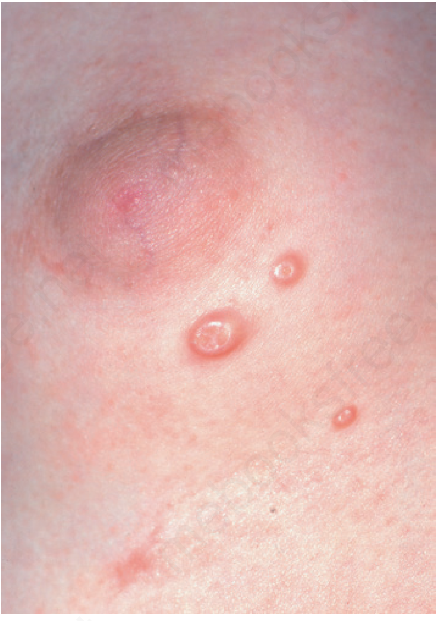
Key point: The large size (5-12 mm, one up to 12 mm) and foot location with yellowish core slightly favor tungiasis or inflamed/giant MC. The "BOTE sign" (beginning of the end - inflammation before spontaneous resolution) can explain the crusted/pustular central appearance.
Treatment: Often self-limiting (resolves in months-years). Options include cryotherapy, curettage, cantharidin, topical imiquimod.
- Andrews' Diseases of the Skin, p. 453-454
- Tintinalli's Emergency Medicine, p. 1065
3. Papular Urticaria / Insect Bite Hypersensitivity
- Intensely pruritic, firm, urticarial papules from insect bites (mosquitoes, fleas, mites)
- Children are particularly susceptible due to lack of prior sensitization
- Siblings in the same household would share the same insect exposure
- Secondary changes (excoriation, secondary infection) can create crusted nodular lesions
- Does NOT typically produce umbilicated papules - less likely here
4. Cutaneous Warts (Verruca plantaris/vulgaris)
- HPV infection causing rough, keratotic papules
- Can affect feet, but typically hyperkeratotic rather than umbilicated/pearly
- Spread by contact - siblings could share
Summary Table
| Feature | Tungiasis | Molluscum Contagiosum |
|---|---|---|
| Age group | Any, children most vulnerable | 1-4 years (peak) |
| Location | Feet (preferred) | Anywhere, feet possible |
| Lesion | Whitish nodule, central dark/white punctum | Pearly papule, central umbilication |
| Transmission | Barefoot soil contact | Skin contact, fomites |
| Both siblings affected | Same soil exposure | Direct contact / fomites |
| Duration 10 days | Matches flea growth (2-3 wks to full size) | Yes (new lesions appear over weeks) |
| Central yellow core | Engorged flea body/eggs | Caseous white core (molluscum body) |
| Treatment | Flea extraction | Self-limiting / cryotherapy |
Recommendation
Please have a dermatologist or pediatrician examine both children in person. The distinction between tungiasis and molluscum contagiosum is important because:
- Tungiasis requires physical removal of the flea and carries risk of serious secondary infection including tetanus if untreated
- Molluscum is generally benign and self-limiting but still benefits from treatment to prevent spread
If either child has been walking barefoot on soil/sand (especially in a tropical region), tungiasis should be ruled out first. A simple dermoscopy examination can confirm the presence of the embedded flea body and egg sacs.
⚠️ Disclaimer: This is general clinical information only and does not replace an in-person medical evaluation. Please see a qualified healthcare professional for diagnosis and treatment.


8ec6abe5-09db-4bae-9e91-aeb3713c5fdf.jpg
+2 more
47 year old female post craniotomy 6 months for brain tumor has such lesions over both legs with mild itching with no history of diabetes and on meds for htn and anti epilepsy..what can be the possible diagnosis and management
Searching sources...
The images show dry, scaly, hyperpigmented, ichthyotic skin over both legs in a post-craniotomy patient on anti-epileptic drugs (AEDs) and antihypertensives. This strongly suggests drug-induced ichthyosis (AEDs are a classic cause) or xerosis/acquired ichthyosis. Let me analyze the images and search the medical library in parallel.
analyse_media
analyse_media
Finding Sources
Finding Sources
The image analysis confirms: diffuse xerosis, fine powdery scaling, hyperpigmentation, lichenification, follicular accentuation, micro-fissuring, excoriations from itching over both legs. Now let me search the library for the specific diagnoses in context.
Finding Sources
Finding Sources
Finding Sources
Finding Sources
Finding Sources
Searching the Web
drug induced ichthyosis antiepileptic drugs phenytoin carbamazepine valproate skin xerosis mechanism management
Let me now read the ichthyosis section from the textbook and extract the Saudi Neurology Journal article on AED skin findings:
Reading File
Reading Web Page
https://nsj.org.sa/content/nsj/10/4/268.full.pdf
Finding Sources
Now let me search for stasis dermatitis + management and the ichthyosis vulgaris management section:
Finding Sources
Searching the Web
acquired ichthyosis post craniotomy immobility leg xerosis antiepileptic drug skin management emollients
I now have all the information needed for a thorough response. Let me compile the complete clinical analysis.
Clinical Analysis
Image Findings
Image 1 (thigh/knee area):
- Diffuse lichenification with exaggerated skin markings (leathery texture)
- Fine whitish-grey scaling
- Post-inflammatory hyperpigmentation (brownish-grey)
- Multiple excoriations with pinpoint hemorrhagic crusts from scratching
- Confluent patches over anterior/lateral thigh extending to knee
Image 2 (leg - close up):
- Widespread fine, powdery, diffuse scaling ("fish-scale" or "dusty" appearance)
- Prominent follicular accentuation with darkened/plugged pores
- Deep mottled hyperpigmentation (golden-brown to ash-grey)
- Micro-fissuring of dry epidermis along tension lines
- Subtle lichenification and pinpoint excoriations
This combination - bilateral, symmetric, diffuse xerosis + ichthyosis-like scaling + hyperpigmentation + pruritus over the legs in a woman on long-term AEDs - is a classic presentation.
Most Likely Diagnoses
1. Acquired Ichthyosis / Drug-Induced Xerosis-Ichthyosis - PRIMARY DIAGNOSIS
This is the most fitting diagnosis given the complete clinical context:
Mechanism via AEDs:
- Phenytoin, carbamazepine, phenobarbital, and valproate are all known to interfere with vitamin D metabolism (CYP450 enzyme induction accelerates vitamin D catabolism), lipid metabolism, and epidermal barrier function
- AEDs reduce skin ceramide synthesis and alter the lipid composition of the stratum corneum, leading to impaired barrier function, increased transepidermal water loss, and resultant compensatory hyperkeratosis and scaling
- Chronic AED use is also associated with xerosis, pruritus, acneiform eruptions, and hyperpigmentation
- The NSJ study on AED skin findings documented xerosis (12.9%) and pruritus (8.1%) among chronic AED users
Additional contributing factors in this patient:
- Post-craniotomy immobility / reduced ambulation - venous stasis and reduced lymphatic flow worsen lower-limb skin changes
- Systemic corticosteroids (commonly given perioperatively/post-craniotomy for cerebral edema) - these reduce skin lipid synthesis and barrier function
- Antihypertensive drugs (certain calcium channel blockers like amlodipine, or beta-blockers) can also cause xerosis/pruritus
- Reduced sun exposure and physical activity post-surgery impairs vitamin D synthesis further
- Age-related (47 years - perimenopausal estrogen decline reduces skin hydration)
From Robbins Pathology: "Acquired (noninherited) variants also exist and may be associated with lymphoid and visceral malignancies" and systemic causes. The pathology shows "buildup of compacted and thickened stratum corneum...defective desquamation." - Robbins Pathologic Basis of Disease, p. 1063
Per Merck Manuals: "Acquired ichthyosis typically presents in adulthood...Some medications also cause ichthyosis (eg, nicotinic acid, triparanol, butyrophenones)."
2. Stasis Dermatitis - Important co-contributor
- Post-craniotomy reduced mobility causes venous insufficiency in the legs
- Presents as bilateral lower limb hyperpigmentation, scaling, pruritus, and lichenification
- The mottled brownish hyperpigmentation seen is consistent with hemosiderin deposition from stasis
- Highly likely to be co-existing with xerosis/ichthyosis here
3. Asteatotic Eczema (Eczema Craquelé) - Differential
- Extreme dryness leading to cracked, fissured skin with superficial eczematous change
- Common in patients with poor mobility, low humidity environments, and altered skin barrier
- The micro-fissuring and scaling pattern seen in image 2 is consistent
4. Rule out: Statin-induced myopathy / Hypothyroidism-related ichthyosis
- Hypothyroidism gives the same pattern (dry, scaly, thickened skin + myxedema); if on any medication post-craniotomy that could affect thyroid, TSH should be checked
- Some antihypertensives (e.g., hydrochlorothiazide, propranolol) are photosensitizers that worsen xerosis
5. Malignancy-related acquired ichthyosis - must be excluded
- This patient has a history of brain tumor - acquired ichthyosis in adults can be a paraneoplastic phenomenon associated with Hodgkin lymphoma, multiple myeloma, and other malignancies
- If tumor is benign (e.g., meningioma), this is less likely but should be noted
Management
Immediate / Topical
| Treatment | Regimen | Notes |
|---|---|---|
| Emollients (white soft paraffin, mineral oil, aqueous cream) | Apply twice daily, especially after bathing while skin is still damp | Mainstay of treatment - seals in moisture |
| Urea-based creams (10-25%) | Once daily on thick/scaly areas | Keratolytic + humectant; avoid on inflamed/fissured skin |
| Lactic acid cream (5-12%) | Once daily | Alpha-hydroxy acid - keratolytic, improves hydration |
| Topical low-potency corticosteroid (hydrocortisone 1%) | Short course to pruritic/eczematous areas | For itch relief; avoid long-term use |
| Soap substitutes / gentle cleansers | Replace soap with emulsifying ointment or aqueous cream | Avoid harsh soaps that strip skin lipids |
| Lukewarm baths (20-30 min) | Daily or every other day | Remove scale; apply emollient immediately after |
Systemic / Underlying Cause
- Vitamin D supplementation - strongly recommended given AED-induced vitamin D deficiency (phenytoin, carbamazepine, phenobarbital are CYP450 inducers); recommended 1000-2000 IU/day
- Antihistamines (cetirizine or loratadine) - for pruritus relief, non-sedating preferred
- Encourage leg elevation and ambulation - addresses venous stasis component
- Compression stockings (class I) - if stasis dermatitis component confirmed
Investigations to Order
| Investigation | Rationale |
|---|---|
| TSH / Free T4 | Rule out hypothyroidism (same clinical picture) |
| CBC, LFT, serum albumin | Screen for systemic disease, nutritional status |
| Serum Vitamin D (25-OH) | AED users frequently have low levels |
| Serum calcium, phosphate | Often abnormal with AED-induced vitamin D deficiency |
| Fasting glucose / HbA1c | Even without known diabetes, post-craniotomy patients may have glucose dysregulation; diabetic xerosis is common |
| AED drug levels | Confirm therapeutic range; toxicity worsens skin changes |
| Review antihypertensive | Identify if calcium channel blocker (amlodipine can cause leg swelling/stasis), diuretic (worsens dryness), or beta-blocker (worsens Raynaud/peripheral circulation) |
Drug Review
- Discuss with the neurologist whether the AED type can be modified:
- Enzyme-inducing AEDs (phenytoin, carbamazepine, phenobarbital) cause the most vitamin D depletion and skin changes
- Newer AEDs (levetiracetam, lamotrigine, lacosamide) have fewer metabolic skin effects
- Do not stop AEDs without neurology input - seizure risk post-craniotomy
Referral
- Dermatology - for formal diagnosis, biopsy if atypical features, and supervised keratolytic therapy
- Neurology / Oncology - ongoing tumor surveillance (rule out paraneoplastic ichthyosis)
- Physiotherapy - mobility improvement to address stasis component
Summary
The most likely diagnosis is acquired ichthyosis/drug-induced xerosis from chronic AED use (accelerated vitamin D catabolism + impaired skin barrier), compounded by post-craniotomy immobility causing stasis dermatitis and possible antihypertensive contribution. The absent diabetes history makes diabetic dermopathy less likely. Hypothyroidism and paraneoplastic ichthyosis (in context of the known brain tumor) must be excluded by investigations.
First-line management: intensive emollient therapy + vitamin D supplementation + AED review + investigations as above.
⚠️ This is clinical guidance for healthcare professionals. All management decisions require in-person clinical assessment.
This is a shared conversation. Sign in to Orris to start your own chat.