Here is a complete, easy guide to the types of interlobular septal thickening with differentials:
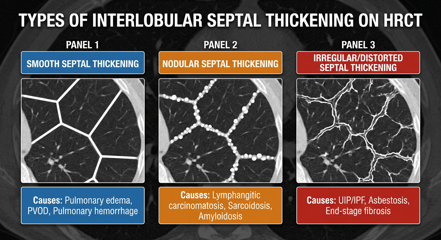
Types of Interlobular Septal Thickening on HRCT
New Educational Figure
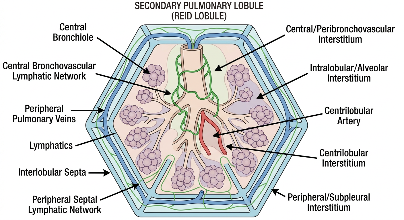
What Are Interlobular Septa?
These are the thin connective tissue walls that separate individual secondary pulmonary lobules. Normally they are invisible or barely visible on HRCT. When thickened, they appear as short, straight white lines at the lung periphery - the classic Kerley B lines on CXR.
The 3 Types - Easy Memory: "S-N-I"
🔵 Type 1: SMOOTH Septal Thickening
Lines are uniform, straight, and even in thickness - like a clean ruler line.
| Feature | Description |
|---|
| Appearance | Uniform thin white lines, regular thickness throughout |
| Distribution | Typically bilateral, basal predominant, subpleural |
| Key sign on CXR | Kerley B lines (short, horizontal, peripheral) |
Differential Diagnosis - "OLPH"
| Cause | Clue |
|---|
| Pulmonary Edema (most common) | Cardiomegaly + pleural effusion + bilateral |
| PVOD (Pulmonary Veno-Occlusive Disease) | No cardiomegaly, PAH features |
| Lymphangitic carcinomatosis | Can be smooth early on |
| Pulmonary hemorrhage | Ground glass + hemoptysis |
Key point: Smooth thickening = think fluid or early infiltration. Pulmonary edema is #1 cause.
🟠 Type 2: NODULAR Septal Thickening ("Beaded Septa")
Lines have small bumps/nodules along them - like a string of pearls or a beaded necklace.
| Feature | Description |
|---|
| Appearance | Irregular beaded/lumpy septa - nodules sit ON the lines |
| Distribution | Often unilateral or asymmetric |
| Pattern | "Beaded septum" sign |
Differential Diagnosis - "SLAB"
| Cause | Clue |
|---|
| Sarcoidosis | Perilymphatic nodules + upper lobe + bilateral hilar LAD |
| Lymphangitic Carcinomatosis | Known malignancy (breast, lung, stomach, colon) |
| Amyloidosis | Rare, systemic features |
| Berylliosis | Occupational history (aerospace/electronics workers) |
Key point: Nodular/beaded septa = think lymphatic disease (tumor or granuloma tracking along lymphatics).
🔴 Type 3: IRREGULAR Septal Thickening
Lines are distorted, broken, non-uniform, and pulled out of shape - like crumpled paper.
| Feature | Description |
|---|
| Appearance | Irregular, coarse, distorted lines with traction |
| Distribution | Basal, subpleural, associated with honeycombing |
| Associated findings | Traction bronchiectasis, honeycombing, volume loss |
Differential Diagnosis - "AURIS"
| Cause | Clue |
|---|
| UIP/IPF | Basal + subpleural + honeycombing, no trigger |
| Asbestosis | Occupational exposure + pleural plaques |
| Rheumatoid lung (UIP pattern) | RA history + joint disease |
| Interstitial fibrosis (any cause) | End-stage fibrosis |
| Scleroderma lung | Skin tightening, Raynaud's |
Key point: Irregular thickening = think fibrosis - the lung architecture is being destroyed and distorted.
Real HRCT Comparison - From Textbooks
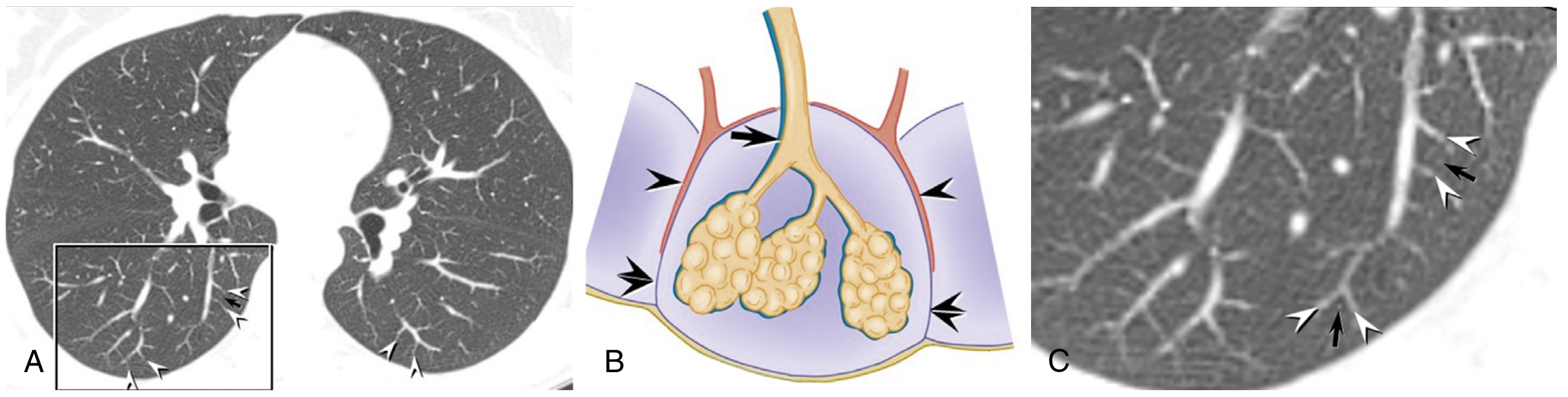
Normal lobular anatomy + septal veins:
(Murray & Nadel, Figure 20.18 - showing normal HRCT secondary pulmonary lobule anatomy)
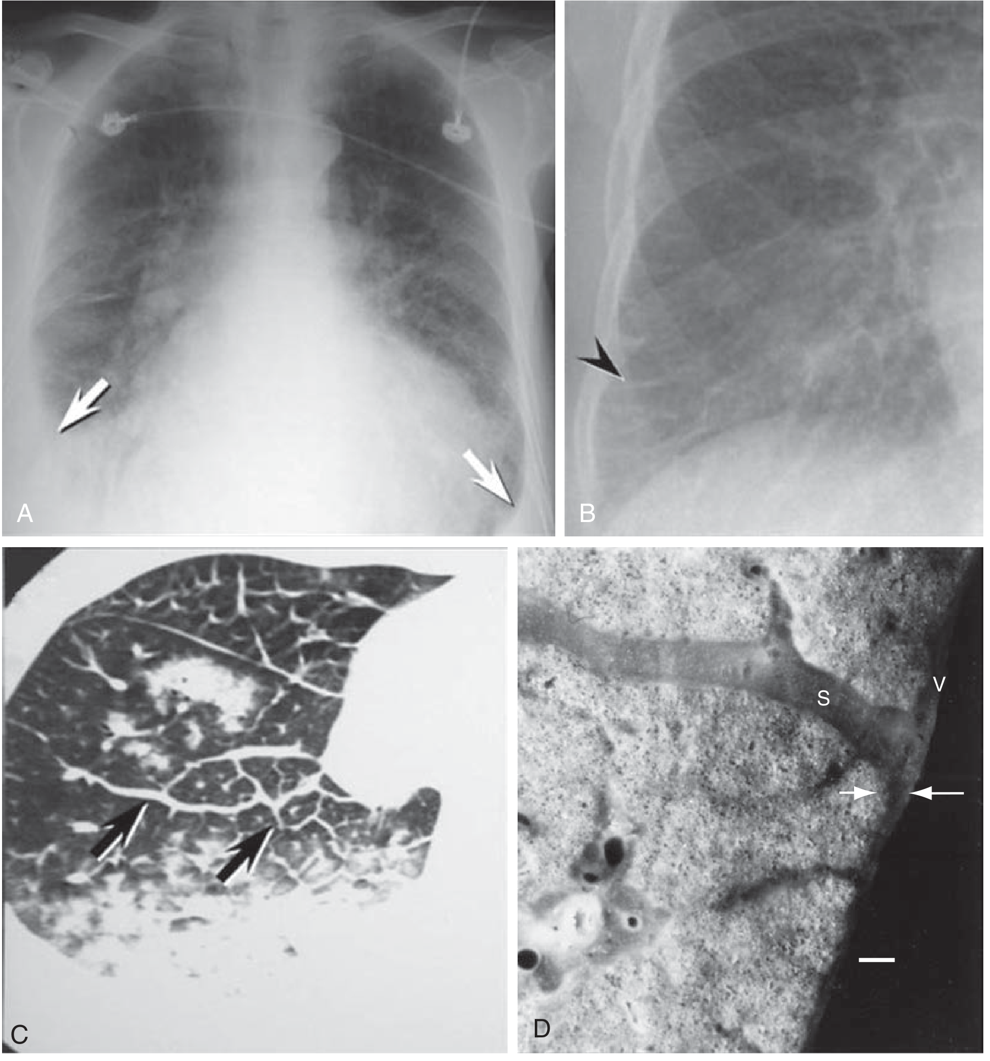
Smooth septal thickening in pulmonary edema (Kerley B lines):
(Murray & Nadel, Figure 108.3 - pulmonary edema CXR + CT with smooth septal thickening)
Panel C (CT) shows the classic smooth peripheral interlobular septal thickening of pulmonary edema.
Quick Summary Table
| Type | Appearance | Top 3 Causes |
|---|
| Smooth | Uniform, straight lines | Pulmonary edema, PVOD, Lymphangitic Ca |
| Nodular | Beaded (string of pearls) | Sarcoidosis, Lymphangitic Ca, Amyloidosis |
| Irregular | Distorted, broken, fibrotic | UIP/IPF, Asbestosis, Scleroderma |
One-Line Memory Rule
"Smooth = Wet (fluid), Nodular = Dirty (lymphatics/tumor/granuloma), Irregular = Scarred (fibrosis)"