
Minoxidil
minoxidil mechanism of action hair loss hypertension 2025
minoxidil androgenetic alopecia
minoxidil hair growth mechanism potassium channel
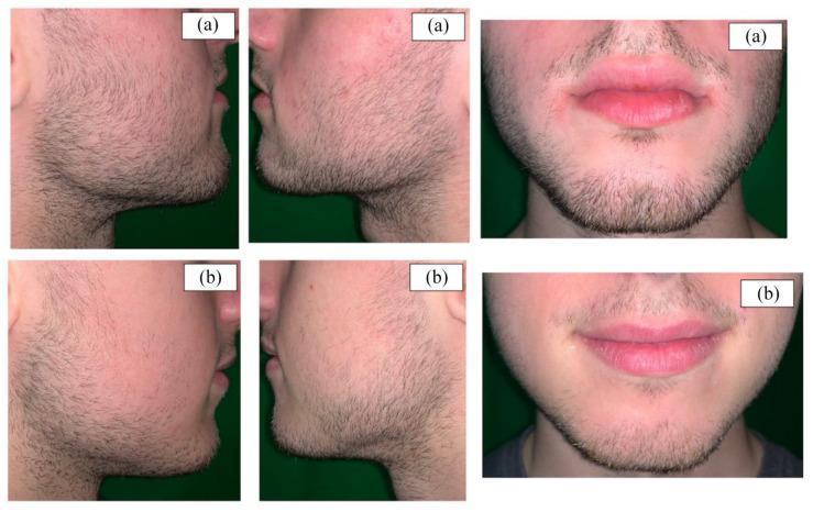
This clinical photograph comparison presents six views of facial hair growth in a study involving identical twins to evaluate the efficacy of topical minoxidil. The images are divided into two rows: (a) shows the treatment subject after 16 months of daily 5% minoxidil foam application, and (b) shows the non-treated control subject. Both individuals are photographed 10 days post-shave. Row (a) demonstrates a significantly higher follicular density, increased hair shaft thickness, and more extensive terminal hair coverage across the beard, mustache, and cheek regions. In contrast, row (b) exhibits sparser, finer, and patchier hair distribution. The comparison highlights the pharmacological effect of minoxidil on prolonging the anagen phase and increasing follicular size in androgen-sensitive facial areas. Key anatomical focuses include the submandibular region, malar area, and perioral region. This visual is intended for dermatological education regarding hair loss treatments and off-label uses of minoxidil for facial hair enhancement.
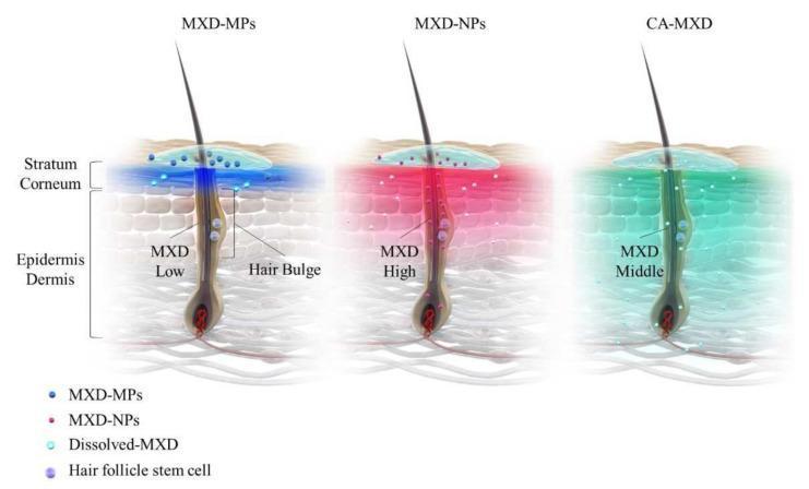
An anatomical diagram illustrating the targeted delivery of minoxidil (MXD) into hair follicles using three different pharmaceutical formulations: MXD microparticles (MXD-MPs), MXD nanoparticles (MXD-NPs), and a commercially available MXD solution (CA-MXD). The diagram depicts a cross-section of human skin, including the stratum corneum, epidermis, and dermis, featuring a central hair follicle with labeled regions for the hair bulge and bulb. Visual coding shows: 1) MXD-MPs (blue) resulting in low drug concentration at the hair bulge and significant accumulation at the stratum corneum; 2) MXD-NPs (pink/red) demonstrating high concentration and deep penetration into the hair bulge, where hair follicle stem cells (lavender spheres) are located; and 3) CA-MXD (teal) showing middle-range concentration levels. The illustration emphasizes the follicular delivery pathway, suggesting that nanoparticle-based formulations facilitate superior drug accumulation in the bulge region, which is critical for activating epithelial stem cells and promoting hair growth, while potentially minimizing systemic absorption through the stratum corneum.
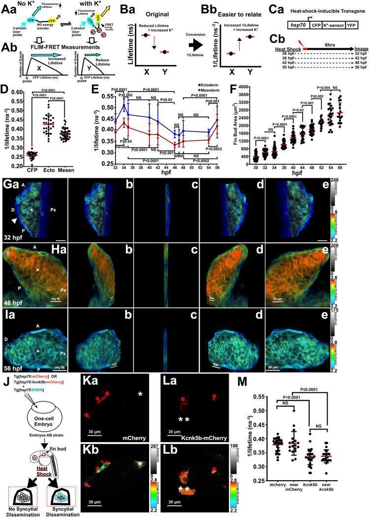
This composite educational image details the use of a FRET-based sensor, KIRIN1, to monitor intracellular potassium (K+) concentrations during zebrafish pectoral fin bud development. Panels A and B explain the mechanism of Fluorescence Lifetime Imaging Microscopy (FLIM), where increased K+ leads to a reduced donor fluorophore lifetime (shorter ns). Panels D-F provide quantitative data showing a significant transient increase in intracellular K+ (as 1/lifetime) in both the ectoderm and mesenchyme peaking around 48 hours post-fertilization (hpf), correlating with rapid fin bud growth. Panels G-I display pseudocolored FLIM z-stacks at 32, 48, and 56 hpf; at 48 hpf, a shift toward red/yellow hues signifies higher K+ levels compared to the blue/green hues at 32 and 56 hpf. Panels J-M investigate the role of the K+ channel kcnk5b using mosaic transgene expression. Confocal planes (Ka, La) and corresponding FLIM images (Kb, Lb) illustrate that cells expressing kcnk5b-mCherry exhibit lower 1/lifetime values, indicating reduced intracellular K+ compared to wild-type neighbors, confirming the channel's functional role in modulating ionic concentration during morphogenesis.
Minoxidil
Chemistry & Prodrug Status
- Goodman & Gilman's, Mechanism of Action section
Mechanism of Action
- Increased K+ permeability causes membrane hyperpolarization
- This reduces the probability of voltage-gated Ca2+ channel opening
- Result: smooth muscle relaxation and vasodilation
- Katzung's Basic and Clinical Pharmacology, 16th Ed., p. 281
- Goodman & Gilman's, Mechanism of Action
Cardiovascular Pharmacology
Hemodynamic Effects
- Potent arteriolar vasodilation → fall in systemic vascular resistance
- Reflex sympathetic activation → tachycardia, increased myocardial contractility
- Cardiac output can rise 3-4-fold
- Renal artery vasodilation, but systemic hypotension can occasionally reduce renal blood flow
- Potent stimulator of renin secretion (via renal sympathetic activation + intrinsic mechanisms)
Cardioprotective Potential
- Goodman & Gilman's, Pharmacological Effects
Pharmacokinetics (Oral Form)
| Parameter | Value |
|---|---|
| Absorption | Well absorbed orally |
| Tmax (blood) | ~1 hour |
| Hypotensive effect peak | Delayed (metabolite formation lag) |
| t½ (plasma) | 3-4 hours |
| Duration of action | ~24 hours |
| Elimination | Bulk as glucuronide conjugate; ~20% excreted unchanged in urine |
- Goodman & Gilman's, ADME
Clinical Use: Hypertension
- A β-blocker (blunts reflex tachycardia)
- A loop diuretic (counters fluid retention)
- Katzung's Basic and Clinical Pharmacology, 16th Ed.
Adverse Effects (Oral)
| Effect | Cause |
|---|---|
| Tachycardia, palpitations, angina | Reflex sympathetic activation |
| Edema, fluid retention | Renal Na/H₂O retention |
| Pericardial effusion | Can occur with prolonged use |
| Headache, sweating | Vasodilation |
| Hypertrichosis | Unknown mechanism; affects ~80% of patients |
| Worsening heart failure | Increased ventricular volume load |
- Goodman & Gilman's, Adverse Effects; Katzung, Toxicity
The Hair Growth Discovery
Topical Minoxidil: Hair Loss
Indication
Efficacy
- Vertex balding responds better than frontal balding
- 5% solution > 2% solution in men
- Women: only the 2% lotion (or 5% foam in some jurisdictions) is approved
- Effect is not permanent - cessation leads to hair loss within 4-6 months
- Long-term data (2025, Kushnir-Grinbaum et al.) show cumulative benefit over 5 years with systemic formulations
Mechanism in Hair Follicles
- Local vasodilation improving nutrient/oxygen delivery to follicles
- Prolonging the anagen (growth) phase
- Direct stimulation of follicular and perifollicular cell proliferation (DNA synthesis)
- Possible counteraction of androgen effects
- Katzung, MINOXIDIL (dermatology section); Fitzpatrick's Dermatology
Available Formulations
- 2% solution (OTC): for men and women (Rogaine)
- 5% solution (OTC): for men (Rogaine Extra Strength)
- 5% foam: Men; 5% recommended over 2% for greater efficacy
- Low-dose oral minoxidil: Increasingly used off-label; as of 2025/2026, a sublingual formulation (by Samson Clinical) is in Phase 3 trials for alopecia
- Harriet Lane Handbook, 23rd Ed.
Adverse Effects (Topical)
- Transient shedding in early weeks - a sign of follicular synchronization/cycle reset; do NOT stop therapy
- Irritant or contact dermatitis
- Minimal percutaneous absorption in normal scalp - but monitor blood pressure in cardiac disease patients
Recent Evidence (2024-2026)
-
Oral vs. topical minoxidil meta-analysis - Sobral et al. (2025, Int J Dermatol, PMID 39425514): Meta-analysis of RCTs comparing oral vs. topical solution in androgenetic alopecia.
-
Minoxidil vs. 5-alpha reductase inhibitors network meta-analysis - Gupta et al. (2025, J Cosmet Dermatol, PMID 40586152): Comparative efficacy of monotherapy options for male pattern hair loss.
-
Microneedling + minoxidil meta-analysis - Ahmed et al. (2025, Arch Dermatol Res, PMID 40056230): Microneedling combined with topical minoxidil outperforms topical minoxidil alone.
Comparison with Related Drugs
| Drug | Mechanism | Route | Use |
|---|---|---|---|
| Minoxidil | K_ATP channel opener | Oral / Topical | Hypertension / Alopecia |
| Hydralazine | Unknown (probable K+ channel / NO) | Oral / IV | Hypertension |
| Diazoxide | K_ATP channel opener | IV | Hypertensive emergency |
| Nicorandil | K_ATP opener + nitrate | Oral | Angina |
| Finasteride | 5α-reductase inhibitor | Oral | Androgenetic alopecia |
Visual: Minoxidil Structure
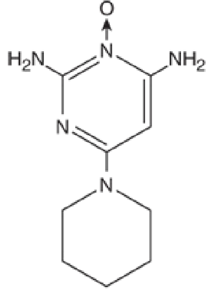
How does minoxidil compare to finasteride for hair loss efficacy and side effects?
minoxidil finasteride androgenetic alopecia efficacy comparison
androgenetic alopecia before after treatment hair loss pattern
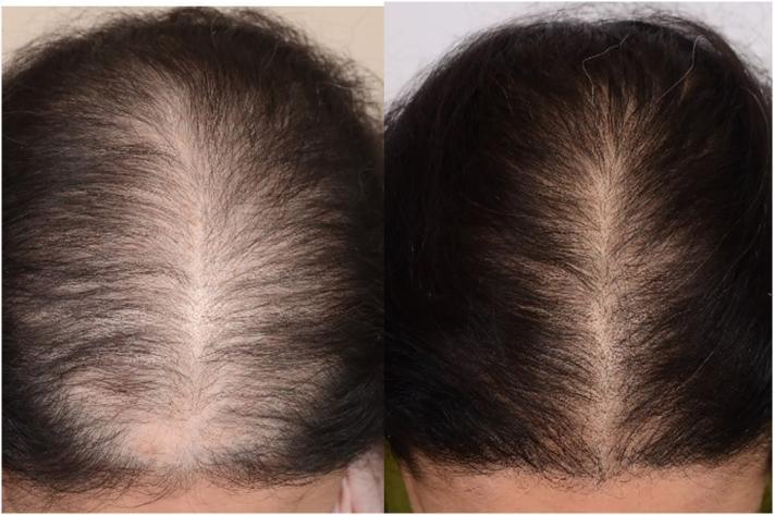
Clinical photograph (top view) demonstrating the scalp of a 31-year-old female with androgenetic alopecia (AGA), showing side-by-side 'before' and 'after' results following five platelet-rich plasma (PRP) treatment sessions over 4 months. The 'before' image illustrates a classic female pattern of hair loss, characterized by significant diffuse thinning across the central scalp, crown, and vertex, with a widened part line and high visibility of the underlying scalp. The hair strands appear fine and miniaturized. The 'after' image demonstrates clinical improvement, evidenced by increased hair density and follicular volume. There is a noticeable narrowing of the central part line and enhanced coverage of the scalp surface. This comparison highlights the efficacy of PRP as a therapeutic modality for non-scarring alopecia, illustrating its role in stimulating hair regrowth and improving hair shaft thickness in patients with hormonally-mediated hair loss.
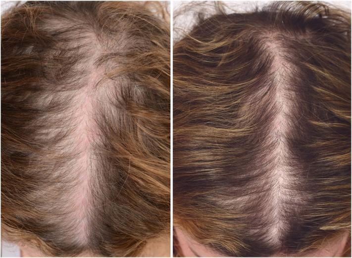
Clinical photograph showing a side-by-side comparison of the vertex and mid-scalp of a 46-year-old female patient with female pattern hair loss (androgenetic alopecia). The 'before' image (left) demonstrates diffuse thinning, characterized by a widened central part and increased scalp visibility, consistent with a Ludwig scale classification of hair loss. The 'after' image (right) displays the clinical outcome three months following three sessions of platelet-rich plasma (PRP) therapy. Visually, the 'after' state shows a significant increase in hair density, a narrowing of the central part line, and improved scalp coverage. The medical specialty is dermatology, focusing on trichology and non-surgical hair restoration. This comparison serves as an educational resource to illustrate the therapeutic potential of PRP in managing follicular miniaturization and increasing hair volume in patients with androgenetic alopecia.
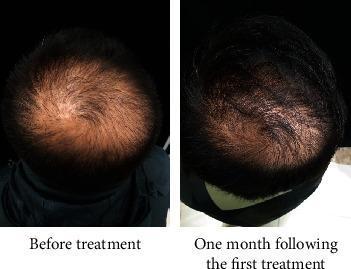
This clinical comparison photograph illustrates the progression of hair growth in a patient with androgenetic alopecia (AGA) before and after therapy. The 'Before treatment' image shows a superior view of the scalp, highlighting significant hair thinning and reduced follicular density primarily at the vertex and mid-scalp regions, resulting in high scalp visibility. The second image, taken 'One month following the first treatment' with Botulinum Toxin Type A (BTA), demonstrates a visible increase in hair density and coverage. The previously exposed patch of scalp is noticeably smaller and less prominent, suggesting improved terminal hair count and follicle activation. This visual evidence supports the clinical application of BTA to improve local blood flow and oxygenation in the scalp to counteract the effects of dihydrotestosterone (DHT) and hypoxia in AGA patients. The comparison serves as an educational tool for evaluating dermatological treatment efficacy for male pattern baldness.
PMID: 38725143
PMID: 37798906
Minoxidil vs. Finasteride for Androgenetic Alopecia
1. Mechanisms - Why They Differ
| Feature | Minoxidil | Finasteride |
|---|---|---|
| Drug class | K_ATP channel opener / vasodilator | 5α-reductase inhibitor (type II) |
| Target | Vascular smooth muscle + hair follicle (unclear) | Enzyme converting testosterone → DHT |
| Active form | Minoxidil sulfate (hepatic conversion) | The parent drug itself |
| Route | Topical (2%, 5%) or oral (low-dose off-label) | Oral 1 mg/day (Propecia) |
| Addresses androgen cause? | No - does not affect DHT | Yes - reduces scalp/serum DHT by ~60-70% |
2. Approved Indications
| Minoxidil | Finasteride | |
|---|---|---|
| Men | Yes (2% and 5% topical; low-dose oral off-label) | Yes (1 mg/day oral) |
| Women | Yes (2% topical; 5% foam in some regions) | No - not approved; teratogenic risk |
| Pattern | Vertex and diffuse thinning | Vertex > frontal (less effective frontally) |
- Katzung's Basic and Clinical Pharmacology, 16th Ed.
3. Efficacy: What the Evidence Shows
Head-to-Head in Men
- Dutasteride 0.5 mg/day ranked #1 (SUCRA 87-98%) for 6-month total and terminal hair density change
- Oral finasteride outperformed oral minoxidil and other newer agents (Botox, microneedling, photobiomodulation)
- Effectiveness is dose-dependent for both drug classes
Practical Efficacy Summary
| Outcome | Minoxidil (5% topical) | Finasteride (1 mg/day oral) |
|---|---|---|
| Hair count increase (vertex) | Moderate | Moderate-good |
| Prevention of further loss | Moderate | Good |
| Frontal hairline | Limited | Limited |
| Time to see effect | 3-6 months | 3-6 months |
| Onset of shedding reversal | Faster (anagen shift) | Slower (gradual DHT reduction) |
In Women
- Goldman-Cecil Medicine, Treatment section
4. Combination Therapy
- Combination group: +81 hair/cm² at 6 months (p < 0.001 vs. baseline); significant vs. both monotherapies
- Finasteride alone: improved but not statistically significant vs. baseline at 3 months
- Minoxidil alone: improved moderately
- 79% of combination group achieved GPAS score ≥2 vs. 8% (finasteride) and 41% (minoxidil) alone
- Tolerability was good in all groups; no hormonal level differences between groups (topical finasteride minimizes systemic DHT suppression)
Topical finasteride spray offers similar local efficacy to oral finasteride but with less systemic DHT reduction - reducing the risk of sexual side effects.
- Goldman-Cecil Medicine
5. Side Effects Comparison
| Side Effect | Minoxidil (Topical) | Minoxidil (Oral low-dose) | Finasteride (Oral 1 mg) |
|---|---|---|---|
| Initial shedding | Common (weeks 2-8); sign of efficacy | Similar | Uncommon |
| Contact/irritant dermatitis | Yes (especially with propylene glycol vehicle) | N/A | No |
| Hypertrichosis | Rare at scalp doses | Possible (unwanted facial/body hair) | No |
| Hypotension | Rare (mostly oral doses) | Monitor in cardiac disease | No |
| Sexual dysfunction | No | No | Yes: decreased libido, erectile dysfunction, ejaculation disorders (~2-4%) |
| Post-finasteride syndrome | No | No | Rare but reported: persistent sexual dysfunction/depression after stopping |
| Teratogenicity | None documented | None | Yes: feminization of male fetus (Category X in pregnancy) |
| Gynecomastia | No | No | Rare (<1%) |
| Mood/depression | No | No | Reported in post-finasteride syndrome |
Post-Finasteride Syndrome
- Goldman-Cecil Medicine
6. Practical Treatment Duration
Both drugs must be continued indefinitely to maintain results.
- Stopping minoxidil: triggers acute telogen efflux (shedding) within 3-4 months; cannot be prevented by concomitant finasteride
- Stopping finasteride: gradual hair loss returns to pre-treatment status within approximately 1 year
- Treatments should be trialed for at least 6 months before assessing efficacy
- Goldman-Cecil Medicine, Treatment
7. Summary Decision Framework
| Scenario | Preferred Agent |
|---|---|
| Men, first-line monotherapy | Either; finasteride may have slight edge in hair count |
| Men wanting maximum efficacy | Combination (topical minoxidil + topical or oral finasteride) |
| Men concerned about sexual side effects | Topical minoxidil; or topical finasteride (less systemic) |
| Women with androgenetic alopecia | Topical minoxidil (2%) - finasteride is not approved |
| Women post-menopause (off-label) | Low-dose oral minoxidil or oral finasteride (with caution, no pregnancy risk) |
| Severe/resistant disease | Add dutasteride 0.5 mg (more potent than finasteride, approved in some countries) |