
Give me the reasons why a bacteria is gram positive and gram negative and also give the reasons for a bacteria being an acid fast.
gram stain gram positive gram negative bacteria cell wall structure
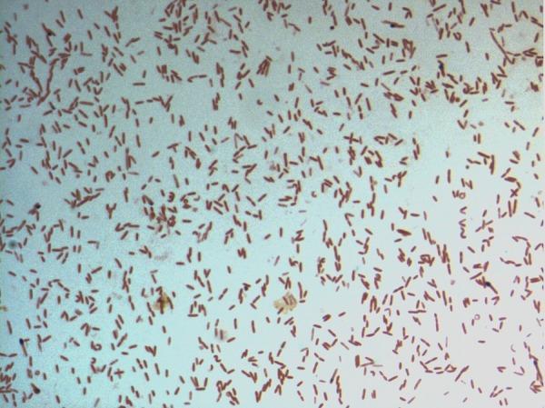
This diagnostic image shows a light microscopy view of a bacterial culture following Gram staining. The specimen displays numerous small, rod-shaped (bacilli) bacteria distributed across the field. The microorganisms exhibit a consistent pink-to-red hue, identifying them as Gram-negative. This staining characteristic indicates a cell wall structure with a thin peptidoglycan layer and an outer membrane that does not retain the crystal violet stain. The arrangement of the bacilli is predominantly individual or scattered, with occasional small clusters and pairs. Specifically identified as D. fastidiosa strain JC13T, this image serves as a primary microbiological reference for the morphological and staining properties of this novel genus within the family Erysipelotrichaceae. The visual demonstrates key diagnostic features including cell morphology, arrangement, and Gram reaction, which are essential for clinical microbiology classification and initial pathogen identification.
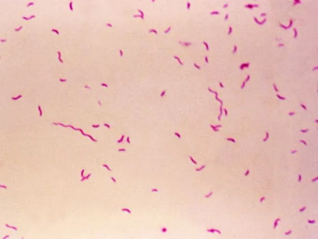
Bright-field light microscopy of a Gram-stained bacterial smear reveals numerous small, slender, curved Gram-negative rods with a characteristic gull-wing appearance consistent with Campylobacter species. The image captures bacteria oriented individually and in small clusters against a pale pink counterstain, illustrating Bacillary morphology typical of enteric pathogens. Gram staining shows purple/blue rods (crystal violet retained by cell wall) with a light pink background from the counterstain (safranin), enabling discrimination from Gram-positive organisms. The organisms appear slender and curved, with a single polar flagellum suggested by motility or alignment in the smear. Specimen type is a bacterial smear obtained from a gastrointestinal sample (feces or culture isolate), prepared for diagnostic microbiology. The imaging modality is bright-field microscopy at high magnification (approximately 1000x with oil immersion), following Gram staining to highlight bacterial cell wall structure. Clinically, detection of Campylobacter species supports infectious gastroenteritis; in the IPSID (immunoproliferative small intestinal disease) context, Campylobacter involvement has been proposed as a pathogenic trigger mirroring Helicobacter pylori's role in gastric MALT lymphoma. Differential diagnoses include Helicobacter, Vibrio, and other curved Gram-negative bacteria. This image serves educational and diagnostic utility for microbiology, clinical pathology, gastroenterology, and infectious disease research.
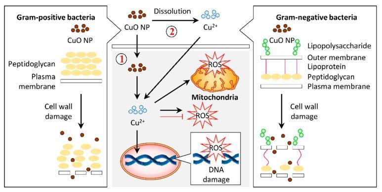
A pathophysiology diagram illustrating the antibacterial mechanisms of copper oxide nanoparticles (CuO NPs) on Gram-positive and Gram-negative bacteria. The left panel shows Gram-positive bacteria with a thick peptidoglycan layer above a plasma membrane. The right panel shows Gram-negative bacteria with a complex cell wall including lipopolysaccharides, an outer membrane, lipoproteins, and a thin peptidoglycan layer. Both panels demonstrate CuO NPs causing cell wall damage and lysis. The central panel details the intracellular bactericidal pathway labeled as mechanism 1 and 2. Mechanism 1 involves the internalization of CuO NPs into the cytosol, while mechanism 2 illustrates the dissolution of CuO NPs into copper ions (Cu2+) which then permeate the cell. Inside the cell, both Cu2+ ions and CuO NPs induce the production of reactive oxygen species (ROS). The resulting oxidative stress leads to mitochondrial disruption and DNA damage, ultimately resulting in cell death. This infographic highlights the role of nanoparticle morphology and ion release in targeting bacterial structural and metabolic integrity.
acid fast staining mycobacterium mycolic acid cell wall Ziehl-Neelsen
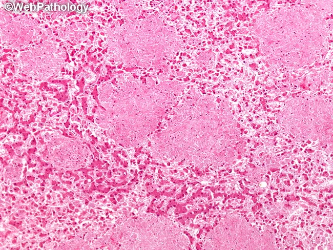
Histopathology of liver tissue from an HIV-positive patient with Mycobacterium avium complex infection. Modality: light microscopy of FFPE liver biopsy; Hematoxylin and Eosin stained section with subsequent Ziehl-Neelsen acid-fast stain. Anatomical location: hepatic parenchyma within the hepatic lobules, sinusoids and portal tracts. Macroscopic pattern: discrete, non-necrotic nodular aggregates of sinusoidal histiocytes (Kupffer cell-derived macrophages) scattered throughout the parenchyma; nodules vary in size. Microscopy shows abundant foamy or epithelioid histiocytes filled with acid-fast bacilli on Ziehl-Neelsen staining; multinucleated giant cells are rare. The granulomatous response is non-necrotizing, with preserved tissue architecture and occasional central macrophages forming small clusters. Inflammatory cells are limited, without caseation. Acid-fast organisms appear as slender red rods within macrophages; bacterial load can be high in disseminated infection. Diagnostic significance: hepatic MAC infection in immunocompromised hosts supports disseminated mycobacterial disease; finding AFB within histiocytes confirms infection and guides antimicrobial therapy. Pathophysiology: intracellular replication of MAC within Kupffer cells leading to granulomatous hepatitis; differential includes M. tuberculosis, other non-tuberculous mycobacteria, fungal granulomas. Clinical correlation: in AIDS, this pattern correlates with low CD4 counts and high mycobacterial burden; management requires combination antimycobacterial therapy and immune reconstitution strategies, with culture and molecular identification. This image exemplifies disseminated hepatic infection patterns clearly.
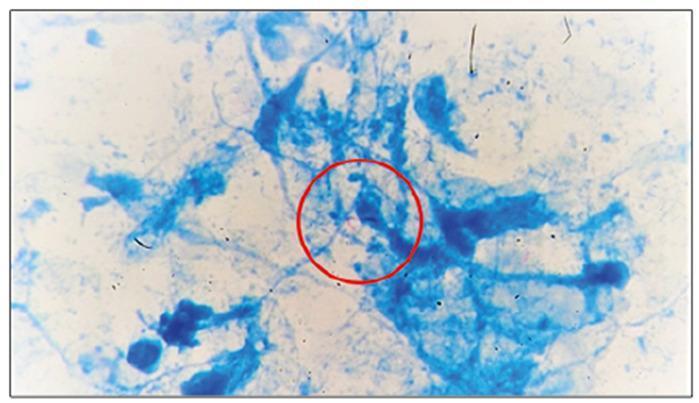
This diagnostic image is a high-power light microscopy field showing a histopathological sample stained with Ziehl-Neelsen or a similar acid-fast stain. The background consists of blue-stained cellular debris, fibrous tissue, and inflammatory infiltrates from a pericardial biopsy. Centrally highlighted within a red circular annotation are several bright red or pink, rod-shaped microorganisms. These characteristics are pathognomonic for acid-fast bacilli (AFB), specifically Mycobacterium tuberculosis. The visual demonstrates the diagnostic findings of tuberculous pericarditis, where the pathogen is directly visualized within the pericardial tissue despite a non-granulomatous histological appearance. This slide serves as an educational example of infectious pathology and the clinical utility of specialized staining in identifying extracellular pathogens in extrapulmonary tuberculosis.
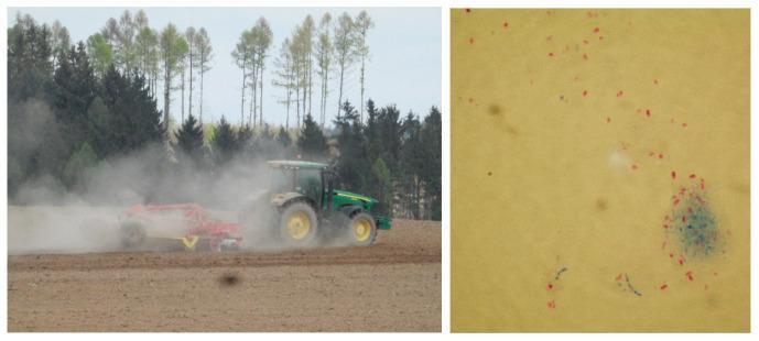
This dual-panel image demonstrates the environmental transmission and microscopic identification of non-tuberculous mycobacteria (NTM). The left panel is a photograph of a tractor tilling soil, illustrating how agricultural activity can generate aerosols and dust that serve as vehicles for environmental mycobacteria. The right panel is a diagnostic light microscopy image (1000x magnification) of a soil sample specimen following Ziehl-Neelsen staining. The micrograph shows numerous bright red, polymorphic, rod-shaped mycobacterial cells, which are characteristic of acid-fast organisms. These acid-fast bacilli (AFB) are visible as individual rods, small groups, and dense aggregates against a pale yellowish-tan background, with occasional blue-stained non-acid-fast material. This visual illustrates the presence of species such as Mycobacterium terrae and Mycobacterium avium complex (MAC) in environmental reservoirs, highlighting the potential for zoonotic or environmental exposure pathways for human infection.
Gram Staining: Why Bacteria Stain Positive, Negative, or Acid-Fast
| Step | Reagent | Role |
|---|---|---|
| 1 | Crystal violet | Primary stain - binds to peptidoglycan |
| 2 | Gram iodine (mordant) | Causes crystal violet to precipitate inside the cell |
| 3 | Acetone-alcohol (decolorizer) | Removes crystal violet from gram-negative organisms |
| 4 | Safranin (counterstain) | Stains gram-negative organisms pink/red |
Why Bacteria Are Gram-Positive (Purple)
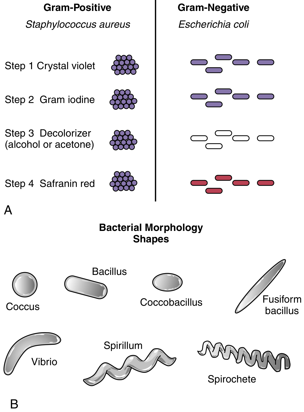
- Thick peptidoglycan layer (20-80 nm): Gram-positive bacteria have a very thick peptidoglycan (murein) cell wall located outside the cytoplasmic membrane. This dense, multilayered network is the fundamental reason for positive staining.
- Crystal violet-iodine complex trapping: Crystal violet enters the cell and Gram iodine (mordant) causes it to precipitate into a large, insoluble crystal violet-iodine (CV-I) complex inside the cell.
- Resistance to decolorization: When acetone-alcohol is applied, it dehydrates and shrinks the thick peptidoglycan, closing the pores and physically trapping the CV-I complex inside. The dye cannot escape.
- No outer membrane: Gram-positive bacteria lack an outer lipid membrane, so there is no lipid layer for the decolorizer to dissolve. The crystal violet remains locked in.
- End result: The cells appear purple/violet even after the safranin counterstain is applied (safranin is masked by the retained crystal violet).
Why Bacteria Are Gram-Negative (Pink/Red)
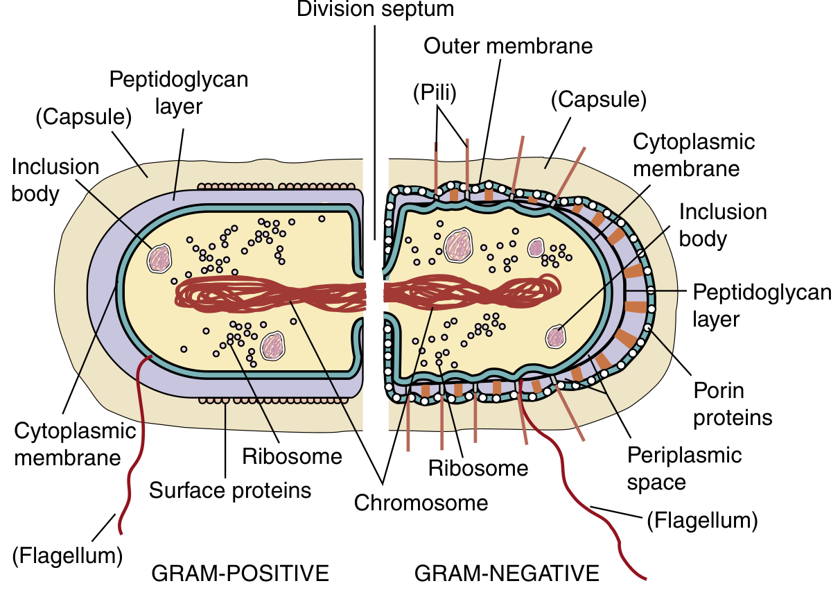
- Thin peptidoglycan layer (2-7 nm): Gram-negative bacteria have a much thinner peptidoglycan layer located in the periplasmic space between the cytoplasmic membrane and the outer membrane. This thin layer holds very little CV-I complex.
- Outer membrane (lipopolysaccharide layer): Gram-negative bacteria have an additional outer membrane made of lipopolysaccharide (LPS), phospholipids, and proteins. This outer membrane initially helps dye entry but is the key to decolorization.
- Outer membrane is dissolved by decolorizer: The acetone-alcohol decolorizer is a lipid solvent. It dissolves the lipid-rich outer membrane and disrupts the thin peptidoglycan underneath, creating large gaps/pores through which the CV-I complex easily washes out.
- Crystal violet is lost: Because the peptidoglycan layer is so thin and the outer membrane is gone, the CV-I complex is not trapped and is washed away by the decolorizer. The cells appear colorless.
- Counterstain uptake: Safranin (pink/red) is then taken up by the now-decolorized cells.
- End result: The cells appear pink/red.
- Outer membrane with LPS (endotoxin)
- Periplasmic space between outer membrane and cytoplasmic membrane
- Porin proteins embedded in the outer membrane for selective permeability
Why Bacteria Are Acid-Fast
- Mycolic acids in the cell wall: Mycobacteria have an extraordinarily lipid-rich cell wall containing very long-chain, branched fatty acids called mycolic acids (60-90 carbon atoms). These are covalently linked to the peptidoglycan layer via arabinogalactan. Mycolic acids form a highly hydrophobic, waxy, nearly impenetrable barrier.
- Lipoarabinomannan (LAM): The cell wall also contains lipoarabinomannan, a lipoglycan anchored in the inner membrane. Along with mycolic acids and other waxes, LAM contributes to the unique staining and biological properties of mycobacteria.
- Other cell wall waxes and fatty acids: Multiple additional lipids (cord factor, sulfolipids, phthiocerol dimycocerosate) further reinforce the waxy envelope.
- Resistance to normal dyes: Because of this high lipid content, mycobacteria do not stain with ordinary aniline dyes (including the regular Gram stain). They require phenol-based arylmethane dyes (e.g., carbolfuchsin) that can penetrate the waxy wall, usually aided by heat.
- Retention after acid-alcohol decolorization: Once carbolfuchsin penetrates the waxy wall and binds to the mycolic acids, the dye is retained even after exposure to strong acid-alcohol (3% HCl in 95% ethanol) or mineral acid. Normal (non-acid-fast) bacteria would be decolorized. The waxy mycolic acid layer physically prevents the acid-alcohol from removing the dye.
- End result: Acid-fast organisms appear bright red/pink (carbolfuchsin is retained) against a blue counterstain (methylene blue).
- Slow growth (nutrients pass slowly through the waxy envelope)
- Resistance to detergents and many common antibiotics
- Resistance to phagocytic killing (survives inside macrophages)
- Causes chronic granulomatous disease (e.g., tuberculosis, leprosy)
Summary Comparison
| Property | Gram-Positive | Gram-Negative | Acid-Fast |
|---|---|---|---|
| Peptidoglycan | Thick (20-80 nm) | Thin (2-7 nm) | Present but thin |
| Outer membrane | Absent | Present (LPS) | Absent |
| Mycolic acids | Absent | Absent | Present (waxy layer) |
| Decolorizer effect | Pores close, dye retained | Outer membrane dissolved, dye lost | Acid-alcohol cannot remove dye |
| Gram stain result | Purple | Pink | Does not stain well |
| Special stain needed | No | No | Yes - Ziehl-Neelsen |
| Example organisms | Staph, Strep | E. coli, Pseudomonas | M. tuberculosis, M. leprae |
Why is counter stain used?
Why a Counterstain is Used
1. Making Decolorized Bacteria Visible
2. Creating Color Contrast for Differentiation
| Stain | Gram-Positive | Gram-Negative |
|---|---|---|
| Gram stain (no counterstain) | Purple | Colorless - invisible |
| Gram stain (with safranin counterstain) | Purple | Pink/Red |
3. Staining Background Structures
- Assess the quality of the specimen (e.g., neutrophils that stain pink confirm proper decolorization)
- Locate bacteria relative to host cells (e.g., intracellular vs. extracellular organisms)
- Identify the type of specimen
The Same Principle in Acid-Fast Staining
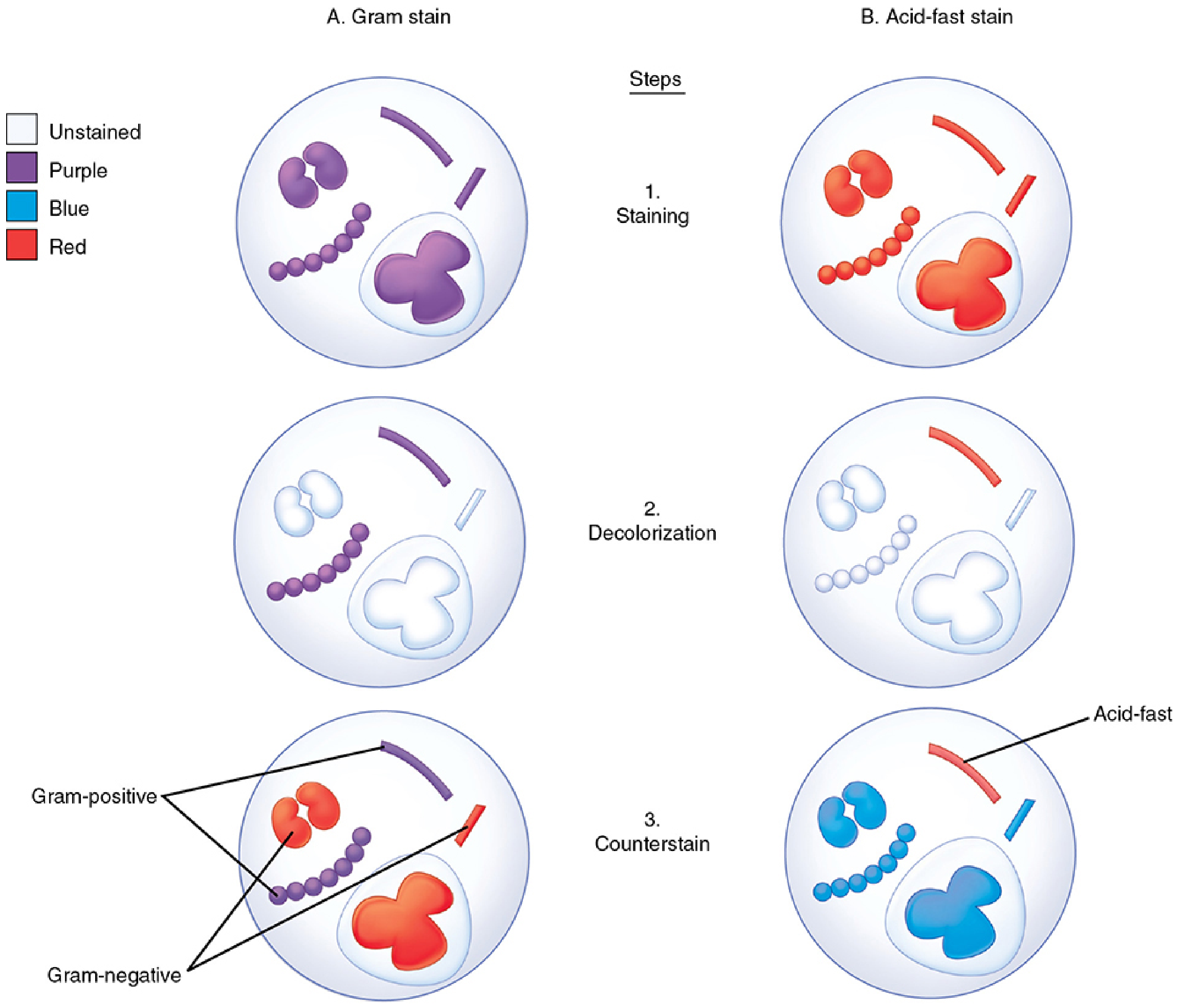
- After decolorization with acid-alcohol, only the acid-fast bacillus retains the red carbolfuchsin
- Everything else (non-acid-fast bacteria, white blood cells, debris) is colorless
- Methylene blue is applied as the counterstain, turning all non-acid-fast organisms and background structures blue
- The acid-fast organism now stands out as a bright red rod against a blue background - impossible to miss
In Short - Three Roles of a Counterstain
- Visibility - Makes decolorized (colorless) organisms visible
- Differentiation - Contrasting color separates two groups of organisms instantly
- Background staining - Colors host cells/debris to assess specimen quality and locate organisms in context