
Dandy walker syndrome
Reading File
Dandy-Walker syndrome is a congenital malformation of the posterior fossa characterized by cerebellar vermis hypoplasia/agenesis, cystic dilation of the fourth ventricle, and enlargement of the posterior fossa. It often presents with hydrocephalus and developmental delay.
Searching sources...
Finding Sources
Searching PubMed
Dandy-Walker syndrome
Good - I have 4 textbook sections. Let me read them all in parallel for maximum coverage.
Reading File
Reading File
Reading File
Reading File
Good content gathered. Let me also fetch the MRI image and search for more clinical details (genetics, associated anomalies, management).
Loading Image
Finding Sources
Let me get the surgical/management details from Bailey & Love and the maternal-fetal medicine definition.
Reading File
Reading File
Excellent - let me get the rest of the associated anomalies and prognosis section.
Reading File
Reading File
Loading Image
I now have comprehensive information from multiple authoritative textbooks. Let me compile the full answer.
Dandy-Walker Syndrome (DWS)
Definition and Spectrum
Dandy-Walker malformation (DWM) describes a spectrum of cystic posterior fossa malformations. The classic (most severe) form has three cardinal features:
- Cystic dilation of the fourth ventricle (ballooning of the posterior half of the fourth ventricle, often with failure of patency of the foramen of Magendie)
- Aplasia or hypoplasia of the cerebellar vermis (with vermian rotation and splaying of the cerebellar hemispheres)
- Enlargement of the posterior fossa with elevation of the tentorium, torcular herophili, and straight sinus above the lambdoid suture
- Bradley and Daroff's Neurology in Clinical Practice
- Creasy & Resnik's Maternal-Fetal Medicine
The Posterior Fossa Spectrum (Mildest to Severest)
| Entity | Key Feature |
|---|---|
| Mega cisterna magna | Normal cerebellum + fourth ventricle; infracerebellar CSF expansion; incidental, no significance |
| Blake pouch cyst | Failure of foramen of Magendie to perforate; normal vermis; obstructive hydrocephalus may develop |
| DW Variant | Partial vermian hypoplasia, mild posterior fossa enlargement |
| Classic DWM | Full triad: fourth ventricle cyst, vermis aplasia, posterior fossa enlargement |
Posterior fossa arachnoid cysts are not in continuity with the fourth ventricle and are distinguished by mass effect and absence of vermian dysplasia.
- Grainger & Allison's Diagnostic Radiology
Epidemiology and Pathogenesis
-
Incidence: 1 in 30,000 births
-
Accounts for 4-12% of all infantile hydrocephaly
-
Results from developmental arrest in the hindbrain between 4-6 weeks of gestation
-
Various incomplete forms (DW variants) are described; classification remains debated
-
Creasy & Resnik's Maternal-Fetal Medicine
Clinical Presentation
Infants typically present with:
- Macrocephaly (from progressive hydrocephalus)
- Developmental delay (motor and cognitive)
- Hydrocephalus - present in 70-80% of cases, with progressive postnatal ventriculomegaly
- Seizures
- Signs of cerebellar dysfunction (ataxia, hypotonia)
Neurological handicaps (spastic diplegia, intellectual disability) relate more to associated brain malformations than to the hydrocephalus itself.
- Bailey & Love's Short Practice of Surgery
- Bradley and Daroff's Neurology in Clinical Practice
Key Diagnostic Features (Imaging)
MRI and CT findings:
- Large cisterna magna communicating with the fourth ventricle
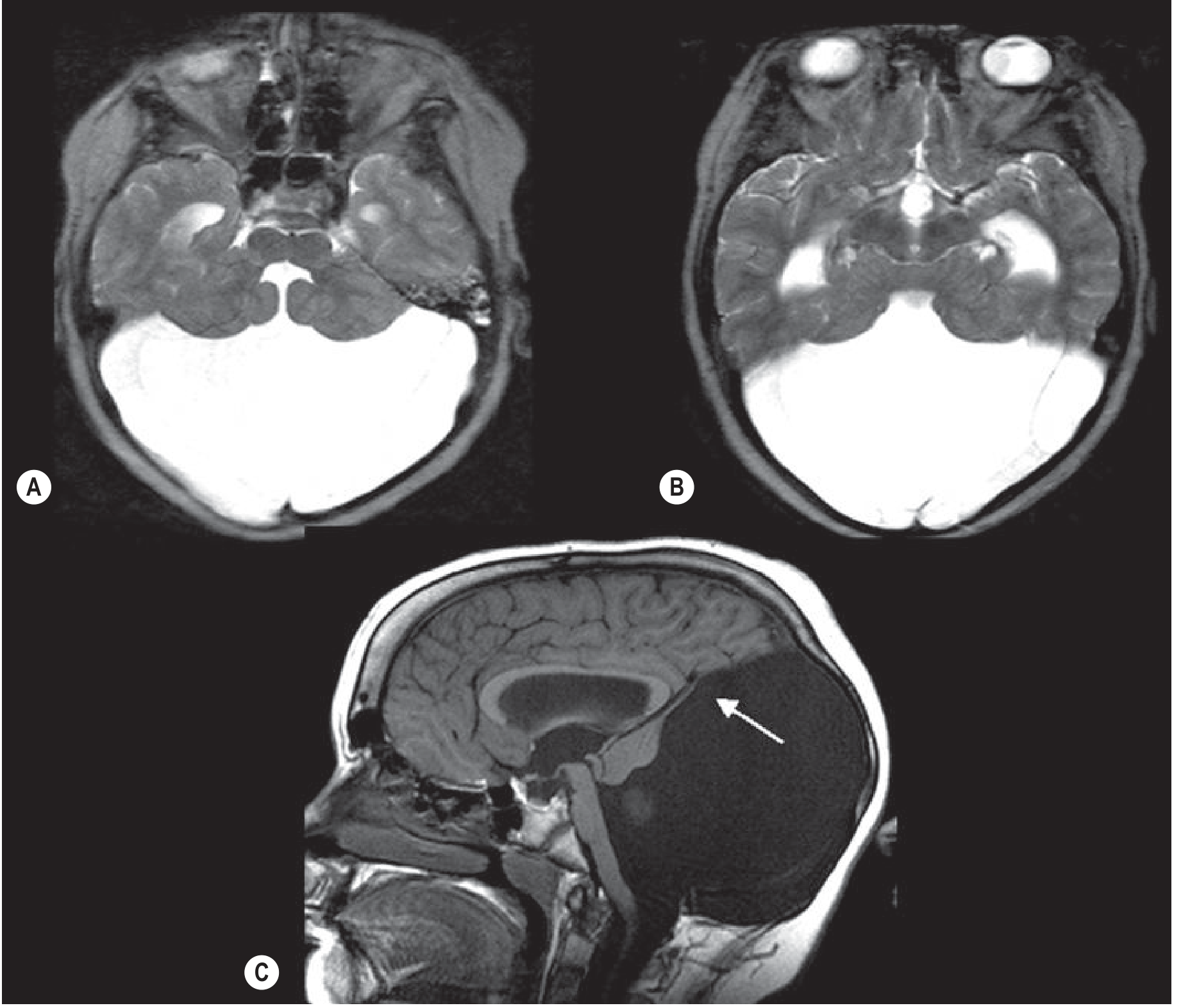
- Absent or hypoplastic vermis
- Cerebellar hemispheres splayed apart
- Elevated tentorium and torcular herophili (torcular-lambdoid inversion - torcular above lambdoid suture, visible on plain X-ray, CT, and MRI)
- Ventriculomegaly / hydrocephalus
The MRI below shows the classic findings - the fourth ventricle opens into a large posterior fossa cyst, the cerebellum is hypoplastic with a thin rim of cerebellar tissue forming the cyst wall (arrow), and the venous structures are elevated:
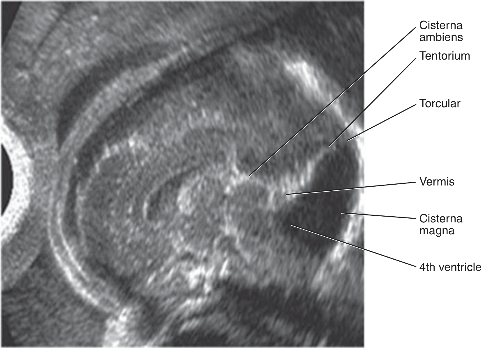
Fetal ultrasound (median view): shows large posterior fossa with elevated tentorium and torcular, communicating fourth ventricle, and hypoplastic upwardly-rotated vermis:
Differential Diagnosis
- Posterior fossa arachnoid cyst (no communication with 4th ventricle)
- Blake pouch cyst
- Mega cisterna magna
- Vermian hypoplasia (isolated)
- Joubert syndrome (molar tooth sign on MRI, ciliopathy)
Associated Anomalies
Found in 50-70% of postnatal cases and ~60% of prenatally diagnosed cases.
CNS anomalies (most common):
- Agenesis or dysgenesis of the corpus callosum
- Holoprosencephaly
- Cephaloceles
- Cortical migrational disorders (pachygyria, heterotopias)
- Inferior olivary heterotopia
Non-CNS anomalies:
- Congenital heart disease
- Craniofacial anomalies
- Renal and limb anomalies
- Abdominal wall defects, diaphragmatic hernia
- Ambiguous genitalia
- Fetal growth restriction
Chromosomal anomalies:
- Trisomy 9, 21, 18, 13
- Triploidy
- 6p and 3q22-q24 deletions
Genetic syndromes:
-
Walker-Warburg syndrome (congenital muscular dystrophy)
-
Joubert syndrome (ciliopathy)
-
Meckel-Gruber syndrome (ciliopathy)
-
Lissencephaly (tubulopathies)
-
Blake pouch cyst: ZIC2 and ZIC5 gene haploinsufficiency (distal 13q deletion)
-
Creasy & Resnik's Maternal-Fetal Medicine
Management
Postnatal
- Shunt placement is the primary treatment for hydrocephalus (ventriculoperitoneal shunt or cyst-peritoneal shunt)
- Multidisciplinary care: neurology, neurosurgery, developmental pediatrics
- Long-term developmental support and rehabilitation
Antenatal (Prenatal Diagnosis)
When identified on fetal ultrasound:
-
Detailed anatomic survey
-
Fetal neurosonography
-
Fetal echocardiography
-
Genetic counseling; karyotype, subtelomeric deletion testing, chromosomal microarray
-
Fetal MRI (to identify associated anomalies more precisely)
-
Consultation with maternal-fetal medicine, neonatology, pediatric neurology
-
Offer termination of pregnancy
-
For ongoing pregnancies: serial scans to monitor ventriculomegaly progression
-
Vaginal delivery is not contraindicated
-
Creasy & Resnik's Maternal-Fetal Medicine; Bailey & Love's Short Practice of Surgery
Prognosis
- If hydrocephalus is treated promptly, prognosis may be good
- Outcome depends heavily on the extent and severity of associated brain malformations, not hydrocephalus alone
- Isolated DWM without associated anomalies carries a better prognosis
- Intellectual disability, spastic diplegia, and cerebellar dysfunction are common long-term sequelae
Recent Evidence (2025)
Two recent systematic reviews are relevant:
- A 2025 systematic review (PMID 41396485) examined DWM with occipital encephalocele - a particularly severe association
- A 2025 meta-analysis (PMID 40785453) on outcomes of isolated fetal posterior fossa anomalies provides updated prognostic data that may refine counseling for prenatally diagnosed cases
Sources: Bradley and Daroff's Neurology in Clinical Practice | Grainger & Allison's Diagnostic Radiology | Creasy & Resnik's Maternal-Fetal Medicine | Bailey & Love's Short Practice of Surgery | Adams and Victor's Principles of Neurology
This is a shared conversation. Sign in to Orris to start your own chat.