
Development of interatrial septum in flow chart with diagrams
Development of the Interatrial Septum
Overview
Flowchart of Development
┌─────────────────────────────────────────────────────────────────────────┐
│          COMMON ATRIUM (single undivided chamber, ~Week 4)              │
└───────────────────────────┬─────────────────────────────────────────────┘
                            │
                            ▼
┌─────────────────────────────────────────────────────────────────────────┐
│  STEP 1: SEPTUM PRIMUM FORMS (~Day 28 / End of Week 4)                  │
│                                                                         │
│  • Sickle-shaped (crescent) crest grows DOWN from roof of atrium        │
│  • Two limbs extend toward endocardial cushions in AV canal             │
│  • Gap between lower rim of septum primum and endocardial              │
│    cushions = OSTIUM PRIMUM (foramen primum)                            │
│  • Allows right-to-left blood flow during fetal life                    │
└───────────────────────────┬─────────────────────────────────────────────┘
                            │
                            ▼
┌─────────────────────────────────────────────────────────────────────────┐
│  STEP 2: OSTIUM PRIMUM CLOSES + OSTIUM SECUNDUM FORMS (~Day 33, 9mm)   │
│                                                                         │
│  • Extensions of superior and inferior endocardial cushions grow        │
│    along the edge of septum primum → OSTIUM PRIMUM CLOSES               │
│  • BEFORE closure is complete:                                          │
│    - Programmed cell death (apoptosis) creates perforations in          │
│      the UPPER portion of septum primum                                 │
│    - Perforations coalesce → OSTIUM SECUNDUM (foramen secundum)         │
│  • Ostium secundum ensures CONTINUED free right-to-left blood flow      │
└───────────────────────────┬─────────────────────────────────────────────┘
                            │
                            ▼
┌─────────────────────────────────────────────────────────────────────────┐
│  STEP 3: SEPTUM SECUNDUM GROWS (~Day 37, 14mm / ~Week 5-6)              │
│                                                                         │
│  • New crescent-shaped fold appears to the RIGHT of septum primum       │
│    as the right atrium expands by incorporating the sinus horn          │
│  • Septum secundum grows downward (like a window shade)                 │
│  • Its anterior limb extends to the AV canal septum                    │
│  • Left venous valve + septum spurium fuse with right side of           │
│    septum secundum                                                      │
│  • NEVER forms a complete partition - leaves a gap                      │
└───────────────────────────┬─────────────────────────────────────────────┘
                            │
                            ▼
┌─────────────────────────────────────────────────────────────────────────┐
│  STEP 4: FORAMEN OVALE ESTABLISHED (Fetal Life)                         │
│                                                                         │
│  • Septum secundum overlaps but does NOT cover the ostium secundum     │
│    completely → opening = FORAMEN OVALE                                 │
│  • Upper part of septum primum gradually disappears                     │
│  • Remaining inferior part of septum primum =                           │
│    VALVE OF THE FORAMEN OVALE                                           │
│  • Blood flows through an obliquely elongated cleft:                    │
│    RIGHT atrium → LEFT atrium (right-to-left shunt)                     │
│  • This is NORMAL in fetal life (bypasses non-functional lungs)         │
└───────────────────────────┬─────────────────────────────────────────────┘
                            │
                            ▼
┌─────────────────────────────────────────────────────────────────────────┐
│  STEP 5: FUNCTIONAL CLOSURE AT BIRTH                                    │
│                                                                         │
│  • Lungs expand → pulmonary vascular resistance falls                   │
│  • Pulmonary blood flow ↑ → left atrial pressure RISES above right     │
│  • Valve of foramen ovale is PRESSED against septum secundum            │
│  • Foramen ovale functionally sealed → no more right-to-left shunt      │
└───────────────────────────┬─────────────────────────────────────────────┘
                            │
                            ▼
┌─────────────────────────────────────────────────────────────────────────┐
│  STEP 6: ANATOMIC CLOSURE (by ~3rd postnatal month)                     │
│                                                                         │
│  • Tissue proliferation + adhesion of septum primum to left margin      │
│    of septum secundum → permanent fusion                                │
│  • Septum primum → floor of OVAL FOSSA (fossa ovalis)                  │
│  • Free edge of septum secundum → border of oval fossa                  │
│    (LIMBUS FOSSAE OVALIS) in adult heart                                │
└─────────────────────────────────────────────────────────────────────────┘
Key Developmental Stages - Diagram
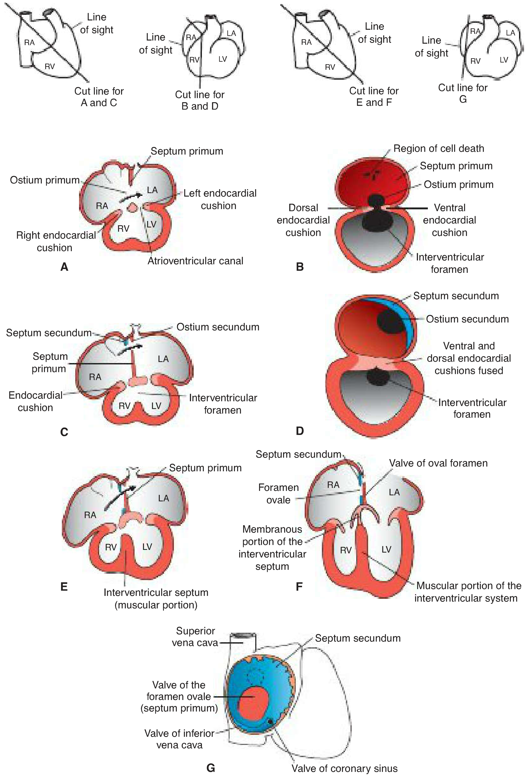
| Panel | Stage | Key Features |
|---|---|---|
| A (30 days, 6mm) | Septum primum growing down | Ostium primum open; endocardial cushions forming |
| B (same stage, right view) | Apoptosis begins in upper septum primum | Region of cell death visible; ostium primum still present |
| C (33 days, 9mm) | Ostium secundum formed; septum secundum begins | Endocardial cushions fused closing ostium primum |
| D (same stage, right view) | Both septa visible | Ostium secundum in septum primum; septum secundum growing |
| E (37 days, 14mm) | Septum secundum overlapping ostium secundum | Foramen ovale established |
| F (Newborn) | Post-birth anatomy | Foramen ovale, membranous IVS, muscular IVS |
| G (Newborn, right view) | Final atrial septum | Septum secundum, valve of foramen ovale (= septum primum), valves of IVC and coronary sinus |
Adult Anatomy Diagram (Normal vs. Patent Foramen Ovale)
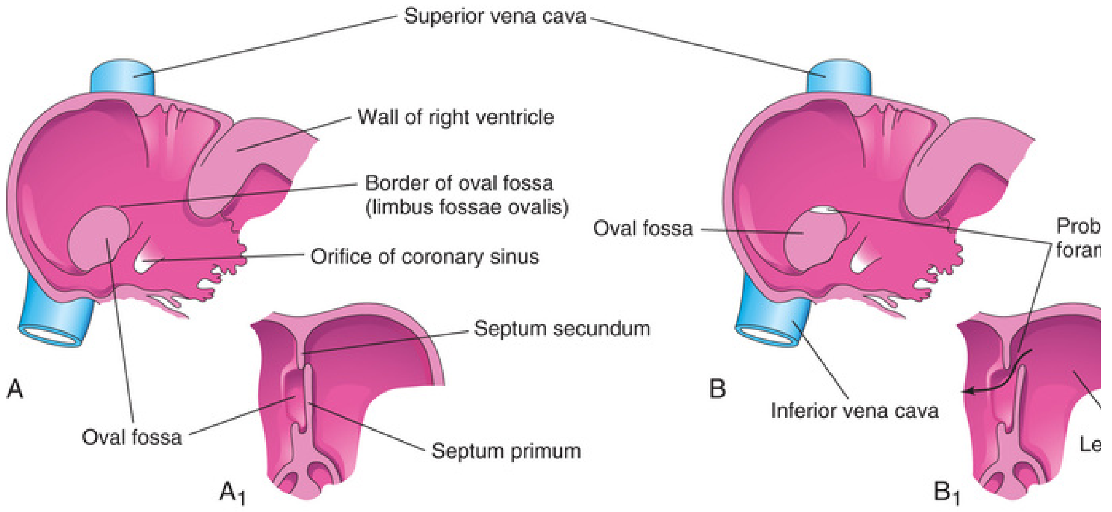
- The oval fossa (fossa ovalis) = depression on right atrial wall
- Its floor = septum primum (thin, fibrous)
- Its border (limbus fossae ovalis) = free edge of septum secundum
Summary Table: Structures and Their Adult Derivatives
| Fetal Structure | Role During Fetal Life | Adult Derivative |
|---|---|---|
| Septum primum | First partition; forms valve of foramen ovale | Floor of fossa ovalis |
| Ostium primum | Transient gap (right-to-left flow) | Obliterated by endocardial cushions |
| Ostium secundum | Formed by apoptosis; maintains right-to-left flow | Covered by septum secundum |
| Septum secundum | Second (thicker) partition growing from right | Border of fossa ovalis (limbus) |
| Foramen ovale | Opening in septum secundum; fetal right-to-left shunt | Fossa ovalis after closure |
| Valve of foramen ovale | Remnant of lower septum primum; opens/closes flap | Fused into floor of fossa ovalis |
Clinical Correlations
Atrial Septal Defects (ASDs)
| Type | % of ASDs | Embryological Basis |
|---|---|---|
| Secundum ASD (most common) | ~90% | Deficient septum secundum formation OR excessive resorption of septum primum near center of atrial septum |
| Primum ASD | ~5% | Failure of endocardial cushions to close ostium primum; often associated with AV valve abnormalities |
| Sinus venosus defect | ~5% | Abnormal absorption of sinus venosus near SVC entrance; may have anomalous pulmonary venous return |
Patent Foramen Ovale (PFO)
- Occurs in ~20% of the population (Langman's)
- Incomplete fusion of septum primum to septum secundum
- Leaves a narrow oblique cleft - a probe can pass between the two septa
- Generally no intracardiac shunting if the opening is small (left atrial pressure keeps it closed in normal adults)
- Clinical significance: potential pathway for paradoxical embolism (cryptogenic stroke)
Key Mnemonic
- Primum appears before Secundum
- Ostium primum closes, then Ostium secundum opens
- Think of it as a relay: each structure hands off to the next before it disappears
Create a 35 slides power point presentation on evaluation, classification and management of show with flow chart and diagrams from latest ESC or ACC guidelines
What is the topic for the 35-slide presentation? 'Show' appears to be a typo - which condition did you mean?
ESC guidelines circulatory shock 2024 2025 cardiogenic septic classification management
https://www.inovanewsroom.org/wp-content/uploads/2025/06/Sin…
mkdir -p /home/daytona/workspace/shock-pptx && ls /home/daytona/workspace/shock-pptx/
Create a 35 slides power point presentation on evaluation, classification and management of show with flow chart and diagrams from latest ESC or ACC guidelines
ls /home/daytona/workspace/shock-pptx/ 2>/dev/null || mkdir -p /home/daytona/workspace/shock-pptx && echo "ready"
Create a 35 slides power point presentation on evaluation, classification and management of show with flow chart and diagrams from latest ESC or ACC guidelines
mkdir -p /home/daytona/workspace/shock-pptx && node -e "require('pptxgenjs'); console.log('pptxgenjs OK')"