
Prepare me seminar for topic cholecystitis including these points 1. Intro 2.Causee 3.examinations 4.majorly incl Lab findings and investigation 5.differential diagnosis 6.treatment
cholecystitis gallbladder inflammation ultrasound diagram
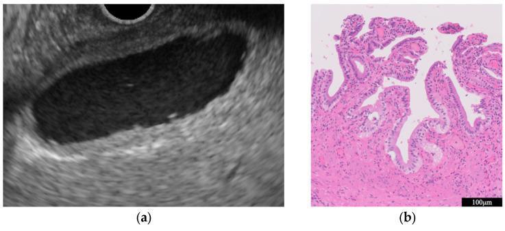
This composite image illustrates the clinical findings of chronic cholecystitis through two modalities: endoscopic ultrasound (EUS) and histopathology. Panel (a) is an ultrasound image of the gallbladder showing diagnostic features of chronic inflammation, characterized by uniform, circumferential thickening of the gallbladder wall while maintaining its layered structural integrity. The lumen is anechoic and free of significant masses. Panel (b) provides a histopathological correlation using Hematoxylin and Eosin (H&E) staining at a 100µm scale. It reveals a dense inflammatory cell infiltrate within the subepithelial layers, predominantly composed of lymphocytes. The gallbladder epithelium appears preserved and benign, specifically lacking atypical or dysplastic cells. This comparison is educationally significant for differentiating benign chronic inflammatory wall thickening from gallbladder carcinoma, where the wall structure is typically disrupted and cellular atypia is present.
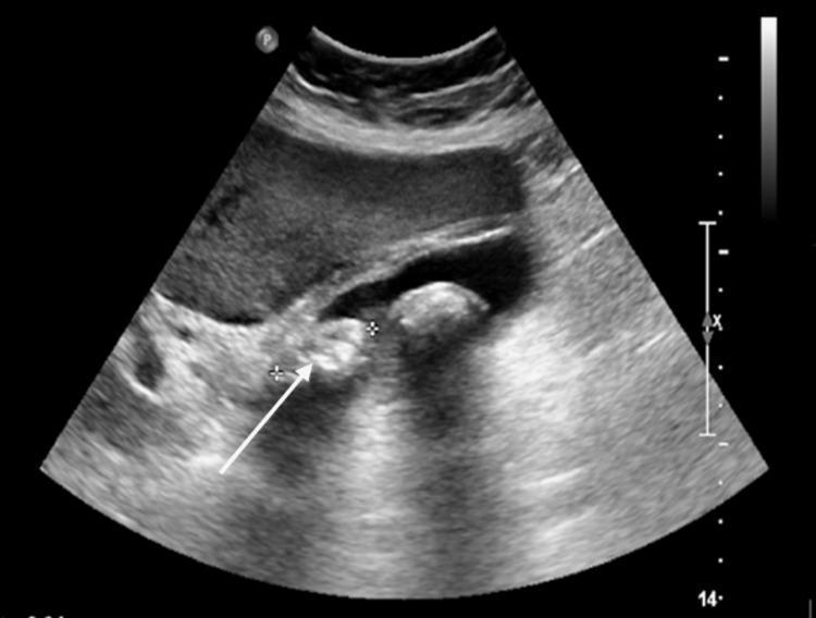
This abdominal ultrasound image displays diagnostic features characteristic of acute cholecystitis. The primary finding is a large, hyperechoic gallstone (indicated by a white arrow) situated within the gallbladder lumen, exhibiting distinct posterior acoustic shadowing. The gallbladder wall is significantly thickened and edematous, appearing as a multi-layered, hypoechoic border around the organ. A thin, anechoic layer of pericholecystic fluid is visible adjacent to the gallbladder wall, further indicating localized inflammation. Clinical correlation provided in the context notes a positive sonographic Murphy's sign, which is an essential bedside diagnostic indicator of gallbladder inflammation. This image is an educational example for radiology and emergency medicine residents to identify the classic triad of gallstones, wall thickening, and pericholecystic fluid in the assessment of right upper quadrant pain.
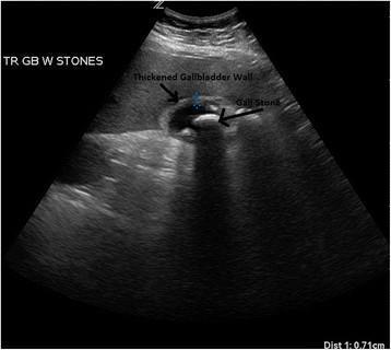
This diagnostic ultrasound image in the transverse plane demonstrates a gallbladder with classic sonographic findings of acute cholecystitis and cholelithiasis. A large, prominent hyperechoic gallstone is visible within the lumen of the gallbladder, exhibiting a characteristic posterior acoustic shadow that extends inferiorly, indicating the dense nature of the calculus. The gallbladder wall is clearly labeled and appears significantly thickened (measuring 0.71 cm as per the distal measurement marker), which is a primary diagnostic criterion for inflammation. Additionally, there is evidence of pericholecystic edema, visualized as a hypoechoic, hazy area surrounding the outer wall of the gallbladder. These features—cholelithiasis, wall thickening, and pericholecystic fluid/edema—collectively support a diagnosis of acute cholecystitis. The image serves as an educational tool for identifying the sonographic hallmarks of biliary disease in a clinical setting.
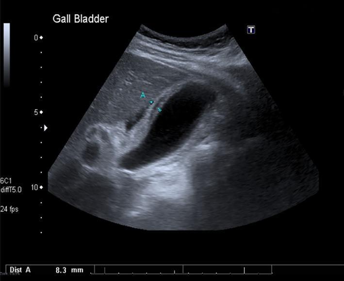
Diagnostic ultrasound of the gallbladder in the right upper quadrant demonstrating signs of acute acalculous cholecystitis. The gallbladder wall is markedly thickened and edematous, showing a striated or layered appearance. An electronic caliper ('Dist A') measures the wall thickness at 8.3 mm, which is significantly above the standard clinical threshold for normalcy (typically <3 mm). The gallbladder lumen is entirely anechoic, indicating clear fluid without evidence of intraluminal cholelithiasis (gallstones), biliary sludge, or shadowing echoes. Pericholecystic fluid is subtly visible adjacent to the wall, suggesting localized inflammation. The surrounding liver parenchyma appears relatively homogeneous. This imaging is characteristic of acalculous cholecystitis, a condition frequently associated with systemic illness or critical care scenarios where gallbladder inflammation occurs in the absence of obstructing stones.
gallstones cholelithiasis gallbladder anatomy pathology
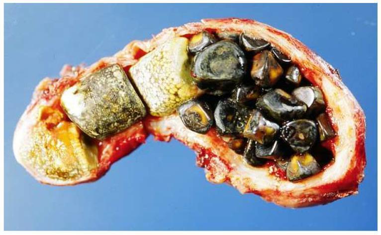
This clinical photograph shows a gross pathology specimen of a human gallbladder that has been surgically incised and opened longitudinally to reveal an internal cavity densely packed with cholelithiasis (gallstones). The gallbladder wall appears thickened and erythematous, consistent with chronic cholecystitis or biliary stasis. The stones present a polymorphic appearance: a subset of larger, faceted, yellowish-tan stones likely composed primarily of cholesterol are visible on the left, while the right portion of the lumen is filled with numerous smaller, dark brown to black, ovoid pigment stones containing bilirubin. The variety in morphology and coloration suggests a mixed-composition stone profile. This image illustrates severe cholelithiasis, demonstrating how calculi can completely fill the gallbladder lumen, potentially leading to obstruction, inflammation, and infection. This visual is suitable for medical education regarding gastrointestinal pathology and surgical anatomy of the biliary system.
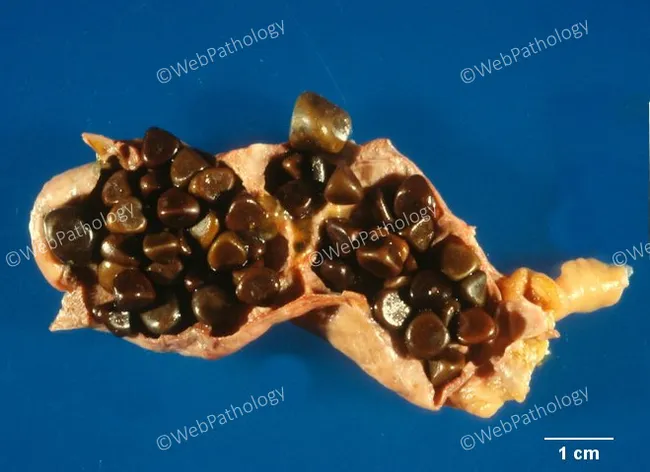
Gross pathology photograph of a gallbladder specimen with cholelithiasis. Modality and technique: Macroscopic imaging of an excised gallbladder, with its lumen largely filled by numerous dark, hard stones. The stones are black to dark brown, variably sized from a few millimeters up to about one centimeter, with smooth to slightly faceted surfaces. The surrounding gallbladder wall appears tan-pink with focal mucosal distortion, consistent with chronic inflammation. The image demonstrates classic features of black pigment gallstones formed primarily from calcium bilirubinate in settings of chronic hemolysis, where accelerated bilirubin turnover promotes pigment precipitation. Surface characteristics are variable; some stones are glossy, others dull. The specimen is presented in longitudinal section showing stones occupying the lumen and adherent to mucosa. The composition is expected to be calcium bilirubinate-enriched pigment; cholesterol is minimal. Radiodensity on gross imaging is not appreciable. Clinically, pigment stones in hemolytic disease are associated with increased bilirubin load, risk of biliary colic, cholecystitis, and possible bile duct obstruction from stone migration. This image supports medical education, pathology training, differential diagnosis of cholelithiasis etiologies, and radiology correlation in hepatobiliary disease, including hemolysis-related pigment stone formation and surgical pathology reporting for teaching.
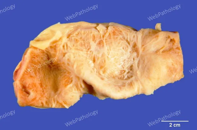
Gross pathology photograph of an excised gallbladder illustrating chronic calculous cholecystitis with gallstone obstruction. Ex vivo specimen photographed with scale; the wall shows marked hypertrophy of the muscularis propria and fibrous thickening consistent with long-standing inflammation. The mucosa is relatively inconspicuous beneath the thickened muscular layer. Gallstones are present within the lumen, contributing to increased intraluminal pressure and episodic biliary stasis. The external surface may appear pale to tan with irregular contour owing to fibrotic remodeling. This image highlights the pathophysiology of chronic obstruction: progressive mural hypertrophy, submucosal fibrosis, and potential porcelain gallbladder phenotype if calcification is extensive. The specimen represents biliary tract disease related to cholelithiasis and chronic cholecystitis, clarifying how stone burden correlates with wall remodeling observed in radiologic and gross examinations. Clinically this finding correlates with long-standing biliary colic, right upper quadrant discomfort, and risk of gallbladder complications such as empyema or carcinoma in chronic disease. This photograph is educational for pathology training, surgical planning, and radiology-pathology correlation. Suitable for teaching gross anatomy, disease progression, and specimen processing workflow, including fixation, sectioning, and paraffin embedding for subsequent histology. The scale bar (2 cm) in the image provides quantitative context for wall thickness and lumen caliber assessment precisely.
CHOLECYSTITIS - Seminar Presentation
1. INTRODUCTION
Types
| Type | Description |
|---|---|
| Acute Calculous Cholecystitis | ~90% of cases; caused by gallstone impaction at the cystic duct |
| Acute Acalculous Cholecystitis | ~10% of cases; occurs without gallstones, predominantly in critically ill patients |
| Chronic Cholecystitis | Longstanding gallstone disease causing repeated low-grade inflammation and fibrosis |
| Complicated Forms | Gangrenous, emphysematous, hemorrhagic, and perforated cholecystitis |
Epidemiology
- Gallstone disease affects approximately 10-15% of adults in Western countries
- Acute calculous cholecystitis is a disease predominantly of young, otherwise healthy women
- Risk factors (the "5 Fs"): Female, Fat, Fertile, Forty, Fair
- Acalculous cholecystitis carries significantly higher morbidity and mortality and occurs in the elderly, critically ill, and post-surgical patients
2. CAUSES (ETIOLOGY & PATHOPHYSIOLOGY)
A. Calculous Cholecystitis (Gallstone-Related)
- Cholesterol stones (75-80%): form when bile becomes supersaturated with cholesterol, which then crystallizes. Associated with obesity, female sex, pregnancy, rapid weight loss, and fibrates.
- Pigment stones (black): form from excess bilirubin, seen in hemolytic anemias (sickle cell disease, hereditary spherocytosis)
- Pigment stones (brown): associated with biliary infections and parasitic infestations
- A gallstone becomes impacted in the cystic duct or gallbladder neck
- Persistent obstruction causes bile stasis within the gallbladder lumen
- Stasis leads to mucosal damage and release of intracellular enzymes
- Activation of inflammatory cascade - prostaglandins, phospholipase A release lysolecithin from lecithin
- Edema, vascular congestion, and subserosal hemorrhage of the gallbladder wall develop
- Secondary bacterial infection (E. coli, Klebsiella, Enterococcus) may follow
- Continued obstruction can lead to ischemia, necrosis, gangrene, or perforation
B. Acalculous Cholecystitis
- Prolonged fasting / total parenteral nutrition (TPN)
- Major surgery, trauma, burns
- Mechanical ventilation / critical illness
- Sepsis, multi-organ failure
- Diabetes mellitus
- Vasculitis, medications
C. Chronic Cholecystitis
3. CLINICAL EXAMINATION
History
| Feature | Detail |
|---|---|
| Pain | Right upper quadrant (RUQ) or epigastric; constant, severe, lasting >4-6 hours (distinguishes from biliary colic which is <6 hours) |
| Radiation | To right shoulder or right scapular region (diaphragmatic irritation via phrenic nerve) |
| Onset | Often following a fatty meal; may wake patient from sleep |
| Associated symptoms | Nausea, vomiting, anorexia, low-grade fever |
| History of prior episodes | Often elicited - previous shorter, milder, self-resolving attacks of biliary colic |
Physical Examination
- Fever (typically low-grade, 38-38.5°C)
- Patient appears uncomfortable, lying still (unlike renal colic where patient is restless)
- Mild jaundice may be visible (sclerae first, frenulum of tongue)
- RUQ tenderness to palpation - the most common physical finding
- Guarding and rigidity over RUQ in severe cases
- An inflammatory mass (Hartmann pouch or Calot's triangle inflammation) may be palpable
Murphy's Sign - Inspiratory arrest when the examiner's fingers are pressed beneath the right costal margin while the patient takes a deep breath. The descending diaphragm forces the inflamed gallbladder against the examiner's fingers, causing sharp pain and inspiratory arrest. It has a positive predictive value of 92% when gallstones are also present on US.
- Gangrenous cholecystitis (denervated wall)
- Diabetic patients (autonomic neuropathy)
- Elderly patients
- Prior administration of analgesics
- Empyema / perforation (temporarily relieves pressure on stretch receptors)
Severity Grading - Tokyo Guidelines (2018)
| Grade | Criteria |
|---|---|
| Grade I (Mild) | No organ dysfunction, mild local inflammation |
| Grade II (Moderate) | WBC >18,000/mm³; palpable tender RUQ mass; duration >72 hours; marked local inflammation |
| Grade III (Severe) | Organ dysfunction (cardiovascular, neurological, respiratory, renal, hepatic, hematological) |
4. LAB FINDINGS & INVESTIGATIONS
A. Laboratory Findings
1. Complete Blood Count (CBC)
- Leukocytosis - hallmark finding; WBC elevation reflects systemic inflammation
- WBC >18,000/mm³ = Grade II (moderate) cholecystitis per Tokyo Guidelines
- WBC >15,000/mm³ with high fever, chills = suspect empyema or perforation
- Neutrophil predominance
- Elevated CRP (C-Reactive Protein) - correlates with severity
2. Liver Function Tests (LFTs) / Hepatic Function Panel
| Parameter | Finding | Significance |
|---|---|---|
| Serum Bilirubin | Mildly elevated (<3 mg/dL) in severe cholecystitis | Pericholecystic inflammation compressing ducts; significant elevation suggests choledocholithiasis or Mirizzi syndrome |
| Alkaline Phosphatase (ALP) | Elevated | Cholestatic pattern - bile duct obstruction |
| ALT / AST (Transaminases) | Mildly elevated | Primarily cholestatic pattern in cholecystitis; marked elevation suggests hepatocellular injury or CBD obstruction |
| GGT (Gamma-Glutamyl Transferase) | Elevated; rises early and persists | Sensitivity 90%, specificity 85% for choledocholithiasis |
Key distinction: In uncomplicated biliary colic, LFTs are usually normal. Significant bilirubin/ALP elevation implies obstruction of the common bile duct (CBD).
3. Serum Amylase and Lipase
- Amylase may be mildly elevated in severe cholecystitis (not specific for pancreatitis)
- Markedly elevated amylase/lipase suggests coexisting gallstone pancreatitis
4. Urine Analysis
- Bilirubin in urine (bilirubinuria) in obstructive jaundice
- Urinalysis also used to rule out pyelonephritis and renal calculi in the differential
5. Blood Cultures
- Indicated when fever >38.5°C, rigors, or sepsis suspected
- Common organisms: E. coli, Klebsiella, Enterococcus, Bacteroides spp.
B. Imaging Investigations
1. Transabdominal Ultrasound (US) - FIRST LINE
| Feature | Diagnostic Significance |
|---|---|
| Gallstones (hyperechoic with acoustic shadowing) | Present in ~90% of acute cholecystitis |
| Gallbladder wall thickening (>3-4 mm) | Primary criterion for inflammation |
| Pericholecystic fluid | Inflammation/edema around gallbladder |
| Sonographic Murphy's sign | PPV >90% for acute cholecystitis with gallstones |
| Gallbladder distension (>8 cm length, >4 cm width) | Suggests obstruction |
| Color Doppler: hyperemia of wall | Active inflammation |
- Sensitivity 81-85%, Specificity 83-95% for acute cholecystitis
- Sensitivity >95% for detecting gallstones
- Negative predictive value of 95% when no stones and no Murphy's sign
2. HIDA Scan (Hepatobiliary Iminodiacetic Acid / Cholescintigraphy) - SECOND LINE
- Most sensitive test for confirming acute cholecystitis
- Principle: Radiolabeled tracer is taken up by hepatocytes and excreted into bile
- Normal result: Radiotracer visualizes gallbladder, bile duct, and small intestine within 30-60 minutes - virtually excludes cholecystitis
- Abnormal result (non-filling of gallbladder): Confirms cystic duct obstruction - diagnosis of cholecystitis
- Sensitivity ~94%, Specificity ~94%
- False positives occur in: prolonged fasting, TPN, liver disease, alcohol use
- Best used as secondary study when ultrasound is inconclusive
3. CT Abdomen (with IV contrast)
- Indicated when US is inconclusive or complications are suspected
- Best modality for detecting complications:
- Emphysematous cholecystitis (gas in gallbladder wall or lumen)
- Gallbladder perforation and abscess formation
- Mirizzi syndrome
- Also excludes other intra-abdominal pathologies (pancreatitis, appendicitis, bowel obstruction)
- CT findings: wall thickening, pericholecystic stranding, intraluminal stones, intramural gas
4. MRI / MRCP (Magnetic Resonance Cholangiopancreatography)
- Highly sensitive for cholecystitis
- Gold standard for detecting choledocholithiasis (CBD stones)
- Reserved for cases where:
- CBD stone is suspected from labs (elevated bilirubin + ALP)
- Need to rule out biliary malignancy
- CT/US inconclusive
- More expensive and less expeditious than US/CT
5. Plain Abdominal X-Ray (AXR)
- Limited diagnostic value in cholecystitis
- Only 15-20% of gallstones are radio-opaque (calcium-containing pigment stones)
- Role is mainly to exclude other diagnoses: pneumoperitoneum (perforated viscus), bowel obstruction, right lower lobe pneumonia
- Rigler's triad (pneumobilia + bowel obstruction + RLQ stone) = pathognomonic for gallstone ileus
6. Endoscopic Ultrasound (EUS)
- Useful when tranabdominal US is inconclusive
- Emerging role in EUS-guided drainage for acute cholecystitis in high-risk surgical patients
- Highly sensitive for small stones and CBD stones
| Modality | 1st/2nd Line | Best For |
|---|---|---|
| US | 1st line | Initial diagnosis, gallstones, wall thickening |
| HIDA Scan | 2nd line | Confirming cystic duct obstruction |
| CT Abdomen | 2nd line | Complications, unclear diagnosis |
| MRCP | 3rd line | CBD stones, malignancy exclusion |
| AXR | Limited | Excluding pneumoperitoneum |
5. DIFFERENTIAL DIAGNOSIS
| Condition | Key Distinguishing Features |
|---|---|
| Biliary Colic | Pain <6 hours, no fever, no leukocytosis, normal LFTs; resolves spontaneously |
| Acute Pancreatitis | Epigastric pain radiating to back; elevated amylase/lipase; tenderness more diffuse; CT shows pancreatic inflammation |
| Appendicitis | Pain migrates to RLQ (McBurney's point); nausea/vomiting; no biliary history; US/CT confirms appendix inflammation |
| Choledocholithiasis | CBD stone causing obstructive jaundice; marked bilirubin + ALP elevation; MRCP confirms CBD stone |
| Acute Cholangitis | Charcot's triad (fever + jaundice + RUQ pain); associated with CBD obstruction; more systemic illness |
| Peptic Ulcer Disease | Epigastric pain, history of NSAIDs/H. pylori, may have free air on AXR (perforation); endoscopy confirms |
| Acute Hepatitis | Diffuse hepatomegaly; markedly elevated ALT/AST; viral serology positive |
| Pyelonephritis / Renal Colic | Costovertebral angle tenderness; urinalysis shows pyuria/hematuria; CT urogram confirms |
| Right Lower Lobe Pneumonia | Pleuritic chest pain, cough, fever; reduced breath sounds; CXR confirms infiltrate |
| Hepatic Abscess or Tumor | Gradual onset, weight loss, hepatomegaly; CT/MRI defines lesion |
| Gonococcal / Chlamydial Perihepatitis (Fitz-Hugh-Curtis syndrome) | Young women; RUQ pain; history of PID; violin-string adhesions on laparoscopy |
| Myocardial Infarction (Inferior) | RUQ pain atypically; ECG changes; elevated troponin - always consider in high-risk patients |
| Xanthogranulomatous Cholecystitis | Rare; grayish-yellow nodules in gallbladder wall (lipid-laden macrophages); may mimic malignancy |
6. TREATMENT
A. Initial Management (All Patients)
- Hospital admission and NPO (nothing by mouth)
- IV fluid resuscitation - correct dehydration and electrolyte imbalance
- Nasogastric tube - if persistent vomiting
- Analgesia:
- NSAIDs (diclofenac, ketorolac) - reduce prostaglandin-mediated inflammation; also provide analgesia
- Opioids (morphine, meperidine) - for severe pain; concern about sphincter of Oddi spasm with morphine is not clinically significant at standard doses
- Antiemetics (ondansetron, metoclopramide)
B. Antibiotics
- Indicated for moderate to severe cholecystitis, fever, leukocytosis, elderly, immunocompromised, diabetic
- Cover gram-negative enteric organisms and anaerobes:
- 1st line: Piperacillin-tazobactam, ampicillin-sulbactam
- Alternatives: Ceftriaxone + metronidazole, or fluoroquinolone + metronidazole
- Severe/sepsis: Meropenem or imipenem
C. Definitive Treatment - SURGICAL
Laparoscopic Cholecystectomy (Lap Chol) - GOLD STANDARD
- Ideal treatment for acute cholecystitis is cholecystectomy
- Early laparoscopic cholecystectomy (within 24-72 hours of diagnosis) is preferred over delayed/interval cholecystectomy:
- Shorter hospital stay
- Less morbidity
- Avoids risk of recurrent attacks while waiting
- More recent evidence strongly supports early surgery
- Advantages over open: smaller incisions, less pain, faster recovery, fewer complications
Open Cholecystectomy
- Reserved for cases where laparoscopic approach fails (conversion)
- Complicated anatomy, dense adhesions, bleeding, bile duct injury
- Historically was the standard of care
Timing Based on Severity (Tokyo Guidelines):
| Grade | Treatment Approach |
|---|---|
| Grade I (Mild) | Early laparoscopic cholecystectomy (within 72 hours) |
| Grade II (Moderate) | Early lap chol when safely possible; if high surgical risk, percutaneous cholecystostomy, then interval chol |
| Grade III (Severe) | Stabilize with ICU care + antibiotics; percutaneous cholecystostomy first; delayed cholecystectomy after stabilization |
D. Non-Operative Options (High-Risk / Non-Surgical Candidates)
-
Percutaneous Cholecystostomy (PC-tube):
- Image-guided percutaneous drainage of gallbladder contents
- Indicated when patient is too high-risk for surgery
- Provides temporary decompression; definitive surgery planned later
-
EUS-Guided Transmural Drainage (emerging therapy):
- Endoscopic drainage of gallbladder via stomach or duodenum using stent
- Used in poor surgical candidates
- Increasingly popular for acalculous and calculous cholecystitis in ICU patients
-
ERCP (Endoscopic Retrograde Cholangiopancreatography):
- Not directly for cholecystitis itself
- Used to remove CBD stones if choledocholithiasis coexists
- Sphincterotomy + stone extraction before cholecystectomy
E. Management of Specific Complications
| Complication | Management |
|---|---|
| Empyema of gallbladder | Urgent cholecystectomy or cholecystostomy; broad-spectrum antibiotics |
| Gangrenous cholecystitis | Urgent surgery; high risk of perforation |
| Emphysematous cholecystitis | Emergency surgery; gas-forming organisms (Clostridium, E. coli); diabetics at risk |
| Gallbladder perforation | Emergency surgery + peritoneal lavage; high mortality |
| Mirizzi syndrome | ERCP + stenting or surgery depending on type |
| Gallstone ileus | Surgery - enterotomy to remove stone; cholecystectomy at same or later sitting |
F. Chronic / Recurrent Cholecystitis
- Elective laparoscopic cholecystectomy is the treatment of choice
- Medical dissolution with ursodeoxycholic acid (UDCA): reserved for non-operative candidates with small, uncalcified cholesterol stones; high recurrence rate on stopping
- Extracorporeal Shock Wave Lithotripsy (ESWL): largely obsolete for biliary stones
SUMMARY TABLE
| Aspect | Key Points |
|---|---|
| Definition | Inflammation of gallbladder, usually from cystic duct obstruction by gallstone |
| Cause | 90% calculous; 10% acalculous (critically ill patients) |
| Classic Presentation | RUQ pain >4-6 hrs, fever, nausea/vomiting, positive Murphy's sign |
| Key Lab Findings | Leukocytosis, elevated ALP/bilirubin (mild), elevated GGT |
| Best Initial Imaging | Transabdominal Ultrasound |
| Confirmatory Test | HIDA Scan (cholescintigraphy) |
| Gold Standard Tx | Early laparoscopic cholecystectomy (within 24-72 hrs) |
| Key Differentials | Pancreatitis, appendicitis, cholangitis, peptic ulcer, pyelonephritis |
Can u consise it to imp points and convert it to pdf form
ls -la /tmp/workspace/ 2>/dev/null || mkdir -p /tmp/workspace && echo "created"
mkdir -p /tmp/workspace/cholecystitis && cat > /tmp/workspace/cholecystitis/generate_pdf.py << 'PYEOF' from reportlab.lib.pagesizes import A4 from reportlab.lib.styles import getSampleStyleSheet, ParagraphStyle from reportlab.lib.units import cm from reportlab.lib import colors from reportlab.platypus import (SimpleDocTemplate, Paragraph, Spacer, Table, TableStyle, HRFlowable, KeepTogether) from reportlab.lib.enums import TA_CENTER, TA_LEFT, TA_JUSTIFY # ── Document setup ────────────────────────────────────────────────────────── doc = SimpleDocTemplate( "/tmp/workspace/cholecystitis/Cholecystitis_Seminar.pdf", pagesize=A4, leftMargin=1.8*cm, rightMargin=1.8*cm, topMargin=2*cm, bottomMargin=2*cm ) W = A4[0] - 3.6*cm # usable width # ── Colour palette ─────────────────────────────────────────────────────────── DARK_BLUE = colors.HexColor("#1B3A6B") MID_BLUE = colors.HexColor("#2E6DA4") LIGHT_BLUE = colors.HexColor("#D6E8F7") ACCENT = colors.HexColor("#E8F4FD") WHITE = colors.white RED_WARN = colors.HexColor("#C0392B") GOLD = colors.HexColor("#F39C12") LIGHT_GREY = colors.HexColor("#F4F6F9") DARK_GREY = colors.HexColor("#2C3E50") # ── Styles ─────────────────────────────────────────────────────────────────── styles = getSampleStyleSheet() title_style = ParagraphStyle("Title", fontName="Helvetica-Bold", fontSize=22, textColor=WHITE, alignment=TA_CENTER, spaceAfter=4) subtitle_style = ParagraphStyle("Subtitle", fontName="Helvetica", fontSize=11, textColor=colors.HexColor("#BDD9F2"), alignment=TA_CENTER) sec_style = ParagraphStyle("Section", fontName="Helvetica-Bold", fontSize=13, textColor=WHITE, alignment=TA_LEFT, leftPadding=8, spaceAfter=2) body = ParagraphStyle("Body", fontName="Helvetica", fontSize=9.5, textColor=DARK_GREY, leading=14, spaceAfter=4, alignment=TA_JUSTIFY) bullet_style = ParagraphStyle("Bullet", fontName="Helvetica", fontSize=9.5, textColor=DARK_GREY, leading=13, leftIndent=14, firstLineIndent=-10, spaceAfter=2) bold_body = ParagraphStyle("BoldBody", fontName="Helvetica-Bold", fontSize=9.5, textColor=DARK_BLUE, leading=13, spaceAfter=2) small_note = ParagraphStyle("Note", fontName="Helvetica-Oblique", fontSize=8.5, textColor=colors.HexColor("#5D6D7E"), leading=12, spaceAfter=3, alignment=TA_JUSTIFY) footer_style = ParagraphStyle("Footer", fontName="Helvetica", fontSize=7.5, textColor=colors.HexColor("#7F8C8D"), alignment=TA_CENTER) # ── Helper: section header banner ──────────────────────────────────────────── def section_header(num, title): label = f" {num} {title}" tbl = Table([[Paragraph(label, sec_style)]], colWidths=[W]) tbl.setStyle(TableStyle([ ("BACKGROUND", (0,0), (-1,-1), DARK_BLUE), ("ROUNDEDCORNERS", [4]), ("TOPPADDING", (0,0), (-1,-1), 5), ("BOTTOMPADDING", (0,0), (-1,-1), 5), ])) return tbl # ── Helper: sub-section title ───────────────────────────────────────────────── def sub_header(text): return Paragraph(f"<b><font color='#2E6DA4'>{text}</font></b>", body) # ── Helper: bullet ──────────────────────────────────────────────────────────── def b(text): return Paragraph(f"• {text}", bullet_style) # ── Helper: two-column info table ───────────────────────────────────────────── def info_table(rows, col_widths=None, header_row=None): if col_widths is None: col_widths = [W*0.38, W*0.62] data = [] if header_row: data.append([Paragraph(f"<b>{h}</b>", bold_body) for h in header_row]) for r in rows: data.append([Paragraph(str(c), body) for c in r]) style = [ ("BACKGROUND", (0,0), (-1,0), LIGHT_BLUE if header_row else ACCENT), ("GRID", (0,0), (-1,-1), 0.4, colors.HexColor("#BDC3C7")), ("ROWBACKGROUNDS", (0, 1 if header_row else 0), (-1,-1), [WHITE, LIGHT_GREY]), ("TOPPADDING", (0,0), (-1,-1), 4), ("BOTTOMPADDING", (0,0), (-1,-1), 4), ("LEFTPADDING", (0,0), (-1,-1), 6), ("RIGHTPADDING", (0,0), (-1,-1), 6), ("FONTNAME", (0,0), (-1,0), "Helvetica-Bold"), ("FONTSIZE", (0,0), (-1,-1), 9), ("VALIGN", (0,0), (-1,-1), "TOP"), ] t = Table(data, colWidths=col_widths) t.setStyle(TableStyle(style)) return t def sp(n=6): return Spacer(1, n) def hr(): return HRFlowable(width="100%", thickness=0.5, color=colors.HexColor("#D5D8DC"), spaceAfter=4) # ═══════════════════════════════════════════════════════════════════════════════ # TITLE BLOCK # ═══════════════════════════════════════════════════════════════════════════════ def title_block(): title_tbl = Table([ [Paragraph("CHOLECYSTITIS", title_style)], [Paragraph("A Concise Clinical Seminar", subtitle_style)], [Paragraph("Sabiston Surgery | Sleisenger & Fordtran GI | Tintinalli EM | Tokyo Guidelines 2018", subtitle_style)], ], colWidths=[W]) title_tbl.setStyle(TableStyle([ ("BACKGROUND", (0,0), (-1,-1), DARK_BLUE), ("TOPPADDING", (0,0), (-1,0), 14), ("BOTTOMPADDING", (0,-1), (-1,-1), 14), ("TOPPADDING", (0,1), (-1,2), 3), ("BOTTOMPADDING", (0,0), (-1,1), 3), ("ROUNDEDCORNERS", [6]), ])) return title_tbl # ═══════════════════════════════════════════════════════════════════════════════ # BUILD CONTENT # ═══════════════════════════════════════════════════════════════════════════════ story = [] story.append(title_block()) story.append(sp(12)) # ────────────────────────────────────────────────────────────────────────────── # 1. INTRODUCTION # ────────────────────────────────────────────────────────────────────────────── story.append(section_header("1", "INTRODUCTION")) story.append(sp(5)) story.append(Paragraph( "<b>Cholecystitis</b> is inflammation of the gallbladder, most commonly from obstruction of the " "cystic duct by a gallstone. It is the most frequent complication of gallstone disease and a " "leading cause of acute abdominal pain.", body)) story.append(sp(5)) types_data = [ ["Acute Calculous", "~90% of cases; gallstone impacted at cystic duct / neck"], ["Acute Acalculous", "~10%; no stones; critically ill, ICU, post-surgical patients; higher mortality"], ["Chronic Cholecystitis", "Recurrent attacks or mechanical irritation → fibrosis, wall thickening"], ["Complicated Forms", "Gangrenous, emphysematous, haemorrhagic, perforated cholecystitis"], ] story.append(info_table(types_data, col_widths=[W*0.30, W*0.70], header_row=["Type", "Key Feature"])) story.append(sp(4)) story.append(Paragraph( "<b>Epidemiology:</b> Affects 10-15% of Western adults. Calculous type = young healthy women " "(5 Fs: <b>F</b>emale, <b>F</b>at, <b>F</b>ertile, <b>F</b>orty, <b>F</b>air). " "Acalculous type = elderly/critically ill, high morbidity.", body)) story.append(sp(10)) # ────────────────────────────────────────────────────────────────────────────── # 2. CAUSES # ────────────────────────────────────────────────────────────────────────────── story.append(section_header("2", "CAUSES & PATHOPHYSIOLOGY")) story.append(sp(5)) story.append(sub_header("A. Gallstone Types")) gs_data = [ ["Cholesterol stones (75-80%)", "Bile supersaturated with cholesterol → crystallisation. Associated with obesity, female sex, pregnancy, rapid weight loss, fibrates"], ["Pigment (Black) stones", "Excess bilirubin; haemolytic anaemias (sickle cell, spherocytosis)"], ["Pigment (Brown) stones", "Biliary stasis + infection; parasitic infestations (Ascaris, Clonorchis)"], ] story.append(info_table(gs_data, col_widths=[W*0.32, W*0.68])) story.append(sp(5)) story.append(sub_header("B. Pathophysiology of Acute Calculous Cholecystitis")) for step in [ "Gallstone impacted in cystic duct or gallbladder neck", "Bile stasis → mucosal damage → release of intracellular enzymes", "Inflammatory cascade: prostaglandins, phospholipase A → lysolecithin", "Oedema, vascular congestion, subserosal haemorrhage of gallbladder wall", "Secondary bacterial infection: E. coli, Klebsiella, Enterococcus, Bacteroides", "Continued obstruction → ischaemia, necrosis, gangrene, perforation", ]: story.append(b(step)) story.append(sp(5)) story.append(sub_header("C. Acalculous Cholecystitis — Risk Factors")) risk_cols = [ ["Prolonged fasting / TPN", "Major surgery or trauma"], ["Burns, mechanical ventilation", "Sepsis, multi-organ failure"], ["Diabetes mellitus", "Vasculitis / medications"], ] for row in risk_cols: story.append(Paragraph(f"• {row[0]} • {row[1]}", bullet_style)) story.append(sp(10)) # ────────────────────────────────────────────────────────────────────────────── # 3. CLINICAL EXAMINATION # ────────────────────────────────────────────────────────────────────────────── story.append(section_header("3", "CLINICAL EXAMINATION")) story.append(sp(5)) story.append(sub_header("History")) hx_data = [ ["Pain", "RUQ / epigastric; constant; >4-6 hrs (biliary colic <6 hrs, self-resolving)"], ["Radiation", "Right shoulder / right scapula (diaphragmatic irritation via phrenic nerve)"], ["Onset", "Often after fatty meal; may wake patient from sleep"], ["Associated Sx", "Nausea, vomiting, anorexia, low-grade fever"], ["Prior episodes", "History of shorter, milder, self-resolving attacks (biliary colic)"], ] story.append(info_table(hx_data, col_widths=[W*0.22, W*0.78])) story.append(sp(6)) story.append(sub_header("Physical Examination")) for pt in [ "Fever (38–38.5°C); patient lies still (vs. renal colic = restless)", "<b>Murphy's Sign</b> — inspiratory arrest on palpation of RUQ under right costal margin. PPV >92% when gallstones also present on US", "RUQ tenderness — most common finding; guarding and rigidity in severe cases", "Mild jaundice (sclera first at bilirubin >2.5 mg/dL; skin at >5 mg/dL)", "Palpable inflammatory mass (Hartmann's pouch or Calot's triangle region)", ]: story.append(b(pt)) story.append(sp(4)) story.append(Paragraph( "<b>Murphy's sign may be absent in:</b> gangrenous cholecystitis (denervated wall), " "diabetes (autonomic neuropathy), elderly patients, prior analgesic use, perforation.", small_note)) story.append(sp(6)) story.append(sub_header("Severity Grading — Tokyo Guidelines 2018")) tg_data = [ ["Grade I (Mild)", "No organ dysfunction; mild local inflammation; no systemic signs"], ["Grade II (Moderate)", "WBC >18,000/mm³; palpable RUQ mass; duration >72 hrs; marked local inflammation"], ["Grade III (Severe)", "Organ dysfunction — cardiovascular, neurological, respiratory, renal, hepatic, or haematological"], ] story.append(info_table(tg_data, col_widths=[W*0.28, W*0.72])) story.append(sp(10)) # ────────────────────────────────────────────────────────────────────────────── # 4. LAB FINDINGS & INVESTIGATIONS # ────────────────────────────────────────────────────────────────────────────── story.append(section_header("4", "LAB FINDINGS & INVESTIGATIONS")) story.append(sp(5)) story.append(sub_header("A. Laboratory Findings")) lab_data = [ ["CBC — WBC", "Leukocytosis (neutrophilia); WBC >18,000 = Grade II; WBC >15,000 + high fever = suspect empyema / perforation"], ["CRP", "Elevated; correlates with severity and complications"], ["Serum Bilirubin", "Mildly elevated (<3 mg/dL) in severe cholecystitis. Marked elevation → suspect CBD stone (choledocholithiasis) or Mirizzi syndrome"], ["ALP (Alkaline Phosphatase)", "Elevated in cholestatic pattern. Disproportionate rise relative to transaminases"], ["ALT / AST", "Mildly elevated; marked elevation → hepatocellular injury or CBD obstruction"], ["GGT", "Elevated; sensitivity 90%, specificity 85% for choledocholithiasis; rises early, persists longer"], ["Amylase / Lipase", "Markedly elevated → coexisting gallstone pancreatitis. Mild amylase rise can occur in severe cholecystitis alone"], ["Blood cultures", "Indicated with fever >38.5°C, rigors, sepsis. Organisms: E. coli, Klebsiella, Enterococcus, Bacteroides"], ["Urinalysis", "Exclude pyelonephritis and renal calculi; bilirubinuria in obstructive jaundice"], ] story.append(info_table(lab_data, col_widths=[W*0.28, W*0.72], header_row=["Test", "Key Finding / Significance"])) story.append(sp(4)) story.append(Paragraph( "<b>Note:</b> In uncomplicated biliary colic all LFTs are <i>normal</i>. Significant bilirubin / ALP " "elevation implies CBD obstruction.", small_note)) story.append(sp(7)) story.append(sub_header("B. Imaging Investigations")) img_data = [ ["Ultrasound (US)\n1st LINE", "Sensitivity 81-85%, Specificity 83-95%\n• Gallstones: hyperechoic + posterior acoustic shadow\n• Wall thickening >3-4 mm\n• Pericholecystic fluid\n• Sonographic Murphy's sign (PPV >90%)\n• Gallbladder distension (>8 cm length)\n• Colour Doppler: wall hyperaemia"], ["HIDA Scan\n2nd LINE", "Most sensitive overall (~94% sensitivity & specificity)\n• Non-filling of gallbladder = cystic duct obstruction = cholecystitis confirmed\n• Normal filling virtually excludes cholecystitis\n• Used when US inconclusive; false +ve in fasting, TPN, liver disease"], ["CT Abdomen\n2nd LINE", "Best for complications:\n• Emphysematous cholecystitis (intramural gas)\n• Gallbladder perforation + abscess\n• Mirizzi syndrome\nAlso excludes: pancreatitis, appendicitis, bowel obstruction"], ["MRCP\n3rd LINE", "Gold standard for CBD stones; excludes biliary malignancy\nReserved when: elevated bilirubin + ALP, CT/US inconclusive"], ["Plain X-Ray\nLimited role", "Only 15-20% gallstones radio-opaque\nUse: exclude pneumoperitoneum, bowel obstruction, chest pneumonia\nRigler's triad = pathognomonic for gallstone ileus"], ] img_tbl = Table( [[Paragraph(f"<b>{r[0]}</b>", bold_body), Paragraph(r[1].replace("\n", "<br/>"), body)] for r in img_data], colWidths=[W*0.22, W*0.78] ) img_tbl.setStyle(TableStyle([ ("BACKGROUND", (0,0), (0,-1), LIGHT_BLUE), ("BACKGROUND", (1,0), (1,-1), WHITE), ("ROWBACKGROUNDS", (1,0), (1,-1), [WHITE, LIGHT_GREY]), ("GRID", (0,0), (-1,-1), 0.4, colors.HexColor("#BDC3C7")), ("VALIGN", (0,0), (-1,-1), "TOP"), ("TOPPADDING", (0,0), (-1,-1), 5), ("BOTTOMPADDING", (0,0), (-1,-1), 5), ("LEFTPADDING", (0,0), (-1,-1), 6), ("RIGHTPADDING", (0,0), (-1,-1), 6), ("FONTSIZE", (0,0), (-1,-1), 9), ])) story.append(img_tbl) story.append(sp(10)) # ────────────────────────────────────────────────────────────────────────────── # 5. DIFFERENTIAL DIAGNOSIS # ────────────────────────────────────────────────────────────────────────────── story.append(section_header("5", "DIFFERENTIAL DIAGNOSIS")) story.append(sp(5)) dd_data = [ ["Biliary Colic", "Pain <6 hrs; no fever; no leukocytosis; normal LFTs; self-resolves"], ["Acute Pancreatitis", "Epigastric → back radiation; markedly elevated amylase/lipase; CT confirms"], ["Appendicitis", "Migratory pain to RLQ (McBurney's); no biliary history; US/CT confirms"], ["Choledocholithiasis", "Obstructive jaundice; markedly elevated bilirubin + ALP; MRCP confirms CBD stone"], ["Acute Cholangitis", "Charcot's triad (RUQ pain + fever + jaundice); Reynolds' pentad adds shock + confusion"], ["Peptic Ulcer / Perforation", "Epigastric; NSAID/H. pylori history; free air on AXR if perforated"], ["Acute Hepatitis", "Diffuse hepatomegaly; markedly elevated ALT/AST; viral serology +ve"], ["Pyelonephritis / Renal Colic", "Costovertebral angle tenderness; urinalysis: pyuria/haematuria; CT urogram"], ["Right Lower Lobe Pneumonia", "Pleuritic chest pain, cough; reduced breath sounds; CXR infiltrate"], ["Hepatic Abscess / Tumour", "Gradual onset; weight loss; CT/MRI defines lesion"], ["Fitz-Hugh-Curtis Syndrome", "Young women; PID history; RUQ pain; 'violin-string' adhesions on laparoscopy"], ["Inferior MI", "Atypical RUQ pain; ECG changes + elevated troponin — always consider in high-risk"], ] story.append(info_table(dd_data, col_widths=[W*0.32, W*0.68], header_row=["Condition", "Key Distinguishing Features"])) story.append(sp(10)) # ────────────────────────────────────────────────────────────────────────────── # 6. TREATMENT # ────────────────────────────────────────────────────────────────────────────── story.append(section_header("6", "TREATMENT")) story.append(sp(5)) story.append(sub_header("A. Initial / Supportive Management (All Patients)")) for pt in [ "Hospital admission + NPO (nil by mouth)", "IV fluid resuscitation — correct dehydration and electrolytes", "Analgesia: NSAIDs (diclofenac/ketorolac) ± opioids (morphine/meperidine) for severe pain", "Antiemetics: ondansetron / metoclopramide", "Nasogastric tube if persistent vomiting", ]: story.append(b(pt)) story.append(sp(5)) story.append(sub_header("B. Antibiotics")) abx_data = [ ["Indication", "Moderate-severe disease, fever, leukocytosis, elderly, diabetic, immunocompromised"], ["1st Line", "Piperacillin-tazobactam OR Ampicillin-sulbactam"], ["Alternatives", "Ceftriaxone + Metronidazole OR Fluoroquinolone + Metronidazole"], ["Severe / Sepsis", "Meropenem or Imipenem (carbapenem cover)"], ["Target organisms", "E. coli, Klebsiella, Enterococcus, Bacteroides spp."], ] story.append(info_table(abx_data, col_widths=[W*0.28, W*0.72])) story.append(sp(6)) story.append(sub_header("C. Definitive Treatment — Surgery")) story.append(Paragraph( "<b>Laparoscopic Cholecystectomy = GOLD STANDARD</b>. Early surgery (within 24-72 hrs of diagnosis) " "is preferred over delayed/interval approach — shorter stay, lower morbidity, avoids recurrent attacks.", body)) story.append(sp(4)) sev_data = [ ["Grade I (Mild)", "Early laparoscopic cholecystectomy within 72 hours"], ["Grade II (Moderate)", "Early lap chol when safely possible; if high surgical risk → percutaneous cholecystostomy then interval cholecystectomy"], ["Grade III (Severe)", "ICU stabilisation + broad-spectrum antibiotics → percutaneous cholecystostomy first → delayed cholecystectomy after stabilisation"], ] story.append(info_table(sev_data, col_widths=[W*0.26, W*0.74], header_row=["Severity", "Surgical Approach"])) story.append(sp(6)) story.append(sub_header("D. Non-Operative Options (High-Risk / Non-Surgical Candidates)")) for pt in [ "<b>Percutaneous Cholecystostomy (PC-tube):</b> Image-guided drainage of gallbladder; temporary decompression; definitive surgery later", "<b>EUS-Guided Transmural Drainage:</b> Endoscopic drainage via stomach or duodenum using stent; growing role in ICU patients", "<b>ERCP:</b> Not for cholecystitis itself — used to remove coexisting CBD stones before cholecystectomy", "<b>Ursodeoxycholic Acid (UDCA):</b> Medical dissolution of small uncalcified cholesterol stones; non-operative candidates only; high recurrence on stopping", ]: story.append(b(pt)) story.append(sp(6)) story.append(sub_header("E. Management of Complications")) comp_data = [ ["Empyema of gallbladder", "Urgent cholecystectomy or cholecystostomy + broad-spectrum antibiotics"], ["Gangrenous cholecystitis", "Urgent surgery; high perforation risk; Murphy's sign +ve in only 33%"], ["Emphysematous cholecystitis", "Emergency surgery; gas-forming organisms (Clostridium, E. coli); diabetics at highest risk"], ["Gallbladder perforation", "Emergency surgery + peritoneal lavage; high mortality"], ["Mirizzi syndrome", "ERCP + stenting or surgery depending on type"], ["Gallstone ileus", "Surgery: enterotomy to remove stone; cholecystectomy same or delayed sitting"], ] story.append(info_table(comp_data, col_widths=[W*0.32, W*0.68], header_row=["Complication", "Management"])) story.append(sp(12)) # ────────────────────────────────────────────────────────────────────────────── # SUMMARY TABLE # ────────────────────────────────────────────────────────────────────────────── summary_banner = Table([[Paragraph(" QUICK SUMMARY", sec_style)]], colWidths=[W]) summary_banner.setStyle(TableStyle([ ("BACKGROUND", (0,0), (-1,-1), MID_BLUE), ("TOPPADDING", (0,0), (-1,-1), 5), ("BOTTOMPADDING", (0,0), (-1,-1), 5), ("ROUNDEDCORNERS", [4]), ])) story.append(summary_banner) story.append(sp(5)) summary_data = [ ["Definition", "Gallbladder inflammation; mostly cystic duct obstruction by gallstone"], ["Cause", "90% calculous | 10% acalculous (critically ill)"], ["Classic Presentation", "RUQ pain >4-6 hrs, fever, nausea/vomiting, +ve Murphy's sign"], ["Key Lab Findings", "Leukocytosis | mild ALP/bilirubin rise | elevated GGT | blood cultures if septic"], ["Best Initial Imaging", "Transabdominal Ultrasound (sensitivity 81-85%, specificity 83-95%)"], ["Confirmatory Test", "HIDA Scan — non-filling confirms cystic duct obstruction"], ["Gold Standard Tx", "Early laparoscopic cholecystectomy (within 24-72 hours)"], ["Key Differentials", "Pancreatitis, appendicitis, cholangitis, peptic ulcer, pyelonephritis, inferior MI"], ] story.append(info_table(summary_data, col_widths=[W*0.30, W*0.70])) story.append(sp(8)) story.append(hr()) story.append(Paragraph( "Sources: Sabiston Textbook of Surgery | Sleisenger & Fordtran's GI and Liver Disease | " "Yamada's Textbook of Gastroenterology (7th ed.) | Tintinalli's Emergency Medicine | " "Robbins Pathologic Basis of Disease | Tokyo Guidelines 2018", footer_style)) # ── Build ───────────────────────────────────────────────────────────────────── doc.build(story) print("PDF generated successfully.") PYEOF python /tmp/workspace/cholecystitis/generate_pdf.py
Cholecystitis Seminar
PDF Document · PDF
| Section | What's in it |
|---|---|
| 1. Introduction | Types (calculous, acalculous, chronic, complicated) + epidemiology & 5 Fs |
| 2. Causes | Gallstone types, step-by-step pathophysiology, acalculous risk factors |
| 3. Examination | History table, Murphy's sign, when it's absent, Tokyo severity grading |
| 4. Lab & Investigations | Full lab table (WBC, LFTs, GGT, amylase, cultures, urine) + imaging table (US, HIDA, CT, MRCP, AXR) with sensitivity/specificity |
| 5. Differential Diagnosis | 12 conditions with key distinguishing features |
| 6. Treatment | Supportive care, antibiotics (first-line + alternatives), surgery by Tokyo grade, non-operative options, complications management |
| Summary | One-page quick-reference table at the end |